

Review
doi: 10.1016/j.drudis.2007.09.009.
Epub 2007 Oct 30.
Kinase packing defects as drug targets
Affiliations
- PMID: 17993409
- PMCID: PMC2136416
- DOI: 10.1016/j.drudis.2007.09.009
Item in Clipboard
Review
Kinase packing defects as drug targets
Drug Discov Today.
2007 Nov.
Erratum in
- Drug Discov Today. 2014 Apr;19(4):520
Abstract
Protein kinases constitute major targets in molecular cancer therapy. The structural conservation of kinases causes specificity problems in most drug inhibitors, often resulting in dangerous side effects. Here we survey recent approaches in drug design that exploit a molecular marker for specificity: the pattern of packing defects. These packing defects are solvent-exposed intramolecular hydrogen bonds that may be protected by drugs upon association. In this light, we review design strategies to achieve paralogue discrimination, to control cross reactivity and to overcome drug resistance induced by target mutations.
Figures
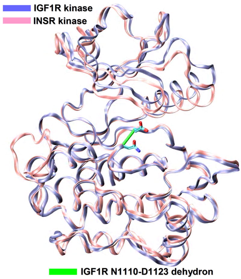
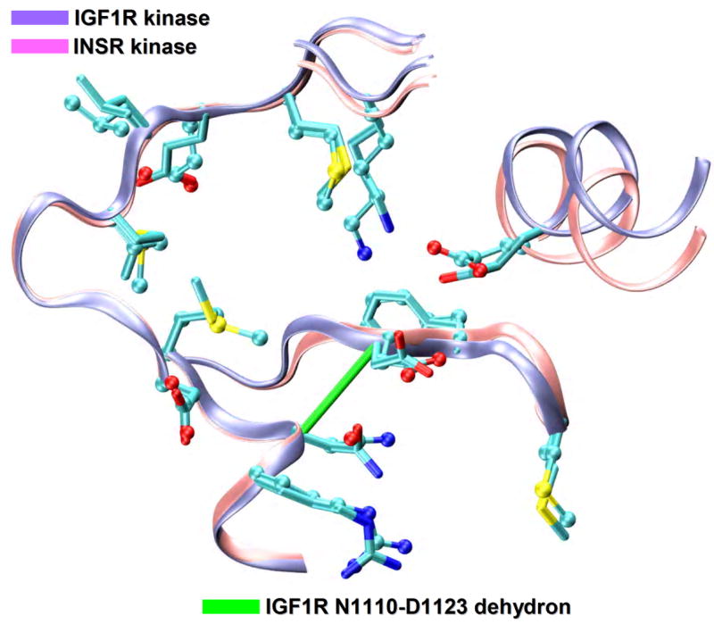
Specificity problems in kinase inhibition. Structural alignment (ribbon representation) of the IGF1R kinase (PDB.1K3A, ice blue), a proposed cancer target and the INSR kinase (PDB.1GAG, pink), a related target to be avoided. The degree of structural similarity between the two paralogs is staggeringly high (RMSD=1.2Å) as expected from the 80% sequence identity. The IGF1R nonconserved N1110-D1123 dehydron is also shown as a green virtual bond joining α-carbons (ρ=18 nonpolar groups for desolvation radius of 6.2Å).
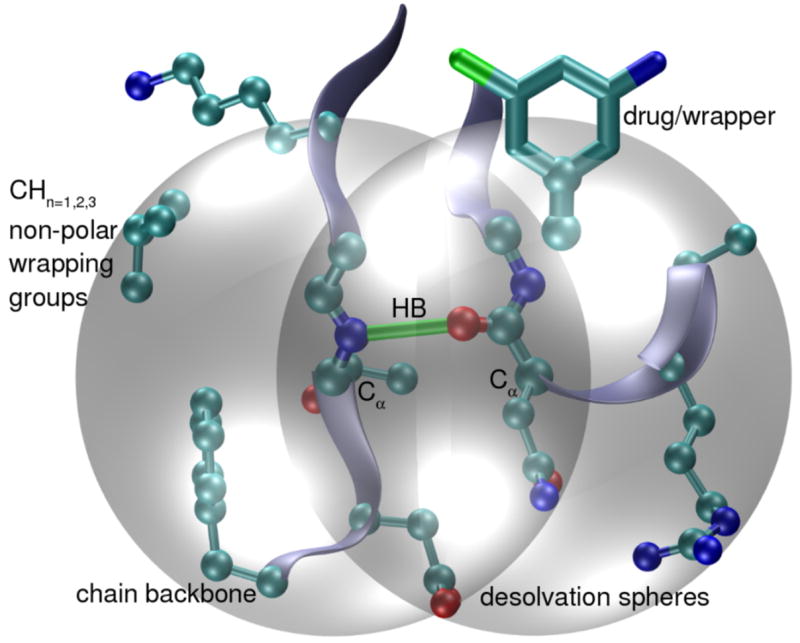
Dehydron concept. Intramolecular backbone hydrogen bonds in target proteins (green bond) prevail only if protected from water attack. Their extent of intramolecular wrapping, ρ, is defined by counting the nonpolar groups (carbonaceous atoms, cyan) contained within the desolvation domain (grey spheres). Poorly wrapped hydrogen bonds (dehydrons) are defined with 19 or fewer hydrophobic groups in their desolvation domains (desolvation radius of 6.2Å). These packing defects consist of solvent-exposed backbone hydrogen bonds and are targetable features because of their inherent stickiness, since they promote the removal of surrounding water molecules as a mean to strengthen and stabilize the underlying electrostatic interaction.
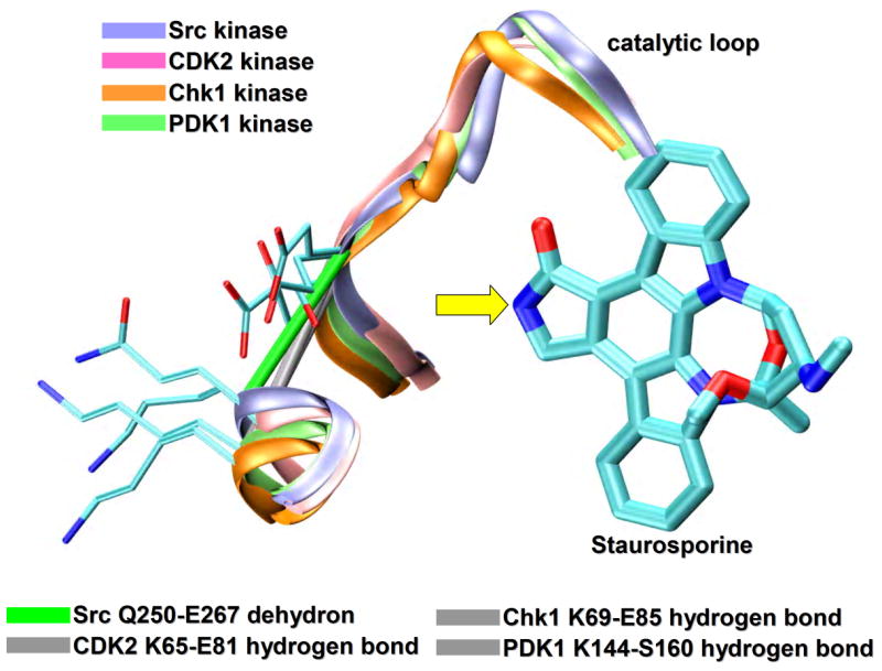
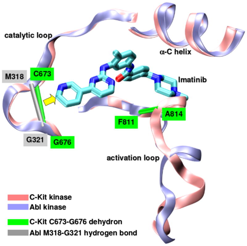
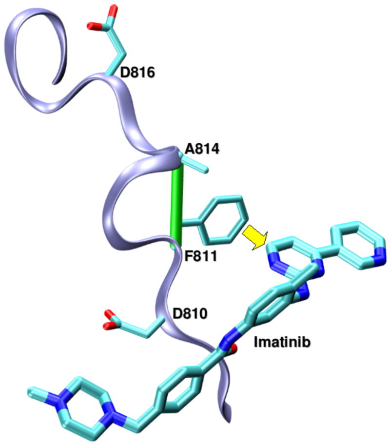
Targeting non-conserved kinase packing defects. (a) Aligned backbones (ribbon representation) of Src (PDB.1BYG, ice blue), CDK2 (PDB.1AQ1, pink), Chk1 (PDB.1NVR, orange) and PDK1 (PDB.1OKY, lime) kinases complexed with staurosporine. The Src Q250-E267 dehydron (green virtual bond joining α-carbons) maps into well-wrapped backbone hydrogen bonds (grey virtual bond joining α-carbons): K65-E81 in CDK2, K69-E85 in Chk1 and K144-S160 in PDK1. Methylation at the position indicated by the yellow arrow would turn the otherwise promiscuous ligand into a wrapper of the nonconserved packing defect. (b) Aligned backbones (ribbon representation) of Abl (PDB.1FPU, ice blue) and c-Kit (PDB.1T46, pink) kinases in the induced-fit conformation generated by co-crystallyzation with imatinib. The nonconserved dehydron C673-G676 (green virtual bond joining α-carbons) in c-Kit, which aligns with the well wrapped M318-G321 hydrogen bond (grey virtual bond joining α-carbons) in Abl, could be targeted by a methylation modification of imatinib in a specific position (yellow arrow) to achieve specificity. (c) Ribbon representation of the c-Kit (PDB.1T46, ice blue) kinases in the induced-fit conformation generated by co-crystallyzation with imatinib. The F811-A814 dehydron (green virtual bond joining α-carbons) present close to the D816V mutation site could be targeted by a methylation modification of imatinib in a specific position (yellow arrow) to overcome drug resistance. (d) Aligned ATP-binding pockets (ribbon representation) of IGF1R (PDB.1K3A, backbone: ice blue, sidechains: licorice representation) and INSR (PDB.1GAG, backbone: pink, sidechains: ball-and-stick representation) kinases. The degree of structure and sequence identity is very high at the ATP binding site. However, the nonconserved dehydron N1110-D1123 (green virtual bond joining α-carbons) in IGF1R, which does not align with any well wrapped hydrogen-bond or dehydron in the INSR, could be targeted to discriminate these closely related paralogs.
Targeting non-conserved kinase packing defects. (a) Aligned backbones (ribbon representation) of Src (PDB.1BYG, ice blue), CDK2 (PDB.1AQ1, pink), Chk1 (PDB.1NVR, orange) and PDK1 (PDB.1OKY, lime) kinases complexed with staurosporine. The Src Q250-E267 dehydron (green virtual bond joining α-carbons) maps into well-wrapped backbone hydrogen bonds (grey virtual bond joining α-carbons): K65-E81 in CDK2, K69-E85 in Chk1 and K144-S160 in PDK1. Methylation at the position indicated by the yellow arrow would turn the otherwise promiscuous ligand into a wrapper of the nonconserved packing defect. (b) Aligned backbones (ribbon representation) of Abl (PDB.1FPU, ice blue) and c-Kit (PDB.1T46, pink) kinases in the induced-fit conformation generated by co-crystallyzation with imatinib. The nonconserved dehydron C673-G676 (green virtual bond joining α-carbons) in c-Kit, which aligns with the well wrapped M318-G321 hydrogen bond (grey virtual bond joining α-carbons) in Abl, could be targeted by a methylation modification of imatinib in a specific position (yellow arrow) to achieve specificity. (c) Ribbon representation of the c-Kit (PDB.1T46, ice blue) kinases in the induced-fit conformation generated by co-crystallyzation with imatinib. The F811-A814 dehydron (green virtual bond joining α-carbons) present close to the D816V mutation site could be targeted by a methylation modification of imatinib in a specific position (yellow arrow) to overcome drug resistance. (d) Aligned ATP-binding pockets (ribbon representation) of IGF1R (PDB.1K3A, backbone: ice blue, sidechains: licorice representation) and INSR (PDB.1GAG, backbone: pink, sidechains: ball-and-stick representation) kinases. The degree of structure and sequence identity is very high at the ATP binding site. However, the nonconserved dehydron N1110-D1123 (green virtual bond joining α-carbons) in IGF1R, which does not align with any well wrapped hydrogen-bond or dehydron in the INSR, could be targeted to discriminate these closely related paralogs.
Targeting non-conserved kinase packing defects. (a) Aligned backbones (ribbon representation) of Src (PDB.1BYG, ice blue), CDK2 (PDB.1AQ1, pink), Chk1 (PDB.1NVR, orange) and PDK1 (PDB.1OKY, lime) kinases complexed with staurosporine. The Src Q250-E267 dehydron (green virtual bond joining α-carbons) maps into well-wrapped backbone hydrogen bonds (grey virtual bond joining α-carbons): K65-E81 in CDK2, K69-E85 in Chk1 and K144-S160 in PDK1. Methylation at the position indicated by the yellow arrow would turn the otherwise promiscuous ligand into a wrapper of the nonconserved packing defect. (b) Aligned backbones (ribbon representation) of Abl (PDB.1FPU, ice blue) and c-Kit (PDB.1T46, pink) kinases in the induced-fit conformation generated by co-crystallyzation with imatinib. The nonconserved dehydron C673-G676 (green virtual bond joining α-carbons) in c-Kit, which aligns with the well wrapped M318-G321 hydrogen bond (grey virtual bond joining α-carbons) in Abl, could be targeted by a methylation modification of imatinib in a specific position (yellow arrow) to achieve specificity. (c) Ribbon representation of the c-Kit (PDB.1T46, ice blue) kinases in the induced-fit conformation generated by co-crystallyzation with imatinib. The F811-A814 dehydron (green virtual bond joining α-carbons) present close to the D816V mutation site could be targeted by a methylation modification of imatinib in a specific position (yellow arrow) to overcome drug resistance. (d) Aligned ATP-binding pockets (ribbon representation) of IGF1R (PDB.1K3A, backbone: ice blue, sidechains: licorice representation) and INSR (PDB.1GAG, backbone: pink, sidechains: ball-and-stick representation) kinases. The degree of structure and sequence identity is very high at the ATP binding site. However, the nonconserved dehydron N1110-D1123 (green virtual bond joining α-carbons) in IGF1R, which does not align with any well wrapped hydrogen-bond or dehydron in the INSR, could be targeted to discriminate these closely related paralogs.
Targeting non-conserved kinase packing defects. (a) Aligned backbones (ribbon representation) of Src (PDB.1BYG, ice blue), CDK2 (PDB.1AQ1, pink), Chk1 (PDB.1NVR, orange) and PDK1 (PDB.1OKY, lime) kinases complexed with staurosporine. The Src Q250-E267 dehydron (green virtual bond joining α-carbons) maps into well-wrapped backbone hydrogen bonds (grey virtual bond joining α-carbons): K65-E81 in CDK2, K69-E85 in Chk1 and K144-S160 in PDK1. Methylation at the position indicated by the yellow arrow would turn the otherwise promiscuous ligand into a wrapper of the nonconserved packing defect. (b) Aligned backbones (ribbon representation) of Abl (PDB.1FPU, ice blue) and c-Kit (PDB.1T46, pink) kinases in the induced-fit conformation generated by co-crystallyzation with imatinib. The nonconserved dehydron C673-G676 (green virtual bond joining α-carbons) in c-Kit, which aligns with the well wrapped M318-G321 hydrogen bond (grey virtual bond joining α-carbons) in Abl, could be targeted by a methylation modification of imatinib in a specific position (yellow arrow) to achieve specificity. (c) Ribbon representation of the c-Kit (PDB.1T46, ice blue) kinases in the induced-fit conformation generated by co-crystallyzation with imatinib. The F811-A814 dehydron (green virtual bond joining α-carbons) present close to the D816V mutation site could be targeted by a methylation modification of imatinib in a specific position (yellow arrow) to overcome drug resistance. (d) Aligned ATP-binding pockets (ribbon representation) of IGF1R (PDB.1K3A, backbone: ice blue, sidechains: licorice representation) and INSR (PDB.1GAG, backbone: pink, sidechains: ball-and-stick representation) kinases. The degree of structure and sequence identity is very high at the ATP binding site. However, the nonconserved dehydron N1110-D1123 (green virtual bond joining α-carbons) in IGF1R, which does not align with any well wrapped hydrogen-bond or dehydron in the INSR, could be targeted to discriminate these closely related paralogs.
Similar articles
-
Anticipating drug resistance in the MAP kinase pathway.Pigment Cell Melanoma Res. 2010 Feb;23(1):7-9. doi: 10.1111/j.1755-148X.2009.00657.x. Epub 2009 Dec 2. Pigment Cell Melanoma Res. 2010. PMID: 19968818 Free PMC article. No abstract available.
-
Synergistic interactions between imatinib mesylate and the novel phosphoinositide-dependent kinase-1 inhibitor OSU-03012 in overcoming imatinib mesylate resistance.Blood. 2005 May 15;105(10):4021-7. doi: 10.1182/blood-2004-07-2967. Epub 2005 Jan 21. Blood. 2005. PMID: 15665113 Free PMC article.
-
Rational drug redesign to overcome drug resistance in cancer therapy: imatinib moving target.Cancer Res. 2007 May 1;67(9):4028-33. doi: 10.1158/0008-5472.CAN-07-0345. Cancer Res. 2007. PMID: 17483313 Free PMC article.
-
Mechanisms of resistance to tyrosine kinase inhibitors in chronic myeloid leukemia and recent therapeutic strategies to overcome resistance.Hematology Am Soc Hematol Educ Program. 2009:461-76. doi: 10.1182/asheducation-2009.1.461. Hematology Am Soc Hematol Educ Program. 2009. PMID: 20008232 Review.
-
Imatinib-refractory gastrointestinal stromal tumors: the clinical problem and therapeutic strategies.Curr Oncol Rep. 2006 May;8(3):192-7. doi: 10.1007/s11912-006-0019-3. Curr Oncol Rep. 2006. PMID: 16618383 Review.
References
-
- Dancey J, Sausville EA. Issues and progress with protein kinase inhibitors for cancer treatment. Nat Rev Drug Discov. 2003;2:296–313. - PubMed
-
- Levitski A, Gazit A. Tyrosine kinase inhibition: an approach to drug development. Science. 1995;267:1782–1788. - PubMed
-
- Donato NJ, Talpaz M. Clinical use of tyrosine kinase inhibitors: Therapy for chronic myelogenous leukemia and other cancers. Clin Cancer Res. 2000;6:2965–2966. - PubMed
-
- Tibes R, et al. Tyrosine kinase inhibitors and the dawn of molecular cancer therapeutics. Annu Rev Pharmacol Toxicol. 2005;45:357–384. - PubMed
-
- Gibbs J, Oliff A. Pharmaceutical research in molecular oncology. Cell. 1994;79:193–198. - PubMed
Publication types
MeSH terms
Substances
Grants and funding
LinkOut - more resources
Full Text Sources
Other Literature Sources
