. 2008 Jan 11;283(2):977-87.
doi: 10.1074/jbc.M707158200. Epub 2007 Nov 8.

Delta-catenin-induced dendritic morphogenesis. An essential role of p190RhoGEF interaction through Akt1-mediated phosphorylation

Affiliations

Delta-catenin-induced dendritic morphogenesis. An essential role of p190RhoGEF interaction through Akt1-mediated phosphorylation

Hangun Kim et al. J Biol Chem. .

Abstract

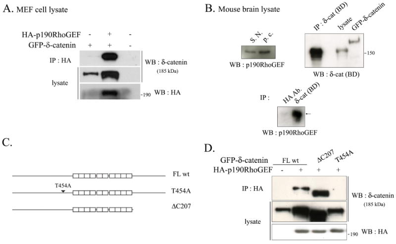
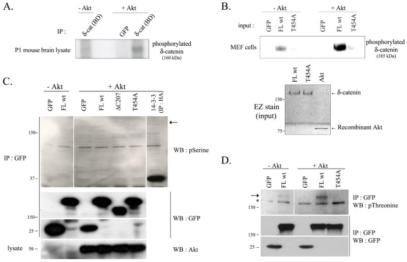
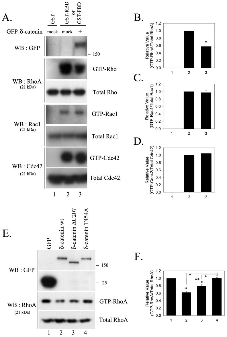
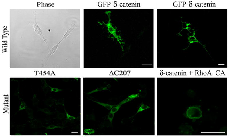
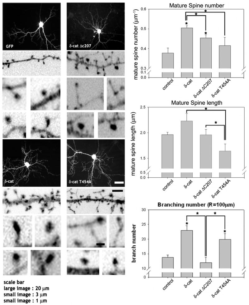
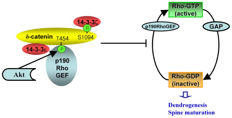
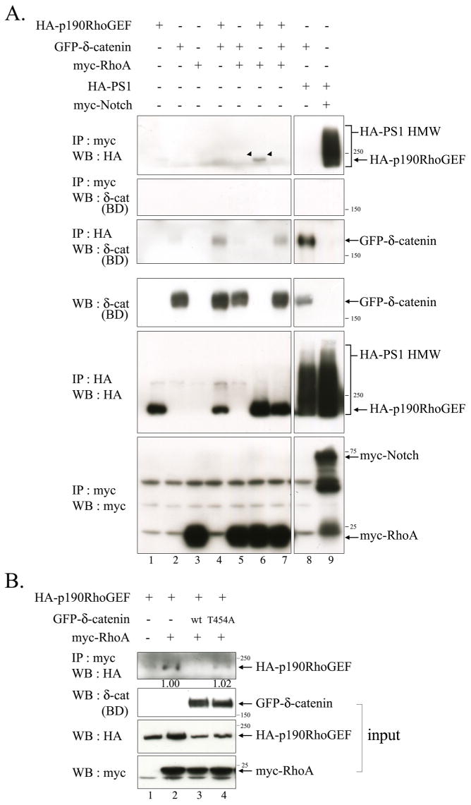
Delta-catenin was first identified through its interaction with Presenilin-1 and has been implicated in the regulation of dendrogenesis and cognitive function. However, the molecular mechanisms by which delta-catenin promotes dendritic morphogenesis were unclear. In this study, we demonstrated delta-catenin interaction with p190RhoGEF, and the importance of Akt1-mediated phosphorylation at Thr-454 residue of delta-catenin in this interaction. We have also found that delta-catenin overexpression decreased the binding between p190RhoGEF and RhoA, and significantly lowered the levels of GTP-RhoA but not those of GTP-Rac1 and -Cdc42. Delta-catenin T454A, a defective form in p190RhoGEF binding, did not decrease the binding between p190RhoGEF and RhoA. Delta-catenin T454A also did not lower GTP-RhoA levels and failed to induce dendrite-like process formation in NIH 3T3 fibroblasts. Furthermore, delta-catenin T454A significantly reduced the length and number of mature mushroom shaped spines in primary hippocampal neurons. These results highlight signaling events in the regulation of delta-catenin-induced dendrogenesis and spine morphogenesis.


Figures

Fig. 1
Fig. 1. δ-Catenin interacts with p190RhoGEF, for which Thr-454 residue of δ-catenin is indispensable
(A) The MEF cells were transfected with GFP-tagged δ-catenin with or without HA-p190RhoGEF. The binding of δ-catenin and p190RhoGEF was examined by immunoprecipitation with the anti-HA antibody, and western blotting was performed with the anti-δ-catenin antibody (upper panel). The expression of δ-catenin or p190RhoGEF in the cell lysates were detected using the anti-δ-catenin antibody (Middle panel), or the anti-HA antibody (bottom panel), respectively. (B) Adult mouse brain lysates were used to examine the endogenous interaction between δ-catenin and p190RhoGEF (Bottom panel). HA antibody was used as a negative control for immunoprecipitation assay. Endogenous expression of p190RhoGEF in the adult mouse brain region of substantia nigra (indicated as S.N.) and pars compacta (indicated as p.c.) were shown (Upper left panel). Endogenous expression of δ-catenin in the whole adult mouse brain lysate was shown, and the efficiency of immunoprecipitation using the anti-δ-catenin antibody was determined with the same adult mouse brain lysates (Upper right panel). (C) Schematic illustration of δ-catenin constructs used throughout experiments. (D) The MEF cells were transfected with either GFP-δ-catenin FL wt or a mutant of δ-catenin (ΔC207 and T454A) together with or without HA-p190RhoGEF as indicated in the figure. The binding of δ-catenin and p190RhoGEF was examined by immunoprecipitation with the anti-HA antibody, and bloted with the anti-δ-catenin antibody (upper panel). The expression of δ-catenin or p190RhoGEF in the cell lysates was detected with the anti-δ-catenin antibody (middle panel) or the anti-HA antibody (bottom panel), respectively.
Fig. 2
Fig. 2. δ-Catenin undergoes Akt1-mediated phosphorylation at Thr-454 residue
An Akt1 kinase assay was performed as described in Materials and Methods. GFP, mock transfected proteins were used as a negative control in the assay, and GFP-immunocomplex was obtained by immunoprecipitating with GFP antibody. (A) Purified immunocomplex of 1 day post-natal mouse brain lysates with δ-catenin (BD) antibody was used as a substrate for an Akt kinase assay. [γ-32P]-ATP was supplemented with a reaction buffer, and phosphorylation status was detected by autoradiograph. (B) GFP-tagged δ-catenin wt (FL wt) and T454A mutant were transfected in MEF cells, and purified immunocomplex with δ-catenin (BD) antibody was used as a substrate for an Akt kinase assay. [γ-32P]-ATP was supplemented with a reaction buffer, and phosphorylation status was detected by autoradiograph (Upper panel). The level of δ-catenin FL wt and T454A mutant existing in immunocomplex and of recombinant Akt1 were confirmed by EZ staining kit (Bottom panel). (C-D) GFP-tagged full length (FL wt) and mutant δ-catenin (ΔC207 and T454A) were overexpressed in Bosc23 cells, and purified immunocomplex with GFP antibody was used as a substrate for Akt kinase assay. Radio-inactive cold ATP was supplemented with a reaction buffer, and phosphorylation status on serine and threonine residue in the wild type or mutant type δ-catenin was detected using anti-phosphoserine antibody (C) or with anti-phosphothreonine antibody (D) (upper panel). HA-14-3-3 was immunoprecipitated with HA antibody and was used as a positive control (C). An arrow indicates the predicted site (C) or an actual band (D) of phosphorylated δ-catenin, and an asterisk indicates a nonspecific band. The immunocomplexes were sub sequentially reprobed with anti-GFP antibody (middle panels), and the added recombinant Akt1 was also confirmed using anti-Akt antibody (C, bottom panel).
Fig. 3
Fig. 3. Overexpression of δ-catenin results in a decrease in the level of GTP-RhoA, while the Rac1 and Cdc42 activity are unaffected
(A) The levels of active GTP-bound RhoA, Rac1 and Cdc42 were measured in MEF cells transfected with either the mock or GFP-δ-catenin. The RhoA activity was measured using a GST-RBD, and the Rac1, Cdc42 activities were measured using a GST-PBD. The total amounts of RhoA, Rac1, and Cdc42 are also shown to compare the endogenous level of each GTPase in the transfected cells. The relative activity of RhoA (B), Rac1 (C), and Cdc42 (D) were determined as described in Materials and Methods. The data is represented as the mean ± SEM (* p<0.01). (E) The level of active GTP-bound RhoA was measured in the MEF cells transfected with GFP or with wt/mutant GFP-δ-catenin constructs using GST-RBD. (F) The relative activity of RhoA was determined. The data is represented as the mean ± SEM (* p<0.01, ** p<0.05). Results are representative of at least three independent experiments.
Fig. 4
Fig. 4. Effects of the wild type and mutant δ-catenins on the dendrite-like process formation in NIH 3T3 fibroblasts
The NIH 3T3 fibroblast cells were transfected with wild type (GFP-δ-catenin) and mutant δ-catenin (T454A and ΔC207) or co-transfected with a constitutive active mutant of RhoA (RhoA CA) as indicated in each image. After 24 h post-transfection, the cells were fixed, and a fluorescent image was taken sometimes together with its corresponding phase contrast image (left and middle, upper panel). GFP-δ-catenin images on top panel indicate typical δ-catenin-induced dendrite-like processes at different stages (upper middle-early stage; upper right-late stage). Scale bars: 20 μm.
Fig. 5
Fig. 5. Effects of wild type and mutant δ-catenin on the dendrogenesis and spine formation in primary hippocampal neurons
The cultured hippocampal neurons at 16 DIV were transfected with the full-length GFP-δ-catenin or various mutants, fixed and stained with GFP antibody. The high magnification images are inverted for clarity. The average number and length of mature spines, either a cotyloid appearance or flat-apex mushroom appearance, were analyzed and filopodia shaped spines are excluded from analysis (* p<0.01 by ANOVA and Tukey's HSD post hoc test). The number of dendritic branches that intersects a sphere, 100 μm in radius, centered at the soma was added for plotting and statistical analysis using ANOVA and a Tukey's HSD post hoc test. The graphs show the average number of intersections between the dendritic branches and a sphere (* p < 0.01). The study has been repeated with 3 different cultures (3 different litters from 3 different pregnant rats) and from each litter, 3 to 4 coverslips (∼10 neurons per coverslip) have been included in this analysis. Scale bars: 20 μm for low magnification, 3 μm for high magnification, and 1 μm for high magnification of the dendritic spines.
Fig. 6
Fig. 6. A proposed model of δ-catenin-induced morphogenesis
δ-Catenin binds to p190RhoGEF through Akt-mediated Thr-454 phosphorylation, sequesters p190RhoGEF to prevent it from activating RhoA, and thereby reduces the local endogenous RhoA activity. The reduced local GTP-RhoA affects its downstream effectors, which, along with other δ-catenin-induced changes, regulates the dendrogenesis and mature spine formation. The interaction between δ-catenin with the 14-3-3 isoforms occurs through a different binding domain. The C-terminal region of δ-catenin is important for binding to 14-3-3ζ.
Fig. 7
Fig. 7. δ-Catenin Overexpression decreases the interaction between p190RhoGEF and RhoA
(A) Bosc23 cells were transfected with GFP-δ-catenin, HA-p190RhoGEF, myc-RhoA, HA-PS1 wt, and myc-ΔEN1 in a various combination as indicated in the upper side of the figure. The binding between p190RhoGEF and RhoA was examined by immunoprecipitation with the anti-myc antibody, and western blotting was performed with anti-HA antibody (Upper first panel). The binding between RhoA and δ-catenin was examined by immunoprecipitation with the anti-myc antibody, and western blotting was performed with anti-δ-catenin (BD) antibody (Upper second panel). The bindings between Presenilin-1 with δ-catenin or with Notch were examined for positive control experiments (lane 8, 9 in upper first and third panels). Expression of each protein was shown through the bottom three panels. (B) Bosc23 cells were transfected with either wild type or mutant δ-catenin together with HA-p190RhoGEF and/or myc-RhoA. The binding between p190RhoGEF and RhoA was examined by immunoprecipitation with the anti-myc antibody, and western blotting was performed with anti-HA antibody (Upper first panel). 5% volume of each lysate was subjected to western blot analysis to show the input level of each protein. The relative intensity of the immunoprecipitated HA-p190RhoGEF band in lane 2 vs. 4 was determined by normalization against the input band of HA-p190RhoGEF. 3 μg of each plasmid was used to transfect cells plated in 100 mm dish at 50% confluency.

Similar articles

Cited by

References

    1. Zhou J, Liyanage U, Medina M, Ho C, Simmons AD, Lovett M, Kosik KS. Neuroreport. 1997;8:2085–2090. - PubMed
    1. Tanahashi H, Tabira T. Neuroreport. 1999;10:563–568. - PubMed
    1. Paffenholz R, Franke WW. Differentiation. 1997;61:293–304. - PubMed
    1. Lu Q, Paredes M, Medina M, Zhou J, Cavallo R, Peifer M, Orecchio L, Kosik KS. J Cell Biol. 1999;144:519–532. - PMC - PubMed
    1. Israely I, Costa RM, Xie CW, Silva AJ, Kosik KS, Liu X. Curr Biol. 2004;14:1657–1663. - PubMed

Publication types

MeSH terms