. 2007 Dec;134(23):4297-306.
doi: 10.1242/dev.009282.

PAR1 specifies ciliated cells in vertebrate ectoderm downstream of aPKC

Affiliations

PAR1 specifies ciliated cells in vertebrate ectoderm downstream of aPKC

Olga Ossipova et al. Development. 2007 Dec.

Abstract

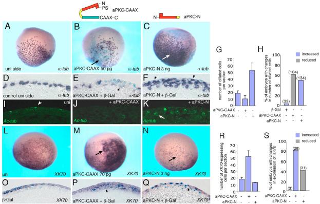
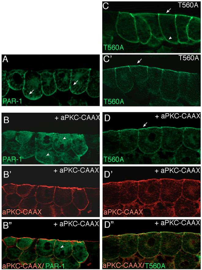
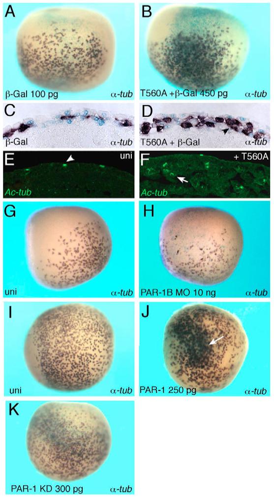
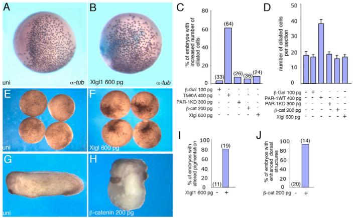
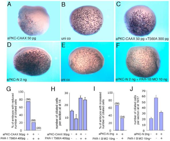
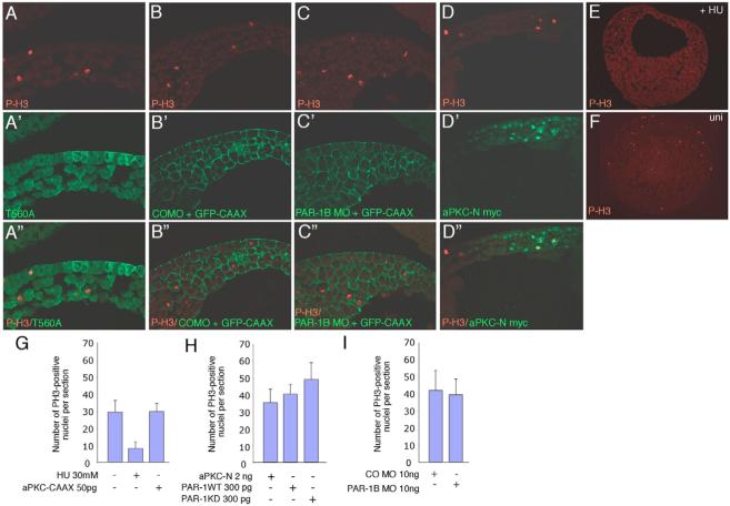
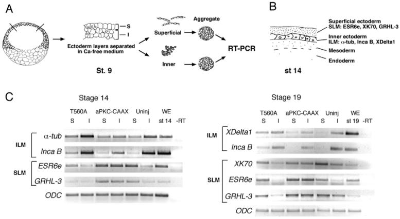
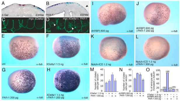
Partitioning-defective 1 (PAR1) and atypical protein kinase C (aPKC) are conserved serine/threonine protein kinases implicated in the establishment of cell polarity in many species from yeast to humans. Here we investigate the roles of these protein kinases in cell fate determination in Xenopus epidermis. Early asymmetric cell divisions at blastula and gastrula stages give rise to the superficial (apical) and the deep (basal) cell layers of epidermal ectoderm. These two layers consist of cells with different intrinsic developmental potential, including superficial epidermal cells and deep ciliated cells. Our gain- and loss-of-function studies demonstrate that aPKC inhibits ciliated cell differentiation in Xenopus ectoderm and promotes superficial cell fates. We find that the crucial molecular substrate for aPKC is PAR1, which is localized in a complementary domain in superficial ectoderm cells. We show that PAR1 acts downstream of aPKC and is sufficient to stimulate ciliated cell differentiation and inhibit superficial epidermal cell fates. Our results suggest that aPKC and PAR1 function sequentially in a conserved molecular pathway that links apical-basal cell polarity to Notch signaling and cell fate determination. The observed patterning mechanism may operate in a wide range of epithelial tissues in many species.


Figures

Fig. 1
Fig. 1. A role of aPKC in epidermal ectoderm development
Four- to eight-cell embryos were unilaterally coinjected with aPKC-CAAX or aPKC-N RNAs and lacZ RNA as a lineage tracer (light blue staining). Injected embryos were cultured until stages 14-16, fixed and subjected to whole mount in situ hybridization with anti-sense probes to α-tubulin (A-H) and XK70 (L-S). aPKC constructs employed are shown above panels B and C. (A-K) Changes in ciliated cell differentiation. aPKC-CAAX decreases (B,E), whereas aPKC-N increases (C,F) ciliated cell differentiation (arrows) on the injected side as compared with the uninjected side (A,D). The same embryo is shown in A and B. (D-F) Sections corresponding to embryos in A-C. (D) A single row of α-tubulin-positive cells in the deep layer of uninjected ectoderm. (F) α-Tubulin-positive cells are found in the superficial layer (arrows). (G) Quantification of the results showing mean numbers of α-tubulin-positive cells per section±s.d. Sections of at least three representative embryos per group were analyzed. (H) Frequency of embryos with altered numbers of ciliated cells (data pooled from several experiments). Embryos were scored positive if the number of ciliated cells per injected area was increased by at least 50%. The data are representative of three independent experiments. (I-K) Cilia differentiation assessed by immunostaining with antibodies to acetylated tubulin in stage 18 embryos. (I) Uninjected control embryos with regular pattern of superficially located ciliated cells, (J) aPKC-CAAX injection inhibits cilia formation, (K) aPKC-N injection results in multiple cells with many differentiated cilia. (L-S) aPKC promotes superficial cell fate. (L,O) Control XK70 staining. (M,P) aPKC-CAAX expands XK70 (arrow) on the injected side. (N,Q) aPKC-N downregulates XK70 (arrow). (O-Q) Sections corresponding to embryos in L-N. Superficial expression of XK70 (O) is extended into the deep cell layer in aPKC-CAAX RNA-injected embryos (P, arrowhead). (Q) Inhibition of XK70 by aPKC-N (arrowhead). In all panels, arrows indicate altered staining as a result of injection. (R,S) Quantification of the effects, shown as average numbers of XK70-expressing cells in affected embryos (R), and frequency of embryos with visible changes in XK70 expression (S). Numbers of embryos per group are given above the bars. The data are from three representative experiments.
Fig. 2
Fig. 2. aPKC regulates subcellular localization of PAR1 in Xenopus ectoderm
(A,B) aPKC inhibits cortical localization of PAR1. (A) PAR1 is predominantly localized to the basolateral cortex of superficial ectoderm cells (arrows). (B) Overexpression of aPKC-CAAX mislocalizes PAR1 to the cytoplasm (arrowheads). (C,D) The apical localization of T560A, the non-phosphorylatable PAR1 mutant (C’) is not affected by aPKC-CAAX (D). At higher doses of injected RNA, T560A was distributed all around the cell cortex, both apically and basolaterally (C). The distribution of coinjected membrane-associated aPKC-CAAX is shown in B’ and D’, the increased apical staining is due to high amounts of endogenous aPKC detected by anti-aPKC antibody. B” and D”, merged images. Embryo injections were as described in Fig. 1. Frozen sections of stage 10.5-11 embryonic ectoderm were stained with anti-Myc (green) to detect PAR1 (B,D) and anti-aPKC antibodies (B’ and D’, red). At least 15 embryos per group were examined and representative sections of three independent experiments are shown.
Fig. 3
Fig. 3. PAR1 promotes ciliated cell differentiation
Four- to eight-cell embryos were unilaterally injected with lacZ RNA (light blue staining) or PAR1 RNAs or MO as indicated and subjected to in situ hybridization for α-tubulin expression at stages 13-14. (A-D) T560A increases the number of α-tubulin-expressing cells in epidermal ectoderm. (C,D) Cross-sections of embryos shown in A,B. A single layer of α-tubulin-positive cells in control ectoderm (C) expanded to a double layer of positive cells in T560A-expressing ectoderm (D, arrowhead). (E,F) Enhanced cilia differentiation in T560A RNA-injected embryos at stage 18 (F), when compared with uninjected controls (E), revealed by immunostaining for acetylated tubulin. Arrowhead in E demarcates ciliated cells that migrated to the surface. Arrow in F indicates ectopic ciliated cells remaining in the inner ectoderm layer. (G,H) PAR1B MO decreases the number of α-tubulin-expressing cells (H) as compared with the uninjected side (G). (I) Uninjected embryo; (J) PAR1 RNA increases ciliated cell number (white arrow); (K) PAR1 KD RNA has no significant effect on ciliated cells. Lateral view is shown in all panels, except I-K (ventral view).
Fig. 4
Fig. 4. Lack of effect of β-catenin and LGL on ciliated cell development
In situ hybridization with α-tubulin probe is shown. For experimental details, see Fig. 1 legend. (A) Uninjected embryo. (B) LGL1 (Xlgl1) RNA has no effect on α-tubulin-expressing cells. (C,D) Quantification of the effects of T560A, PAR1, PAR1-KD, β-catenin and LGL1 RNAs on ciliated cell development, presented as frequencies of affected embryos (C) and numbers of ciliated cells per section (D). In C, numbers of embryos per group are shown above bars. Data are representative of four different experiments. (E,F,I) LGL1 RNA, used in B, altered ectoderm pigmentation in 79% of injected embryos (n=19; F) as compared with uninjected controls (E). (I) Quantification of the results in E and F. (G,H,J) Marginal zone-injected β-catenin RNA dorsalized 92% of injected embryos (n=14), characterized by enlarged head and cement gland and truncated or missing tail (H), as compared with uninjected siblings (G). (J) Quantification of the results in G,H.
Fig. 5
Fig. 5. aPKC functions upstream of PAR1 to specify ectodermal cell fates
Embryos were injected with RNAs or MO as described in Fig. 1. Ciliated cells were detected at stages 14-16 by in situ hybridization with the α-tubulin probe. (A-C) T560A reverses the inhibitory effect of aPKC-CAAX on ciliated cell differentiation. Two sides of the same embryo are shown in B and C. (D-F) PAR1B MO (F) suppresses aPKC-N-mediated expansion of ciliated cells. The injected and uninjected sides of the same embryo are shown in D and E, respectively. (G-J) Quantification of the data shown in A-C (G,H) and D-F (I,J). Numbers of embryos per group are shown above bars. (G,I) Frequencies of embryos showing visible phenotypic changes. (H,J) Mean numbers of α-tubulin-positive cells per section±s.d. are shown. Sections of at least three representative embryos per group were analyzed.
Fig. 6
Fig. 6. PAR1 and aPKC do not significantly alter cell proliferation
(A-F) Embryos injected at the four- to eight-cell stage with the indicated MO and RNAs (green, A’-D’). GFP-CAAX was used as a lineage tracer for MO injections (B’,C’). Embryos were cultured to gastrula stages, fixed, cryosectioned and stained with anti-phospho H3 (P-H3) antibodies (red, A-F; merged images A”-D”). No significant differences were observed in embryos with altered levels of PAR1 or aPKC (compare control MO B with A,C,D), but the number of positive cells was strongly reduced in hydroxyurea (HU)-treated embryos (E) compared with the control embryo (F). No differences in the number of mitotic nuclei were detected between injected and uninjected tissues (marked by lineage tracing) within the same embryo or in different embryos (data not shown). (G-I) Quantification of changes in P-H3-positive cells. Average numbers of P-H3-positive cells per section±s.d. are shown. At least ten embryos were examined for each group.
Fig. 7
Fig. 7. Opposite effects of PAR1 and aPKC on gene expression in separated ectoderm cell layers
(A) Experimental scheme for the layer separation assay. Two-cell embryos were injected with T560A or aPKC-CAAX mRNA. Animal pole explants were dissected from injected or uninjected embryos at stage 9, superficial (S) and inner (I) cell layers were separated based on their different abilities to dissociate in a calcium- and magnesium-free buffer and allowed to reaggregate. Cell aggregates were cultured until sibling embryos reached stage 14 or stage 19. RNA was prepared from aggregate lysates and analyzed by semi-quantitative RT-PCR. (B) A schematic section of the frog embryo at stage 14 shows superficial and inner layers of non-neural ectoderm with distinct sets of molecular markers. SLM, superficial layer markers; ILM, inner layer markers. (C) Stage 14 aggregates: T560A RNA upregulates the inner layer markers α-tubulin and inca B and downregulates the superficial layer markers ESR6e and grhl3 in both inner and outer layer explants. aPKC-CAAX has a complementary effect. Stage 19 aggregates: T560A RNA upregulates the inner layer markers XDelta-1 and inca B and downregulates XK70 in the superficial layer. aPKC-CAAX downregulates XDelta-1 and inca B in the superficial layer and upregulates XK70, ESR6e and grhl3 in inner cell aggregates. ODC is a control for loading. Uninj, no RNA injection; WE, whole embryo; −RT, no reverse transcriptase control. The analysis of two representative sets of cDNAs from several independent experiments is shown.
Fig. 8
Fig. 8. PAR1 synergizes with XDelta-1 to induce ciliated cell differentiation and inhibits the Notch target ESR6e
Four- to eight-cell embryos were unilaterally injected with the indicated RNAs and lacZ RNA as a lineage tracer (light blue staining) and subjected to in situ hybridization with the ESR6e (A,B) or α-tubulin (E-L) probes. (A,B) Superficial staining for ESR6e is unaffected in cross-sections of lacZ RNA-injected control embryos (A, 100 pg), but is inhibited in PAR1 RNA-injected embryos (B, right side, arrowhead, 250 pg). (C) Basolateral localization of XDelta-1 in Xenopus ectoderm. (D) XDelta-1 is detected in multiple cytoplasmic vesicles (arrowheads) in the presence of PAR1. (E) Uninjected embryo. (F,G) XDelta-1 RNA alone (F) or low dose of PAR1 RNA (G) do not significantly alter the number of ciliated cells. (H) The synergistic effect of coinjected PAR1 and XDelta-1 RNAs on ciliated cell development. (I,J) PAR1 does not influence the activity of a dominant intracellular inhibitor of the Notch pathway, dnRBP/j, which can stimulate ciliated cell development. (K) Notch-ICD suppresses ciliated cell differentiation. (L) PAR1 does not alter Notch-ICD activity. (M-O) Quantification of the effects shown in E-J, presented as numbers of ciliated cells per section (M,N) and frequency of embryos with increased α-tubulin staining (O). Numbers of examined embryos are shown above bars. The data are representative of three independent experiments.

Similar articles

Cited by

References

    1. Alonso L, Fuchs E. Stem cells in the skin: waste not, Wnt not. Genes Dev. 2003;17:1189–1200. - PubMed
    1. Bayraktar J, Zygmunt D, Carthew RW. Par-1 kinase establishes cell polarity and functions in Notch signaling in the Drosophila embryo. J. Cell Sci. 2006;119:711–721. - PubMed
    1. Benton R, St Johnston D. Drosophila PAR-1 and 14-3-3 inhibit Bazooka/PAR-3 to establish complementary cortical domains in polarized cells. Cell. 2003;115:691–704. - PubMed
    1. Betschinger J, Knoblich JA. Dare to be different: asymmetric cell division in Drosophila, C. elegans and vertebrates. Curr. Biol. 2004;14:R674–R685. - PubMed
    1. Blanpain C, Lowry WE, Pasolli HA, Fuchs E. Canonical notch signaling functions as a commitment switch in the epidermal lineage. Genes Dev. 2006;20:3022–3035. - PMC - PubMed

Publication types

MeSH terms