. 2008 Mar 1;94(5):1589-99.
doi: 10.1529/biophysj.107.122218. Epub 2007 Nov 9.

Normal-mode flexible fitting of high-resolution structure of biological molecules toward one-dimensional low-resolution data

Affiliations

Normal-mode flexible fitting of high-resolution structure of biological molecules toward one-dimensional low-resolution data

Christian Gorba et al. Biophys J. .

Abstract

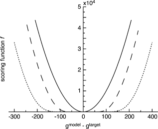
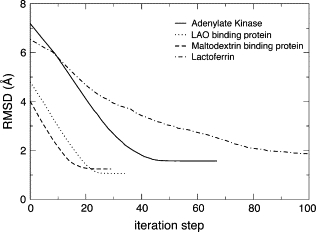
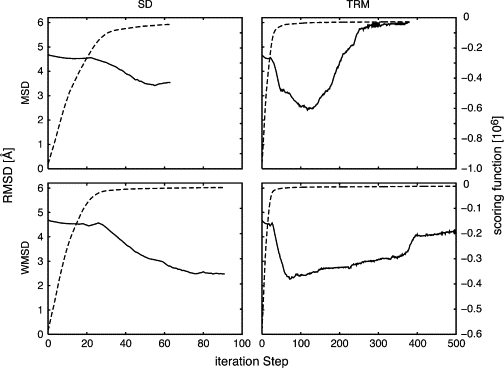
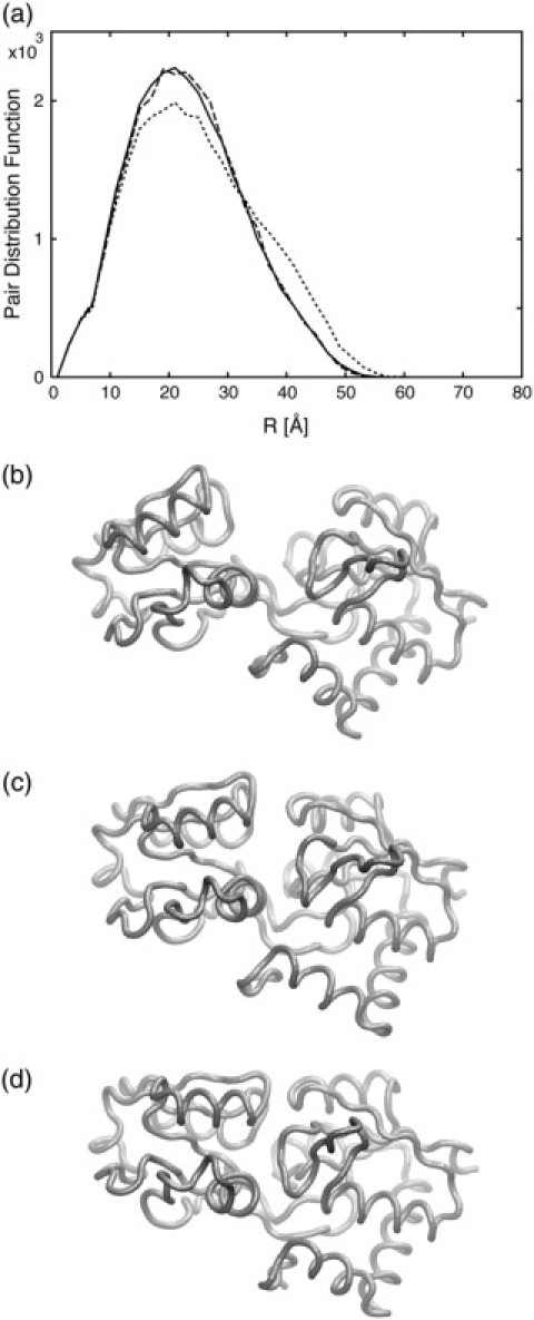
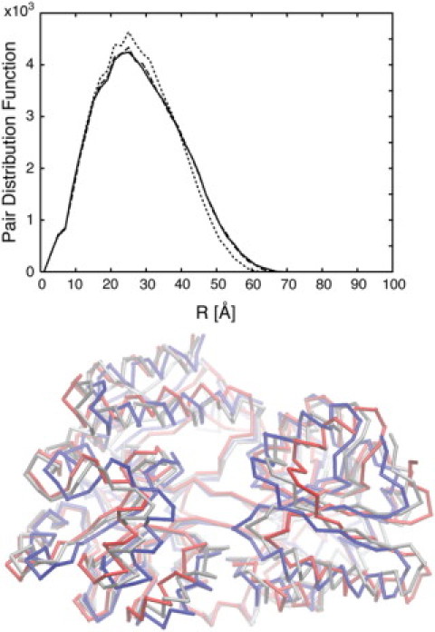
We present a method for reconstructing a 3D structure from a pair distribution function by flexibly fitting known x-ray structures toward a conformation that agrees with the low-resolution data. This method uses a linear combination of low-frequency normal modes from elastic-network description of the molecule in an iterative manner to deform the structure optimally to conform to the target pair distribution function. A simple function, pair distance distribution function between atoms, is chosen as a test model to establish computational algorithms-optimization algorithm and scoring function-that can utilize low-resolution 1D data. To select a correct structural model based on less information, we developed a scoring function that takes into account a characteristic of pair distribution functions. In addition, we employ a new optimization algorithm, the trusted region method, that relies on both first and second derivatives of the scoring function. Illustrative results of our studies on simulated 1D data from five different proteins, for which large conformational changes are known to occur, are presented.


Figures

Figure 1
Figure 1
Examples of scoring functions with different parameters for values found in the PDF of the LAO binding protein (239 residues). (Solid line) simple-MSD scoring function; (dashed line) WMSD scoring function with the parameter ngtar = 150; (dotted line) WMSD scoring function with ngtar = 300. For a target PDF, gtar, with a large value, the scoring function is flattened at the bottom, and thus less sensitive in the optimization process.
Figure 2
Figure 2
Performance of iterative NMA from 3D structure to 3D structure for four proteins. For each protein, the algorithm successfully deforms the structure very close to the target structure.
Figure 3
Figure 3
Progress of the fitting process for the LAO binding protein measured by fitness scoring function and RMSD. Combinations of two scoring functions, simple MSD and WMSD, and two optimization methods, SD and TRM, are tested. The progress of the RMSD is shown as solid lines and the scoring functions as broken lines. The best model, which is closest to the target structure, is obtained by the combination of WMSD and TRM.
Figure 4
Figure 4
Progress of RMSD and relative change in the scoring function over the fitting process for the LAO binding protein. As in Fig. 3, a combination of the two scoring functions, simple MSD and WMSD, and two optimization methods, SD and TRM, are tested. The progress of the RMSD is shown by solid lines and the relative change of scoring functions by dashed lines. The relative change reaches zero when the RMSD is close to the minimum.
Figure 5
Figure 5
(a) The PDFs of initial (dotted line), predicted (dashed line), and target (solid line) structures are shown. (bd). The initial structure of the LAO binding protein (b), the modeled structure predicted from the fitting algorithm (c), and the structure from which the target PDF is created (d). The PDF of the predicted structure is in close agreement with the target PDF and the structures are also very similar.
Figure 6
Figure 6
(Upper) The PDFs of the initial (dotted line), target (solid line), and constructed model (dashed line). (Lower) The initial structure of the maltodextrin binding protein (red), the structure from which the target PDF is created (blue), and the modeled structure predicted from the fitting algorithm (silver) are shown. For this system, since the difference between the initial and target PDFs is small, the modeled structure does not exactly agree with the target structure; however, trends of the conformational change are predicted.

Similar articles

Cited by

References

    1. Saibil H.R. Conformational changes studied by cryo-electron microscopy. Nat. Struct. Biol. 2000;7:711–714. - PubMed
    1. Heller W.T., Vigil D., Brown S., Blumenthal D.K., Taylor S.S., Trewhella J. C subunits binding to the protein kinase A RIα dimer induce a large conformational change. J. Biol. Chem. 2004;279:19084–19090. - PubMed
    1. Priddy T.S., Macdonald B.A., Heller W.T., Nadeau O.W., Trewhella J., Carlson G.M. Ca2+-induced structural changes in phosphorylase kinase detected by small-angle X-ray scattering. Protein Sci. 2005;14:1039–1048. - PMC - PubMed
    1. Vestergaard B., Sanyal S., Roessle M., Mora L., Buckingham R.H., Kastrup J.S., Gajhede M., Svergun D.I., Ehrenberg M. The SAXS solution structure of RF1 differs from its crystal structure and is similar to its ribosome bound cryo-EM structure. Mol. Cell. 2005;20:929–938. - PubMed
    1. Ermolenko D.N., Spiegel P.C., Majumdar Z.K., Hickerson R.P., Clegg R.M., Noller H.F. The antibiotic viomycin traps the ribosome in an intermediate state of translocation. Nat. Struct. Mol. Biol. 2007;14:493–497. - PubMed

LinkOut - more resources