. 2008 Mar 1;94(5):1782-95.
doi: 10.1529/biophysj.107.108175. Epub 2007 Nov 9.

Molecular origin of the self-assembly of lanreotide into nanotubes: a mutational approach

Affiliations

Molecular origin of the self-assembly of lanreotide into nanotubes: a mutational approach

Céline Valéry et al. Biophys J. .

Abstract

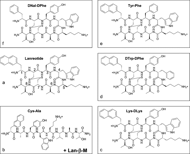
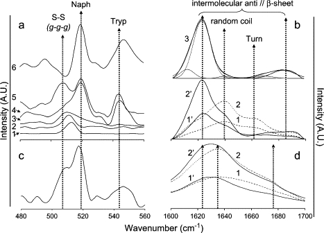
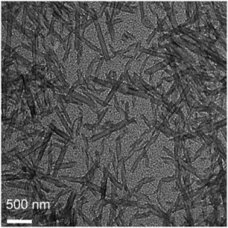
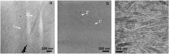
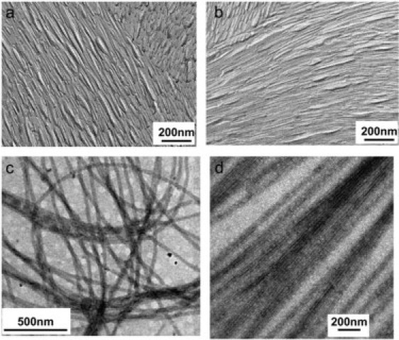
Lanreotide, a synthetic, therapeutic octapeptide analog of somatostatin, self-assembles in water into perfectly hollow and monodisperse (24-nm wide) nanotubes. Lanreotide is a cyclic octapeptide that contains three aromatic residues. The molecular packing of the peptide in the walls of a nanotube has recently been characterized, indicating four hierarchical levels of organization. This is a fascinating example of spontaneous self-organization, very similar to the formation of the gas vesicle walls of Halobacterium halobium. However, this unique peptide self-assembly raises important questions about its molecular origin. We adopted a directed mutation approach to determine the molecular parameters driving the formation of such a remarkable peptide architecture. We have modified the conformation by opening the cycle and by changing the conformation of a Lys residue, and we have also mutated the aromatic side chains of the peptide. We show that three parameters are essential for the formation of lanreotide nanotubes: i), the specificity of two of the three aromatic side chains, ii), the spatial arrangement of the hydrophilic and hydrophobic residues, and iii), the aromatic side chain in the beta-turn of the molecule. When these molecular characteristics are modified, either the peptides lose their self-assembling capability or they form less-ordered architectures, such as amyloid fibers and curved lamellae. Thus we have determined key elements of the molecular origins of lanreotide nanotube formation.


Figures

Figure 1
Figure 1
Chemical structure of lanreotide (a) and its derivatives named Cys-Ala and Lan-β-M (b), Lys-DLys (c), DTrp-DPhe (d), Tyr-Phe (e), and DNal-DPhe (f). The numbers 1–8 on the peptide backbone of lanreotide indicate the positions of the amino acids. All the peptides contain 2 mol of acetate per mole of peptide, the acetate being the counterion of the two positive charges of the peptides.
Figure 2
Figure 2
FT-Raman (a and c) and ATR-FTIR (b and d) spectra. (a) FT-Raman spectra (λ = 1064 nm) of naphthalene powder (trace 1), naphthalene in THF (trace 2), β-mercaptoethanol (β-M) 1 M (trace 3), Lan-β-M 20% w/w in water (trace 4), lanreotide acetate 20% w/w in water (trace 5), and Cys-Ala acetate 15% w/w in water (trace 6). Dashed arrows indicate the position of the vibrations of disulfide bridge in a g-g-g conformation (505 cm−1), the naphthalene group in the peptide (520 cm−1), and tryptophan (545–550 cm−1). (b) ATR-FTIR spectra of the amide I band of lanreotide acetate 5% and 10% w/w in water (traces 1 and 2, respectively) and of Lan-β-M acetate 5% and 10% w/w (traces 1′ and 2′, respectively). ATR-FTIR spectra of Cys-Ala acetate 10% w/w in water (trace 3) together with the decomposition into individual components (thin line peaks under the spectrum; see Table 2 for more details). Arrows indicate the position of the absorption peaks associated with the carbonyl involved in H-bonds in intermolecular β-sheet (1625 and 1685 cm−1), random coil (1640 cm−1), and turn (1660 cm−1) conformations. (c) FT-Raman spectra (λ = 1064 nm) of Lys-DLys acetate 15% w/w in water. For arrow definitions, see legend panel a. (d) ATR-FTIR spectra of Lys-DLys acetate 5% w/w (traces 1 and 1′) and 10% w/w (traces 2 and 2′) in water. The spectra were recorded either just after the dilution of the peptide in water (traces 1 and 2) or 24 h after the dilution (1′ and 2′).
Figure 3
Figure 3
Electron micrographs obtained from negatively stained samples of Lan-β-M (5% w/w) show nanotubes shorter (<1 μm) and wider (100–200 nm) than lanreotide nanotubes.
Figure 4
Figure 4
Electron micrographs obtained either from freeze-fractured (a and b) or negatively stained (c) samples of Cys-Ala (5% w/w in water). In a and b, the arrows indicate either unilamellar (1 and 1′) or multilamellar (2 and 2′) nanotubes
Figure 5
Figure 5
Electron micrographs obtained from negatively stained (a and b) samples of Lys-DLys (10% w/w in water, acetate) and size distribution of the width of the fibers observed (c). The micrographs were taken either just after the preparation of the sample (a) or after 1 week (b), showing slow fiber growth. The size distributions were plotted from ∼200 measurements, indicating an average fiber width of 12 nm.
Figure 6
Figure 6
WAXS patterns of (a) Lanreotide-β-M, (b) Cys-Ala, and (c) Lys-DLys derivatives. (a) WAXS patterns of [Lan-β-M acetate] = 9%, 18%, and 30% w/w (traces 1, 2, and 3, respectively). The position (dashed lines) and the indexing of the Bragg peaks are indicated on the graph. The indexing is detailed in Table 1. (b) WAXS patterns of Cys-Ala acetate 10% w/w in water. The position (dashed lines) and the indexing of the Bragg peaks are indicated on the graph. The indexing is detailed in Table 1. (c) WAXS patterns of Lys-DLys acetate 10% w/w in water.
Figure 7
Figure 7
FT-Raman spectra (a) and ATR-FTIR spectra (b). (a) FT-Raman spectra of DTrp-DPhe acetate 10% w/w in water (trace 1), Tyr-Phe acetate 15% w/w in water (trace 2), and DNal-DPhe acetate 10% w/w in water (trace 3). Arrows indicate the position of scattering mode of the disulfide bridge in a g-g-g conformation (505 cm−1) of naphthalene in the peptides (520 cm−1) and of tryptophan (545–550 cm−1). (b) ATR-FTIR spectra of the amide I vibrations of DTrp-DPhe acetate 5%, 10%, and 15% (traces 1, 2, and 3, respectively) w/w in water of Tyr-Phe acetate 5%, 10%, and 20% (traces 4, 5, and 6, respectively) w/w in water and of DNal-DPhe acetate 5% and 10% (traces 7 and 8, respectively) w/w in water. Arrows indicate the positions of the absorption peaks associated with the carbonyl involved in H-bonds in intermolecular β-sheet (1625 and 1685 cm−1), random coil (1640 cm−1), and turn (1660 cm−1) conformations.
Figure 8
Figure 8
SAXS patterns of DTrp-DPhe samples obtained with a sample to detector distance of 1.5 m (a) and of 6.5 m very small angle x-ray scattering (b) on ID2 beam line (ESRF, Grenoble) and change in the hexagonal parameter with temperature (c). (a) SAXS patterns as a function of DTrp-DPhe acetate concentration (sample to detector distance of 1.5 m). The seven lower patterns were obtained for 2%, 4%, 5%, 8%, 10%, 15%, and 20% (w/w) of peptide acetate in water. The dashed curve is a J0 Bessel function calculated for a radius of 82 nm. The dotted lines i), underline the minima of the Bessel function that fit with the experimental ones, and ii), show that these minima are independent on peptide concentration. The upper curve was obtained for 30% (w/w) of peptide acetate in solution. The arrows underline the diffuse scattering peaks induced by the antiparallel β-sheet network. (b) SAXS patterns obtained for 14% (w/w) of peptide acetate in water at 20°C (sample to detector distance of 6.5 m). The dashed lines are in the theoretical position of the Bragg peaks expected for a hexagonal lattice of 259 Å to underline that the experimental peak positions are in agreement with the theoretical ones. (c) Change in the hexagonal lattice parameter with temperature. These data were obtained for 8%, 10%, 14%, and 20% (w/w) peptide acetate in solution during the heating and cooling process.
Figure 9
Figure 9
Electron micrographs obtained from freeze-fractured solutions of DNal-DPhe acetate 10% w/w in water/glycerol (a) and Tyr-Phe acetate 10% w/w in water/glycerol (b). Arrows in a underline some of the “micellar”-type aggregates. No aggregates were observed in b.
Figure 10
Figure 10
Electron micrographs of replicas of freeze-fractured and etched solution of DTrp-DPhe acetate 10% w/w (a and b) and of negatively stained solution of DTrp-DPhe acetate 8% w/w in water (c and d).

Similar articles

Cited by

References

    1. Chiti F., Calamai M., Taddei N., Stefani M., Ramponi G., Dobson C. Studies of the aggregation of mutant proteins in vitro provide insights into the genetics of amyloide diseases. Proc. Natl. Acad. Sci. USA. 2002;99:16419–16426. - PMC - PubMed
    1. Makin O.S., Serpell L.C. Structures for amyloid fibrils. FEBS J. 2005;272:5950–5961. - PubMed
    1. Makin O.S., Atkins E., Sikorski P., Johansson J., Serpell L.C. Molecular basis for amyloid fibril formation and stability. Proc. Natl. Acad. Sci. USA. 2005;102:315–320. - PMC - PubMed
    1. Papanikolopoulou K., Schoehn G., Forge V., Forsyth V.T., Riekel C., Hernandez J.F., Ruigrok R.W., Mitraki A. Amyloid fibril formation from sequences of a natural β-structured fibrous protein, the adenovirus fiber. J. Biol. Chem. 2005;280:2481–2490. - PubMed
    1. Gao X., Matsui H. Peptide-based nanotubes and their applications in bionanotechnologies. Adv. Mater. 2005;17:2037–2050. - PMC - PubMed

Publication types

MeSH terms

LinkOut - more resources