Comparative Study
. 2008 Jan;190(2):708-17.
doi: 10.1128/JB.01449-07. Epub 2007 Nov 9.

Linking ultrastructure and function in four genera of anaerobic ammonium-oxidizing bacteria: cell plan, glycogen storage, and localization of cytochrome C proteins

Affiliations
Comparative Study

Linking ultrastructure and function in four genera of anaerobic ammonium-oxidizing bacteria: cell plan, glycogen storage, and localization of cytochrome C proteins

Laura van Niftrik et al. J Bacteriol. 2008 Jan.

Abstract

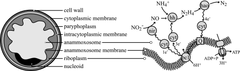
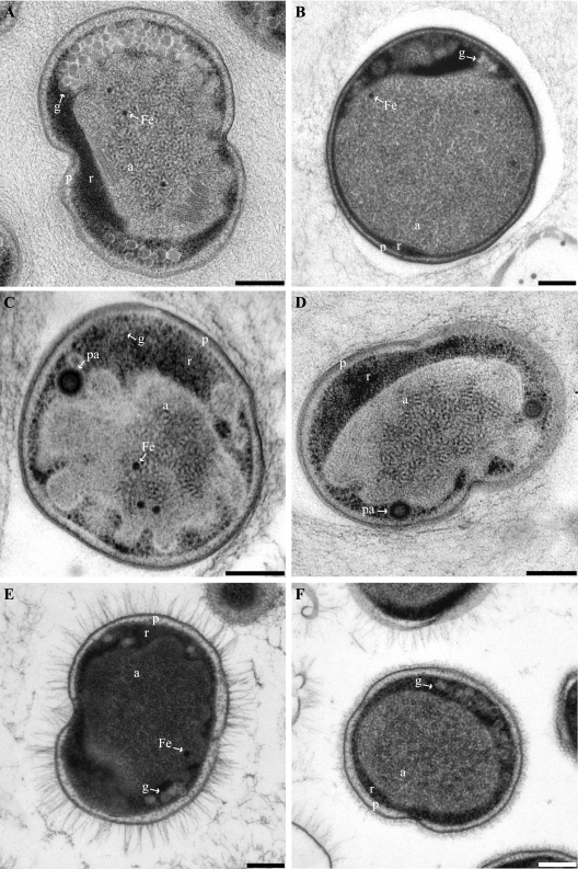
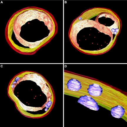
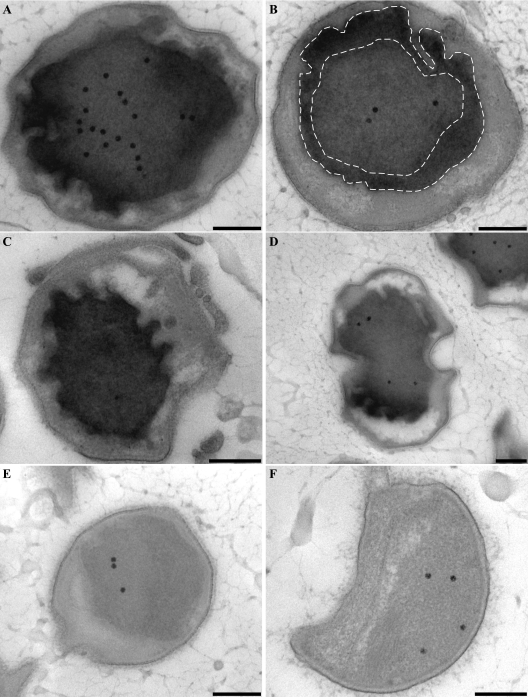
Anaerobic ammonium oxidation (anammox) is an ecologically and industrially important process and is performed by a clade of deeply branching Planctomycetes. Anammox bacteria possess an intracytoplasmic membrane-bounded organelle, the anammoxosome. In the present study, the ultrastructures of four different genera of anammox bacteria were compared with transmission electron microscopy and electron tomography. The four anammox genera shared a common cell plan and contained glycogen granules. Differences between the four genera included cell size (from 800 to 1,100 nm in diameter), presence or absence of cytoplasmic particles, and presence or absence of pilus-like appendages. Furthermore, cytochrome c proteins were detected exclusively inside the anammoxosome. This detection provides further support for the hypothesis that this organelle is the locus of anammox catabolism.


Figures

FIG. 1.
FIG. 1.
Schematic drawing (left), representing the ultrastructure of anammox bacteria and the postulated coupling of the anaerobic oxidation of ammonium (37) to the anammoxosome membrane (right) for the buildup of a proton motive force and subsequent ATP synthesis. nir, nitrite reductase (cytochrome cd1); hh, hydrazine hydrolase; hao, hydrazine/hydroxylamine oxidoreductase (octaheme cyrochrome c) (27, 35); cyt, mono- or diheme cytochrome c electron carriers (8, 11); bc1, cytochrome bc1 complex (complex III); Q, coenzyme Q (ubiquinone).
FIG. 2.
FIG. 2.
Transmission electron micrographs of high-pressure frozen, freeze-substituted, and Epon-embedded thin sections of four anammox genera. All cells are divided into three separate compartments by individual membranes: the paryphoplasm (p), riboplasm (r), and anammoxosome (a) compartments. (A) Dividing “Candidatus Kuenenia stuttgartiensis” cell. (B) “Candidatus Anammoxoglobus propionicus” cell. (C and D) “Candidatus Brocadia fulgida” cells showing riboplasmic particles (pa). (D) Dividing cell. (E and F) Dividing “Candidatus Scalindua spp.” cells with (E) and without (F) pilus-like appendages. Micrographs further show glycogen (g) and putative intra-anammoxosome iron particles (Fe). Scale bars, 200 nm.
FIG. 3.
FIG. 3.
Snapshots of electron tomography models showing the conserved anammox cell plan in three anammox genera. (A) “Candidatus Kuenenia stuttgartiensis” cell. (B) “Candidatus Anammoxoglobus propionicus” cell with riboplasmic particles and with the anammoxosome compartment being divided or lying in a bent configuration in the cell. (C) “Candidatus Brocadia fulgida” cell with (D) riboplasmic particles. Models show (from outside to inside) cell boundary (red), intracytoplasmic membrane (yellow), riboplasmic particles (purple), anammoxosome membrane (pink), and putative intra-anammoxosome iron particles (red). Electron tomography movies are available at http://www.microbiology.science.ruonl/niftrik.
FIG. 4.
FIG. 4.
Transmission electron micrographs showing glycogen staining of high-pressure frozen, freeze-substituted, and Epon-embedded thin sections of four anammox genera. All investigated anammox genera show glycogen staining in the riboplasmic compartment. (A and B) “Candidatus Kuenenia stuttgartiensis.” (B) Negative control incubated with water instead of periodic acid. (C) “Candidatus Anammoxoglobus propionicus.” (D) “Candidatus Brocadia fulgida.” (E and F) “Candidatus Scalindua spp.” cells with (E) and without (F) pilus-like appendages. Scale bars, 200 nm.
FIG. 5.
FIG. 5.
Transmission electron micrographs of chemically fixed and Epon-embedded thin sections of “Candidatus Kuenenia stuttgartiensis” cells showing cytochrome peroxidase staining. This figure is best viewed on screen to avoid change of contrast by printer settings. (A to D) Cytochrome peroxidase staining is observed solely in the anammoxosome compartment. Intense staining occurs within close proximity to the anammoxosome membrane, as outlined by the dashed lines (B) and in places where the membrane is curved (D). (E and F) Negative controls preincubated with KCN and incubated with DAB and H2O2 in the presence of KCN. All sections were poststained for 1 min with Reynolds lead citrate. Scale bars, 200 nm.

References

    1. Anderson, A. J., and E. A. Dawes. 1990. Occurrence, metabolism, metabolic role, and industrial uses of bacterial polyhydroxyalkanoates. Microbiol. Rev. 54450-472. - PMC - PubMed
    1. Arp, D. J., and L. Y. Stein. 2003. Metabolism of inorganic N compounds by ammonium-oxidizing bacteria. Crit. Rev. Biochem. Mol. Biol. 38471-495. - PubMed
    1. Averill, B. A. 1996. Dissimilatory nitrite and nitric oxide reductases. Chem. Rev. 962951-2964. - PubMed
    1. Blattner, F. R., G. Plunkett III, C. A. Bloch, N. T. Perna, V. Burland, M. Riley, J. Collado-Vides, J. D. Glasner, C. K. Rode, G. F. Mayhew, J. Gregor, N. W. Davis, H. A. Kirkpatrick, M. A. Goeden, D. J. Rose, B. Mau, and Y. Shao. 1997. The complete genome sequence of Escherichia coli K-12. Science 2771453-1462. - PubMed
    1. Bonafonte, M. A., C. Solano, B. Sesma, M. Alvarez, L. Montuenga, D. García-Ros, and C. Gamazo. 2000. The relationship between glycogen synthesis, biofilm formation and virulence in Salmonella enteritidis. FEMS Microbiol. Lett. 19131-36. - PubMed

Publication types

MeSH terms

LinkOut - more resources