. 2008 Mar;190(5):1792-811.
doi: 10.1128/JB.01322-07. Epub 2007 Nov 9.

Conditional lethality, division defects, membrane involution, and endocytosis in mre and mrd shape mutants of Escherichia coli

Affiliations

Conditional lethality, division defects, membrane involution, and endocytosis in mre and mrd shape mutants of Escherichia coli

Felipe O Bendezú et al. J Bacteriol. 2008 Mar.

Abstract

Maintenance of rod shape in Escherichia coli requires the shape proteins MreB, MreC, MreD, MrdA (PBP2), and MrdB (RodA). How loss of the Mre proteins affects E. coli viability has been unclear. We generated Mre and Mrd depletion strains under conditions that minimize selective pressure for undefined suppressors and found their phenotypes to be very similar. Cells lacking one or more of the five proteins were fully viable and propagated as small spheres under conditions of slow mass increase but formed large nondividing spheroids with noncanonical FtsZ assembly patterns at higher mass doubling rates. Extra FtsZ was sufficient to suppress lethality in each case, allowing cells to propagate as small spheres under any condition. The failure of each unsuppressed mutant to divide under nonpermissive conditions correlated with the presence of elaborate intracytoplasmic membrane-bound compartments, including vesicles/vacuoles and more-complex systems. Many, if not all, of these compartments formed by FtsZ-independent involution of the cytoplasmic membrane (CM) rather than de novo. Remarkably, while some of the compartments were still continuous with the CM and the periplasm, many were topologically separate, indicating they had been released into the cytoplasm by an endocytic-like membrane fission event. Notably, cells failed to adjust the rate of phospholipid synthesis to their new surface requirements upon depletion of MreBCD, providing a rationale for the "excess" membrane in the resulting spheroids. Both FtsZ and MinD readily assembled on intracytoplasmic membrane surfaces, and we propose that this contributes significantly to the lethal division block seen in all shape mutants under nonpermissive conditions.


Figures
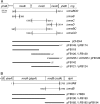
FIG. 1.
FIG. 1.
Genetic constructs. Shown are the E. coli mre (A) and mrd (B) loci, chromosomal deletion-replacements, and inserts present on plasmids and phages. Portions of the chromosome that were replaced with an aph cassette, or an frt scar sequence remaining after eviction of the cassette, are indicated by brackets. Numbers next to brackets refer to the base pairs replaced, counting from the start of mreB (A) or mrdA (B). Inserts were placed downstream of PλR (pFB124, pFB128, and pFB194), PBAD (pFB174), or Plac (all others) control regions of appropriate plasmid/phage vectors (Table 1). LE*, in-frame CTCGAGTAA sequence appended to the end of the gene; M, start codon of mreD changed from GTG to ATG.
FIG. 2.
FIG. 2.
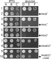
Mre lethality and suppression of MreBCD lethality by extra SdiA. Even-numbered rows show that depletion of MreB (row 2), MreC (row 4), MreD (row 6), or MreBCD (row 8) causes a severe growth defect and that the latter can be suppressed by extra SdiA (row 10). Odd-numbered rows show appropriate wt controls. Cultures were grown to density at 37°C in LB supplemented with appropriate antibiotics and IPTG and were diluted 104 (columns A and D), 105 (B and E), or 106 (C and F)-fold. Aliquots (10 μl) were spotted on LB plates containing either 0.1% glucose (D to F) or IPTG (A to C) at 100 μM (rows 3 to 6) or 250 μM (other rows). Plates were incubated overnight at 37°C and photographed. The strains used were PB103/pFB118/pFB124 and FB17/pFB118/pFB124 (rows 1 and 2), PB103(λFB120)/pFB128 and FB10(λFB120)/pFB128 (rows 3 and 4), PB103(λCH235) and FB11(λCH235) (rows 5 and 6), PB103/pCH244 and FB17/pCH244 (rows 7 and 8), and PB103/pCH244/pCX16 and FB17/pCH244/pCX16 (rows 9 and 10).
FIG. 3.
FIG. 3.
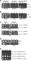
Conditional viability of Mre cells. (A) The MreBCD depletion strain FB30/pFB174 (ΔmreBCD/PBAD::mreBCD) carrying either plasmid pDR3 (Plac::ftsZ) (row 2) or a vector control (row 1) was grown to density at 37°C in LB with 0.5% arabinose. Cultures were diluted and spotted on LB plates containing 0.5% arabinose (columns A to C), 0.1% glucose (columns D to F), or 0.1% glucose plus 100 μM IPTG (columns G to I). Plates were incubated at 37°C. (B) Overnight cultures of strain TB28 (wt) carrying either pDR3 (Plac::ftsZ) (row 2) or a vector control (row 1) were diluted and spotted on LB plates containing the A22-stock solvent methanol (columns A to C), 10 μg/ml A22 (columns D to F), or 10 μg/ml A22 plus 50 μM IPTG (columns G to I). Plates were incubated at 37°C. (C) The MreBCD depletion strain FB21/pFB149 (ΔmreB/Plac::mreBCD) (rows 2 and 4) and its wt parent TB28/pFB149 (rows 1 and 3) were grown overnight at 37°C in LB with 250 μM IPTG. Cultures were diluted and spotted on M9-maltose plates containing 250 μM IPTG (columns A to C) or 0.1% glucose (columns D to F). Plates were incubated at 30°C (rows 1 and 2) or 20°C (RT; rows 3 and 4). (D) An overnight culture of strain TB28 (wt) was diluted and spotted on M9-maltose plates containing methanol (row 1) or 5 μg/ml A22 (row 2). Plates were incubated at RT or 37°C, as indicated. (E) The MreBCD depletion strain FB30/pFB174 (ΔmreBCD/PBAD::mreBCD) (rows 1 and 2) and its wt parent TB28/pFB174 (rows 3 and 4) carrying either plasmid pYT11 (Ptac::relA′) (rows 2 and 4) or pJF118EH (vector) (rows 1 and 3) were grown to density at 30°C in M9-maltose with 0.5% arabinose. Cultures were diluted in LB and spotted on an LB plate with 50 μM IPTG, which was incubated at 30°C. Overnight cultures were serially diluted 104 (columns A, D, and G)-, 105 (columns B, E, and H)-, and 106 (columns C, F, and I)-fold (A to D) or 103 (column A)-, 104 (column B)-, 105 (column C)-, and 106 (column D)-fold (E) in LB, and 10-μl aliquots were spotted in each case. Plates were incubated for 2 days (M9 at RT) or overnight (all others).
FIG. 4.
FIG. 4.
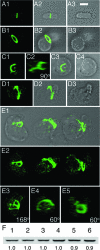
MreB-depleted cells contain normal levels of FtsZ but form aberrant FtsZ structures. (A to E) Immunofluorescence confocal microscopy of MreB-depleted cells with anti-FtsZ antibody, Strain FB17/pFB124/pFB118 (ΔmreBCD/PλR::mreCD/Plac::mreB) was grown in the presence of either 250 μM IPTG (A) or 0.1% glucose (B to E). Samples for staining were taken both early (B) (OD600 = 0.2) and late (A and C to E) (OD600 = 0.6) during growth/depletion. Maximum projection (A1, B1, C1, D1, and E2), DIC (A3, B3, and C4), and merged (A2, B2, C3, D2, and E1) fluorescence images are shown. In panel C2, the image in panel C1 is rotated 90° about the y axis. Panels E3, E4, and E5 show y axis rotations of the left-hand (168°), middle (60°), and right-hand (60°) cell in panel E2, respectively. Bar = 2 μm. (F) Anti-FtsZ immunoblot of whole-cell extracts of MreB-depleted spheres (lanes 3 and 6) and rod-shaped controls (other lanes). Extracts were prepared on strain PB103 (wt) (lanes 1 and 4), and its MreB depletion derivative FB17/pFB124/pFB118 was grown in the presence of either 250 μM IPTG (lanes 2 and 5) or 0.1% glucose (lanes 3 and 6). Cells were harvested both early (OD600 = 0.2) (lanes 1 to 3) and late (OD600 = 0.4 to 0.5) (lanes 4 to 6) during growth/depletion, and each lane received 10 μg total protein. Measured intensity values of FtsZ bands in lanes 2 and 3 were normalized to that in lane 1 and those in lanes 5 and 6 to that in 4. Resulting relative values are shown below the lanes. Cells were grown in LB at 37°C in each case (A to F).
FIG. 5.
FIG. 5.
Aberrant FtsZ assemblies in live MreBCD-depleted spheres. The MreBCD depletion strain FB30(λCH268)/pFB174 [ΔmreBCD(Plac::gfp-zapA)/PBAD::mreBCD] was grown at 37°C in M9-maltose medium lacking arabinose and containing 50 μM IPTG. Live cells were imaged early (A and B) (OD600 = 0.2) and later (C and D) (OD600 = 0.4) during depletion of MreBCD. Cells were mixed with FM4-64 (0.5 μg/ml) immediately prior to imaging. Maximum projection GFP (2), FM4-64 (3), and merged (4) as well as corresponding DIC (1) fluorescence images are shown. Panels D5 to D8 show merged fluorescence images of individual optical slices from the top to the bottom of the cell. The arrows in panel C4 highlight some odd-looking GFP-ZapA accumulations. The arrows in panels D1 and D5 point at small vesicle-like bodies that are both visible by DIC and outlined with FM4-64 fluorescence, while the arrowheads point at much larger bodies that failed to be outlined by the dye. The arrow in panel D8 points at a membrane involution near the bottom of the cell. Bar = 2 μm.
FIG. 6.
FIG. 6.
Intracytoplasmic membrane compartments in MreBCD-depleted spheroids. Shown are live cells of the MreBCD depletion strain FB30/pFB174 (ΔmreBCD/PBAD::mreBCD) producing either transmembrane GFP from lysogenic phage λCH178 [Plac::zipA(1-183)-gfp] (A to C) or periplasmic TTGFP from plasmid pTB6 (Plac::sstorA-gfp) (D to J). Cells were grown to an OD600 of 0.2 at 37°C in LB with 50 μM IPTG and either with (A and D) or without (other panels) 0.5% arabinose. Both DIC (1) and GFP (2) fluorescence images are shown in panels A to I. Panels J show a time lapse series of a spheroid exposed to lysozyme. For this experiment, 2 μl of culture was applied to a slide and covered with a coverslip. A 1-μl aliquot of egg white lysozyme (100 μg/ml in GTE) was then pipetted against the edge of the coverslip, where it was drawn under by capillary action. GFP fluorescence was recorded immediately at 1-s intervals (J1 to J6). The arrowhead in panel J1 points to a compartment that quickly released TTGFP into the medium, in contrast to other compartments that retained the fusion throughout the procedure (arrows). Panel J7 shows a DIC image of the lysozyme-treated sphere a few seconds after the image in panel J6 was taken. Bar = 2 μm.
FIG. 7.
FIG. 7.
Topological separation of internal membrane systems from the external cell membrane. Shown are live MreBCD-depleted spheroids of strain FB30/pFB174 (ΔmreBCD/PBAD::mreBCD) that were grown at 37°C in LB to an OD600 of 0.2. (A to C) Cells were mixed with FM4-64 and imaged immediately. Panels show DIC (1) and FM4-64 (2) fluorescence images. Arrowheads in panels A and B point to large vesicles that were not outlined by FM4-64, while the arrow in panel C points out a vesicle that was. (D to G) Cells were pulse labeled with FM4-64 for 5 min, 30 min prior to imaging. Shown are both DIC (1) and FM4-64 (2 to 6) fluorescence images. Note that all vesicles visible by DIC are now also outlined by FM4-64 fluorescence. In addition, FM4-64 stains structures not readily resolvable by DIC (e.g., arrows in panels D and F). For the cell in panel G, individual z slices from top to bottom (G2 to G5) as well as a maximum projection image (G6) are shown. Note that FM4-64-stained material is present throughout the interior of the cell. (H and I) CellTrace BTME was added 15 min, and FM1-43 immediately, before imaging. DIC (1), FM1-43 (2), and BTME (3) images are shown. Arrowheads point to material labeled with BTME but not with FM1-43. (J to L) The growth medium was supplemented with LY. Cells were gently washed in prewarmed medium lacking LY and imaged immediately. Many vesicles visible by DIC contained trapped LY (e.g., arrowhead in panel K). Some bodies that appeared as a vesicle by DIC did not retain the dye, suggesting that they were still continuous with the external CM and periplasm (arrow in panel L). Panel J illustrates that cells of the wt parent control (TB28) completely failed to retain the dye. Bar = 2 μm.
FIG. 8.
FIG. 8.
Vesicle formation does not require FtsZ polymerization. Shown are spheroids of strain FB30/pTB63/pDR144 (ΔmreBCD/ftsQAZ/Plac::sfiA). (A to D) Cells were inoculated to an OD600 of 0.05 in LP maltose containing no (A) or 0.5 mM (B) IPTG and then grown at 37°C to an OD600 of 0.3, fixed, and labeled with BTME. Shown are DIC (1) and BTME (2) fluorescence. One hundred cells of each culture were further analyzed to determine the average lengths of their long and short axes (C) and the percentages of cells containing internal membrane and/or showing signs of constriction (D). Arrowheads in panel B point to examples of vesicles visible by both DIC and BTME fluorescence. (E to F) Cells were diluted to an OD600 of 0.025 in LB supplemented with 0.5 mM IPTG and either no (E) or 50 μg/ml (F) LY and grown at 37°C to an OD600 of 0.2 to 0.3. For panel E, cells were incubated with BTME for 15 min at 37°C and then treated with FM1-43 immediately before imaging live. Shown are DIC (1), FM1-43 (2), and BTME (3) fluorescence. Note the intracytoplasmic membrane stained by BTME that was inaccessible to FM1-43. For panel F, cells were gently washed in prewarmed growth media prior to imaging. Shown are DIC (1) and LY (2) fluorescence images. Note the trapped LY in intracytoplasmic vesicles. Bar = 2 μm.
FIG. 9.
FIG. 9.
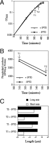
Phospholipid synthesis rates in rods and MreBCD-depleted spheres. The MreBCD depletion strain FB21/pFB149 (ΔmreB/Plac::mreBCD) was grown at 37°C in LP glucose with no or 1 mM IPTG, and the increase in optical density was monitored over time (A). At time points T1 and T2, aliquots were removed to determine phospholipid synthesis rates (B) and cell shape parameters (C).
FIG. 10.
FIG. 10.
Assembly of division proteins on intracytoplasmic membrane. (A to E) Shown are cells of the MreBCD depletion strain FB30(λCH268)/pFB174 [ΔmreBCD (Plac::gfp-zapA)/PBAD::mreBCD]. Cells were grown at 37°C to an OD600 of 0.2 in M9-maltose supplemented with 50 μM IPTG and either no (B to E) or 0.5% (A) arabinose. DIC (1) and GFP-ZapA (2) fluorescence images are shown. Note the accumulation of fluorescence in foci/patches at the cell exteriors as well as surrounding internal vesicles in panels B to E. (F to H) λDR122 (Plac::gfp-minDE) lysogens of TB28 (wt) (F) and the MreBCD depletion strain FB30/pFB174 (ΔmreBCD/PBAD::mreBCD) (G and H) were grown at 37°C to an OD600 of 0.2 to 0.4 in LB supplemented with 50 μM IPTG but lacking arabinose. DIC (1) and GFP-MinD time lapse (2 to 4) fluorescence images at 10-s intervals are shown in panels F and G. Note the transient assembly of GFP-MinD on both the CMs and the surfaces of nearby internal vesicles as it oscillates about the short axis of the spheroid in panel G. The large sphere in panel H was treated with BTME prior to imaging. Shown are DIC fluorescence (1), BTME fluorescence (5), GFP-MinD time-lapse fluorescence at 15 s intervals (2 to 4), and merged GFP-MinD and BTME fluorescence (6 to 8) images. Note the periodic movement of GFP-MinD between BTME-stained sites in the interior of the sphere (H2, H4, H6, and H8) and sites on the CM (H3 and H7).
FIG. 11.
FIG. 11.
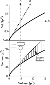
Relationships between volume, surface, and circumference in rods and spheres. The upper panel shows the increase of V/C ratio with increasing volumes of rod-shaped (stippled line) and spherical (solid line) cells. The lower panel shows the increase in surface of rod-shaped and spherical cells with increasing volume. The hatched area highlights the difference in surface requirements between the two cell shapes. See text for details.

Similar articles

Cited by

References

    1. Addinall, S. G., and J. Lutkenhaus. 1996. FtsA is localized to the septum in an FtsZ-dependent manner. J. Bacteriol. 1787167-7172. - PMC - PubMed
    1. Adler, H. I., C. E. Terry, and A. A. Hardigree. 1968. Giant cells of Escherichia coli. J. Bacteriol. 95139-142. - PMC - PubMed
    1. Allison, D. P. 1971. Giant cells of Escherichia coli: a morphological study. J. Bacteriol. 1081390-1401. - PMC - PubMed
    1. Barbour, A. G., L. W. Mayer, and B. G. Spratt. 1981. Mecillinam resistance in Escherichia coli: dissociation of growth inhibition and morphologic change. J. Infect. Dis. 143114-121. - PubMed
    1. Begg, K. J., and W. D. Donachie. 1998. Division planes alternate in spherical cells of Escherichia coli. J. Bacteriol. 1802564-2567. - PMC - PubMed

Publication types

MeSH terms

LinkOut - more resources