. 2008 Jan;146(1):162-77.
doi: 10.1104/pp.107.108902. Epub 2007 Nov 9.

Involvement of polyamine oxidase in wound healing

Affiliations

Involvement of polyamine oxidase in wound healing

Riccardo Angelini et al. Plant Physiol. 2008 Jan.

Abstract

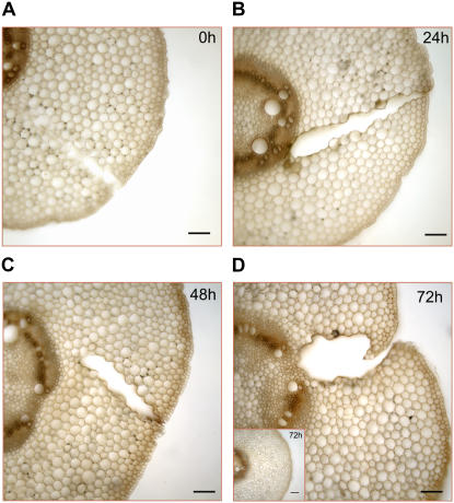
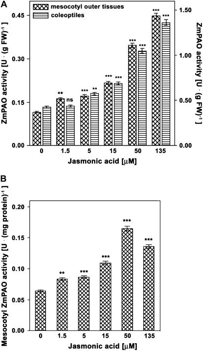
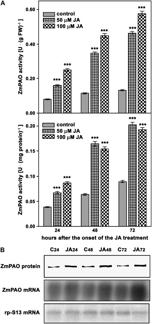
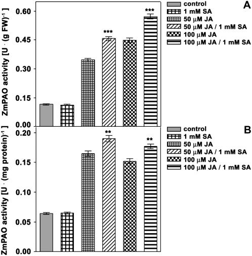
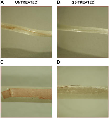
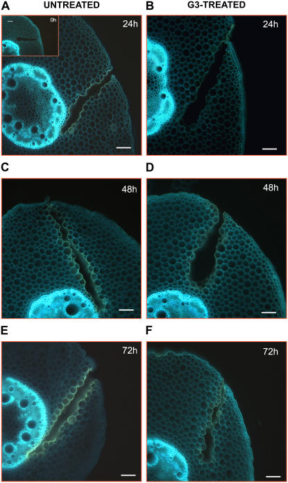
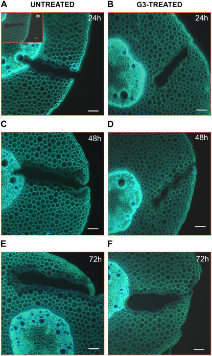
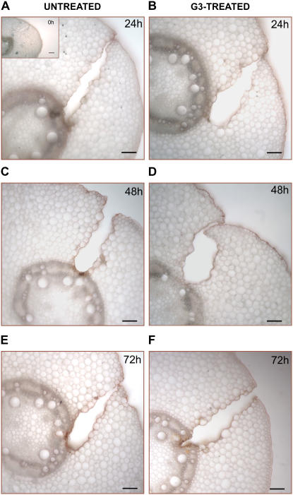
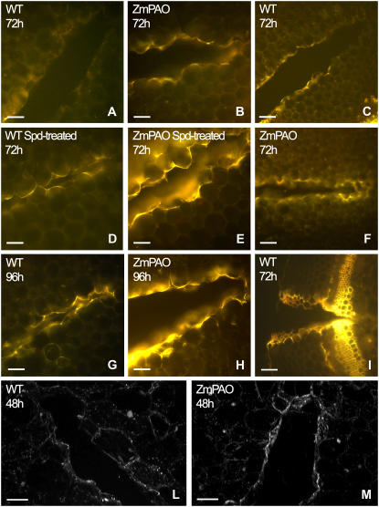
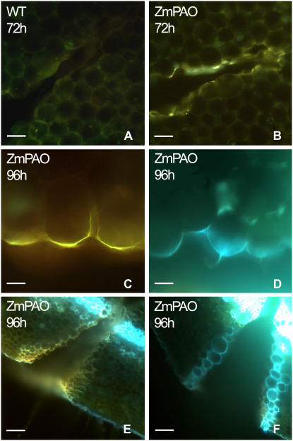
Hydrogen peroxide (H(2)O(2)) is involved in plant defense responses that follow mechanical damage, such as those that occur during herbivore or insect attacks, as well as pathogen attack. H(2)O(2) accumulation is induced during wound healing processes as well as by treatment with the wound signal jasmonic acid. Plant polyamine oxidases (PAOs) are H(2)O(2) producing enzymes supposedly involved in cell wall differentiation processes and defense responses. Maize (Zea mays) PAO (ZmPAO) is a developmentally regulated flavoprotein abundant in primary and secondary cell walls of several tissues. In this study, we investigated the effect of wounding on ZmPAO gene expression in the outer tissues of the maize mesocotyl and provide evidence that ZmPAO enzyme activity, protein, and mRNA levels increased in response to wounding as well as jasmonic acid treatment. Histochemically detected ZmPAO activity especially intensified in the epidermis and in the wound periderm, suggesting a tissue-specific involvement of ZmPAO in wound healing. The role played by ZmPAO-derived H(2)O(2) production in peroxidase-mediated wall stiffening events was further investigated by exploiting the in vivo use of N-prenylagmatine (G3), a selective and powerful ZmPAO inhibitor, representing a reliable diagnostic tool in discriminating ZmPAO-mediated H(2)O(2) production from that generated by peroxidase, oxalate oxidase, or by NADPH oxidase activity. Here, we demonstrate that G3 inhibits wound-induced H(2)O(2) production and strongly reduces lignin and suberin polyphenolic domain deposition along the wound, while it is ineffective in inhibiting the deposition of suberin aliphatic domain. Moreover, ZmPAO ectopic expression in the cell wall of transgenic tobacco (Nicotiana tabacum) plants strongly enhanced lignosuberization along the wound periderm, providing evidence for a causal relationship between PAO and peroxidase-mediated events during wound healing.


Figures

Figure 1.
Figure 1.
Time-course analysis of ZmPAO expression during wound healing in the outer tissues of the nongrowing zone of the maize mesocotyl. Five-day-old seedlings were longitudinally wounded with a blade on the nongrowing zone of the mesocotyl. Two-centimeter-long segments were sampled, after eliminating 1 cm above the seed from unwounded (control) and wounded plants at the indicated time after wounding. Cortical plus epidermal tissues (outer tissues) were obtained by drawing out the stele, and used for determination of extractable ZmPAO activity, western-blot, and northern-blot analysis. A, Extractable ZmPAO activity levels (U: IUs; mean values ± se; n = 5) expressed on a fresh weight (FW) basis. P values indicate statistical significance of differences between ZmPAO activity levels in wounded with respect to control samples for each time. ns, Not significant; *, **, and ***, P values ≤ 0.05, 0.01, and 0.001, respectively. B, Western-blot and northern-blot analyses (samples as in A). Western immunoblotting of extractable ZmPAO fractions was performed after SDS-PAGE loaded on the basis of the total protein content (20 μg/well; top insert). Total RNA was fractionated by agarose/formaldehyde gel electrophoresis, blotted onto a nylon membrane, and hybridized with 32P-labeled maize PAO cDNA probe (middle insert). As a loading control, samples were also hybridized with the cDNA of the S13 ribosomal protein (bottom insert). C, Control; W, wounded; subscripted numbers indicate sampling time.
Figure 2.
Figure 2.
Systemic induction of extractable ZmPAO activity in maize coleoptiles. Five-day-old seedlings were longitudinally wounded with a blade on the nongrowing zone of the mesocotyl and coleoptiles were sampled from unwounded (control) and wounded plants at the indicated time after wounding. Extractable ZmPAO activity levels (U: IUs; mean values ± se; n = 5) are expressed on a fresh weight (FW) basis. P values indicate statistical significance of differences between ZmPAO activity levels in wounded with respect to control samples for each time. ns, Not significant; *, **, and ***, P values ≤ 0.05, 0.01, and 0.001, respectively.
Figure 3.
Figure 3.
Histochemically detected ZmPAO activity in transversal mesocotyl sections from wounded maize seedlings. Hand-cut cross sections (approximately 100 μm thick) obtained from the wounded zone of the mesocotyl and from the corresponding zone from unwounded (control) plants were utilized for light microscopic investigation. ZmPAO activity was histochemically detected at 0 (A), 24 (B), 48 (C), and 72 (D) h after wounding using a peroxidase-coupled assay with DAB as the chromogenic artificial substrate. After washing in distilled water, sections were preincubated in sodium phosphate buffer 10 mm, pH 6.5, containing 60 μg/mL peroxidase and 0.04% DAB for 10 min and then incubated with 3 mm Spd. Reactions were blocked after 3 min by thoroughly washing sections in distilled water. Inset illustrates histochemical ZmPAO activity in section from control plants at 72 h after the onset of the experiment. Bars = 100 μm.
Figure 4.
Figure 4.
Dose-response curve of JA treatment on ZmPAO activity levels in coleoptiles and outer tissues of the nongrowing zone of the maize mesocotyl. Three-day-old seedlings were transferred into an aerated hydroponic culture supplied with a nutrient solution, in the presence or absence of JA at the indicated concentrations. Coleoptiles and mesocotyl segments were sampled after eliminating 0.3 cm above the seed and 0.3 cm below the node from JA-untreated (control) and JA-treated plants 48 h after the onset of the treatments. Mesocotyl cortical plus epidermal tissues (outer tissues) were obtained by drawing out the stele. Coleoptiles and mesocotyl outer tissues were used for determination of extractable ZmPAO activity (U: IUs; mean values ± se; n = 5). P values indicate statistical significance of differences between ZmPAO activity levels in JA-treated with respect to control samples for each concentration tested. ns, Not significant; *, **, and ***, P values ≤ 0.05, 0.01, and 0.001, respectively. A, Extractable ZmPAO activity levels expressed on a fresh weight (FW) basis in the outer tissues of the nongrowing zone of the maize mesocotyl (left y axis) and in coleoptiles (right y axis). B, Extractable ZmPAO activity levels expressed on total protein basis in the outer tissues of the nongrowing zone of the maize mesocotyl.
Figure 5.
Figure 5.
Time-course analyses of ZmPAO expression after JA treatment in the outer tissues of the nongrowing zone of the maize mesocotyl. Three-day-old seedlings were transferred into an aerated hydroponic culture supplied with a nutrient solution in the presence or absence of 50 or 100 μm JA. Mesocotyl segments were sampled, after eliminating 0.3 cm above the seed and 0.3 cm below the node, from JA-untreated (control) and JA-treated plants at the indicated time after the onset of the treatments. Cortical plus epidermal tissues (outer tissues) were obtained by drawing out the stele and used for determination of extractable ZmPAO activity, western-blot, and northern-blot analysis. A, Extractable ZmPAO activity levels (U: IUs; mean values ± se; n = 5) expressed on a fresh weight (FW) basis (top insert) or total protein content (bottom insert). P values indicate statistical significance of differences between ZmPAO activity levels in JA-treated with respect to control samples for each time. ns, Not significant; *, **, and ***, P values ≤ 0.05, 0.01, and 0.001, respectively. B, western-blot and northern-blot analysis (samples as in A). Western immunoblotting of extractable ZmPAO fractions was performed after SDS-PAGE loaded on the basis of the total protein content (20 μg/well; top insert). Total RNA was fractionated by agarose/formaldehyde gel electrophoresis, blotted onto a nylon membrane, and hybridized with 32P-labeled maize PAO cDNA probe (middle insert). As a loading control, samples were also hybridized with the cDNA of the S13 ribosomal protein (bottom insert). C, Control; JA, JA-treated samples; subscripted numbers indicate sampling time.
Figure 6.
Figure 6.
Effects of JA, SA, or JA/SA treatment on ZmPAO activity levels in the outer tissues of the nongrowing zone of the maize mesocotyl. Three-day-old seedlings were transferred into an aerated hydroponic culture supplied with a nutrient solution, in the presence or absence of 50 or 100 μm JA, 1 mm SA, or JA/SA. Mesocotyl segments were sampled, after eliminating 0.3 cm above the seed and 0.3 cm below the node from hormone-untreated (control) and hormone-treated plants after 48 h from the onset of the treatments. Cortical plus epidermal tissues (outer tissues) were obtained by drawing out the stele, and used for extractable ZmPAO enzyme activity analysis at the indicated time after the onset of the treatments (U: IUs; mean values ± se; n = 5). P values indicate statistical significance of differences between ZmPAO activity levels in JA-treated with respect to the respective JA/SA-treated samples for each time. ns, Not significant; *, **, and ***, P values ≤ 0.05, 0.01, and 0.001, respectively. A, Extractable ZmPAO activity levels expressed on a fresh weight (FW) basis. B, Extractable ZmPAO activity levels expressed on total protein content.
Figure 7.
Figure 7.
Inhibition of H2O2 accumulation by G3 in wounded maize mesocotyl. Etiolated seedlings were longitudinally wounded on the mesocotyl with a razor blade (A and B). Alternatively, an epidermal strip was removed (C and D). After root deprivation, wounded seedlings were preincubated for 30 min in the presence or absence of 1 × 10⁻⁵ m G3 (in 10 mm NaH2PO4 solution containing 60 μg/mL peroxidase) prior to be supplied with 1 mg mL−1 DAB for 8 h. Afterward, reactions were stopped by thoroughly washing in distilled water and H2O2 was directly visualized. Photograph magnification: A and B, ×2.0; C and D, ×3.4.
Figure 8.
Figure 8.
UV-induced autofluorescence microscopic analysis in wound-healing maize mesocotyls. Histochemical analysis was performed in hand-cut cross sections (approximately 100 μm thick) obtained from the wounded zone of the mesocotyl of G3-untreated and G3-treated plants, at 0 (inset), 24 (A and B), 48 (C and D), and 72 (E and F) h after wounding. Sections were directly mounted in buffer on slides and observed for autofluorescence under UV light. Bars = 100 μm.
Figure 9.
Figure 9.
UV-induced autofluorescence microscopic analysis after ammonium hydroxide treatment in wound-healing maize mesocotyls. Histochemical analysis was performed in hand-cut cross sections (approximately 100 μm thick) obtained from the wounded zone of the mesocotyl of G3-untreated and G3-treated plants, at 0 (inset), 24 (A and B), 48 (C and D), and 72 (E and F) h after wounding. Sections were incubated for 1 min in 10 mm NH4OH, pH 10, prior to observation under UV light. Bars = 100 μm.
Figure 10.
Figure 10.
Light microscopy detection of suberin aliphatic domain in wound-healing maize mesocotyls. Histochemical analysis was performed in hand-cut cross sections (approximately 100 μm thick) obtained from the wounded zone of the mesocotyl of G3-untreated and G3-treated plants, at 0 (inset), 24 (A and B), 48 (C and D), and 72 (E and F) h after wounding. Sections were preincubated for 10 min in 50% ethanol and then stained for 20 min in a filtered saturated solution of Sudan IV in 70% ethanol. After washing in 50% ethanol (1 min), sections were observed under light microscopy. Bars = 100 μm.
Figure 11.
Figure 11.
Blue-induced autofluorescence and laser scanning confocal microscopy analysis in wound-healing tobacco plants overexpressing ZmPAO in the cell wall. Histochemical analysis under fluorescence microscopy was performed in hand-cut cross sections (approximately 100 μm thick) obtained from the wounded zone of the second internode (numbering from the shoot apex) of Spd-untreated (A–C and F–I) and Spd-treated (D–E) wild-type (WT) as well as ZmPAO transgenic tobacco plants overexpressing ZmPAO in the cell wall (ZmPAO), at 72 and 96 h after wounding. Sections were directly mounted on slides and observed for autofluorescence under blue light (A–I). A, B, D, E, and G to I, bar = 100 μm; C and F, bar = 200 μm. Confocal microscopy analysis was performed on hand-cut cross sections from wild-type and ZmPAO transgenic tobacco plants at 48 h after wounding. L and M sections show a three-dimensional reconstruction of autofluorescence images after blue excitation. L and M, bar = 100 μm.
Figure 12.
Figure 12.
UV-induced autofluorescence microscopic analysis after ammonium hydroxide treatment in wound-healing tobacco plants overexpressing ZmPAO in the cell wall. Histochemical analysis was performed in hand-cut cross sections (approximately 100 μm thick) obtained from the wounded zone of the second internode (numbering from the shoot apex) of Spd-untreated wild type (WT) as well as tobacco plants overexpressing ZmPAO in the cell wall (ZmPAO), at 72 and 96 h after wounding. Some sections were directly mounted on slides and observed for autofluorescence under UV (A–C and E) light. Other sections were incubated for 1 min in 10 mm NH4OH, pH 10, prior to observation under UV light (D and F). A, B, E, and F, bar = 100 μm; C and D, bar = 50 μm.

Similar articles

Cited by

References

    1. Alcázar R, Marco F, Cuevas JC, Patron M, Ferrando A, Carrasco P, Tiburcio AF, Altabella T (2006) Involvement of polyamines in plant response to abiotic stress. Biotechnol Lett 28 1867–1876 - PubMed
    1. Allan AC, Fluhr R (1997) Two distinct sources of elicited reactive oxygen species in tobacco epidermal cells. Plant Cell 9 1559–1572 - PMC - PubMed
    1. Apel K, Hirt H (2004) Reactive oxygen species: metabolism, oxidative stress and signal transduction. Annu Rev Plant Biol 55 373–399 - PubMed
    1. Artiss JD, Entwistle WM (1981) The application of a sensitive uricase-peroxidase couple reaction to a centrifugal fast analyser for the determination of uric acid. Clin Chim Acta 116 301–309 - PubMed
    1. Augeri M, Angelini R, Federico R (1990) Sub-cellular localization and tissue distribution of polyamine oxidase in maize (Zea mays L.) seedlings. J Plant Physiol 136 690–695

Publication types

MeSH terms

LinkOut - more resources