. 2008 Jan;146(1):140-8.
doi: 10.1104/pp.107.107870. Epub 2007 Nov 9.

Diarch symmetry of the vascular bundle in Arabidopsis root encompasses the pericycle and is reflected in distich lateral root initiation

Affiliations

Diarch symmetry of the vascular bundle in Arabidopsis root encompasses the pericycle and is reflected in distich lateral root initiation

Boris Parizot et al. Plant Physiol. 2008 Jan.

Abstract

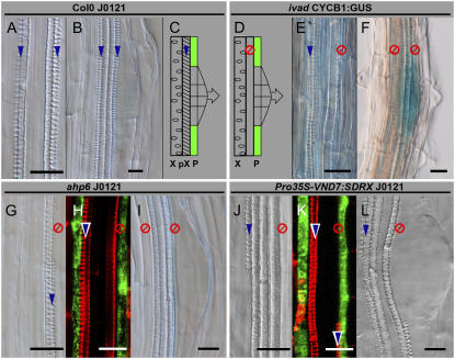
The outer tissues of dicotyledonous plant roots (i.e. epidermis, cortex, and endodermis) are clearly organized in distinct concentric layers in contrast to the diarch to polyarch vascular tissues of the central stele. Up to now, the outermost layer of the stele, the pericycle, has always been regarded, in accordance with the outer tissue layers, as one uniform concentric layer. However, considering its lateral root-forming competence, the pericycle is composed of two different cell types, with one subset of cells being associated with the xylem, showing strong competence to initiate cell division, whereas another group of cells, associated with the phloem, appears to remain quiescent. Here, we established, using detailed microscopy and specific Arabidopsis thaliana reporter lines, the existence of two distinct pericycle cell types. Analysis of two enhancer trap reporter lines further suggests that the specification between these two subsets takes place early during development, in relation with the determination of the vascular tissues. A genetic screen resulted in the isolation of mutants perturbed in pericycle differentiation. Detailed phenotypical analyses of two of these mutants, combined with observations made in known vascular mutants, revealed an intimate correlation between vascular organization, pericycle fate, and lateral root initiation potency, and illustrated the independence of pericycle differentiation and lateral root initiation from protoxylem differentiation. Taken together, our data show that the pericycle is a heterogeneous cell layer with two groups of cells set up in the root meristem by the same genetic pathway controlling the diarch organization of the vasculature.


Figures

Figure 1.
Figure 1.
TEM analysis of pericycle cells. TEM analysis of pericycle cells at the protoxylem pole (A, C, and E) as compared to pericycle cells at the protophloem pole (B, D, and F) after growth on control medium (A and B), NPA (C and D), and NPA followed by 10 h of incubation on NAA (E and F). A newly formed periclinally oriented cell wall in a pericycle cell at the phloem pole on NPA is indicated by two black arrowheads. pc, Pericycle; pP, protophloem pole; pX, protoxylem pole. Bars = 3.5 μm (A–D); 3 μm (E and F).
Figure 2.
Figure 2.
Histological analysis of heterogeneous GFP expression in Rm1007 and J0121 enhancer trap lines. A to H, J0121 analysis. A, Binocular imaging of a J0121 plantlet 5 DAG. B and C, Confocal imaging of mature part and apex, respectively; D, transversal section of mature part; E, enlargement of the protoxylem and the associated pericycle; F, lateral root initiation. G and H, Expression of the GFP in the root tip of a 5-DAG J0121 plantlet germinated on medium supplemented with 10−5 m NPA (G), on nonsupplemented medium for 2 d, and then transferred to medium supplemented with 10−5 m NAA for 24 h (H). Asterisks mark pericycle cells associated with xylem poles. I to N, Rm1007 analysis. I and J, Binocular imaging of a Rm1007 plantlet root tip 5 DAG, the arrow indicating the quiescent center (I); mature zone where lateral root formation is induced by transferring a plantlet germinated on nonsupplemented medium for 2 d to medium supplemented with 10−5 m NAA for 24 h (J). K to N, Confocal longitudinal imaging of Rm1007 in a protoxylem plan (K), in a plan parallel to the phloem in which can be seen three cell files of pericycle expressing GFP (L), during embryo heart-stage formation (M), and in the mature root (N). The eye symbol gives observation axis indication referring to the pericycle cells expressing GFP. Bars = 50 μm (C and I).
Figure 3.
Figure 3.
Histological study of stele alterations in lhw and ivad mutants. A to H, Transversal sections of 5-DAG embedded plants: C24 plants, respectively, in the differentiation zone and in mature parts of the root (A–C); lhw plants, respectively, at the level of the four quiescent center cells and in mature part of the root (D and E); and ivad plant in the mature part of the root (G and H). F and I, Longitudinal confocal imaging of lhw (F; one section) and ivad mutants (I; z-series stack superposition). Black lines on sections delimit scheme enlargement. Green, pericycle; blue and darker blue, xylem and protoxylem; orange, phloem poles. The eye symbol gives observation axis indication referring to the pericycle cells expressing GFP. Bars = 50 μm.
Figure 4.
Figure 4.
Lateral root initiation in lhw mutant. J0121 (A and B) and lhw (C and D) plants 3 DAG on medium supplemented with 10−5 m NPA (A and C) and then transferred 4 d on medium supplemented with 10−5 m NAA (B and D). Asterisks indicate lateral root emergence. The eye symbol gives observation axis indication referring to the pericycle cells expressing GFP. Bars = 100 μm.
Figure 5.
Figure 5.
Lateral root initiation in ivad, ahp6, and Pro35S-VND7:SDRX mutants does not require differentiated protoxylem. Lateral root initiation in relation to the presence or absence of fully developed protoxylem elements has been studied in Col-0 J0121 (A and B) and in three lines showing alteration in protoxylem differentiation: ivad (E and F), ahp6 J0121 (G–I), and Pro35S-VND7:SDRX J0121 (J–L). In Col-0 J0121, metaxylem is surrounded by protoxylem (A and C) and lateral root initiation happens in the pericycle adjacent to the protoxylem. In the three displayed mutants, absence of protoxylem surrounding metaxylem can be noticed (E, G, and J) and this absence prevents GFP expression neither in pericycles associated with xylem poles (H and K) nor in lateral root initiation (D, F, I, and L). Fully differentiated protoxylem presence is indicated with blue arrows; absence is indicated with the red symbol. P, Pericycle; pX, protoxylem; X, metaxylem. Bars = 20 μm.
Figure 6.
Figure 6.
Auxin treatment on wol mutant. Col-0 J0121 (A–C) and wol J0121 (D–F) have been cultivated (A, B, D, and E) 3 DAG on nonsupplemented medium and then transferred (C and F) 48 h on supplemented medium with 10−5 m NAA. Bar = 50 μm.
Figure 7.
Figure 7.
Model for stele determination and differentiation in Arabidopsis. The model (A) proposed previously (Barlow, 1984; Skene, 2000) introduced the diarch organization of the vascular bundle that becomes determined at the level of stele initials. The differentiation of the vascular tissues will later induce the bipolar specification of the pericycle and the differentiation into different cell populations, one of which is associated with the xylem pole and competent for lateral root initiation. Here, we propose an alternative model (B) in which the diarch organization affecting both the vascular bundle and the pericycle is determined from the stele initials onward. The capacity of pericycle cells to give rise to lateral root primordia occurs later and remains associated with vascular tissue differentiation. Blue and orange, respectively, represent xylem and phloem tissues. Green represents the pericycle, with cells associated with the xylem (light green) or phloem (darker green) tissues.

Similar articles

Cited by

References

    1. Barlow P (1984) Positional controls in root development. In PW Barlow, DJ Carr, eds, Positional Controls in Plant Development. Cambridge University Press, Cambridge, UK, pp 281–318
    1. Beeckman T, Burssens S, Inzé D (2001) The peri-cell-cycle in Arabidopsis. J Exp Bot 52 403–411 - PubMed
    1. Casero P, Casimiro I, Knox J (1998) Occurrence of cell surface arabinogalactan-protein and extensin epitopes in relation to pericycle and vascular tissue development in the root apex of four species. Planta 204 252–259
    1. Casero PJ, García-Sánchez C, Lloret PG, Navascués J (1989) Changes in cell length and mitotic index in vascular pattern-related pericycle cell types along the apical meristem and elongation zone of the onion root. Protoplasma 153 85–90
    1. Casimiro I, Beeckman T, Graham N, Bhalerao R, Zhang H, Casero P, Sandberg G, Bennett MJ (2003) Dissecting Arabidopsis lateral root development. Trends Plant Sci 8 165–171 - PubMed

Publication types

MeSH terms