. 2008 Jan;7(1):131-40.
doi: 10.1128/EC.00301-07. Epub 2007 Nov 9.

Exit from host cells by the pathogenic parasite Toxoplasma gondii does not require motility

Affiliations

Exit from host cells by the pathogenic parasite Toxoplasma gondii does not require motility

Mark D Lavine et al. Eukaryot Cell. 2008 Jan.

Abstract

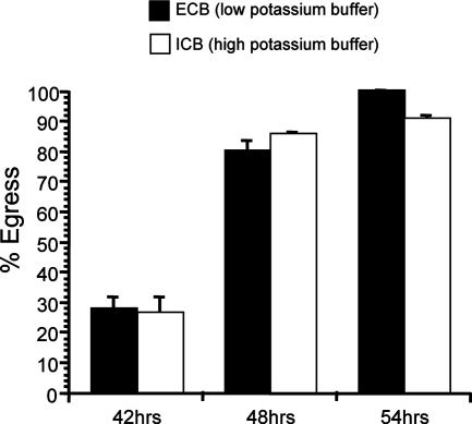
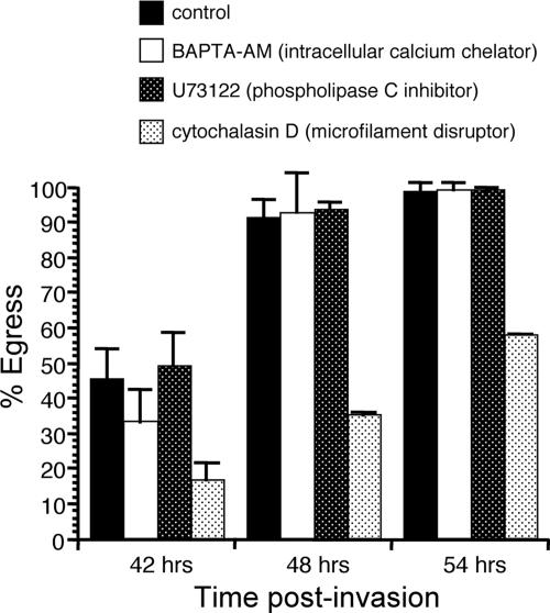
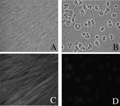
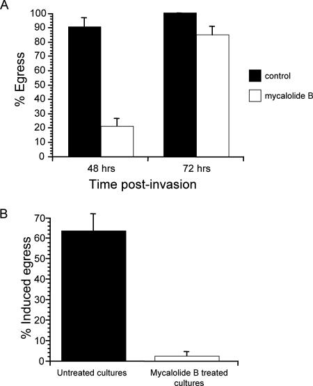
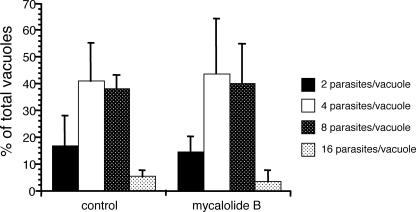
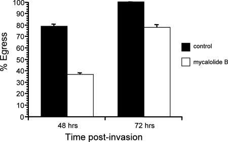
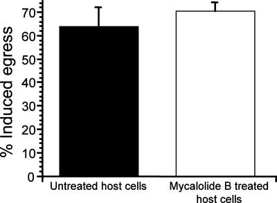
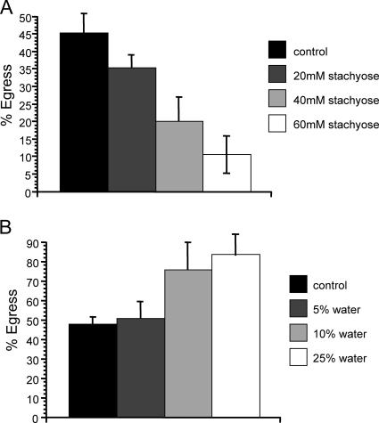
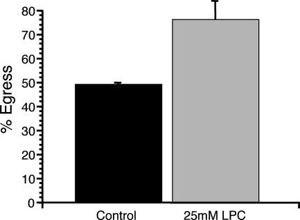
The process by which the intracellular parasite Toxoplasma gondii exits its host cell is central to its propagation and pathogenesis. Experimental induction of motility in intracellular parasites results in parasite egress, leading to the hypothesis that egress depends on the parasite's actin-dependent motility. Using a novel assay to monitor egress without experimental induction, we have established that inhibiting parasite motility does not block this process, although treatment with actin-disrupting drugs does delay egress. However, using an irreversible actin inhibitor, we show that this delay is due to the disruption of host cell actin alone, apparently resulting from the consequent loss of membrane tension. Accordingly, by manipulating osmotic pressure, we show that parasite egress is delayed by releasing membrane tension and promoted by increasing it. Therefore, without artificial induction, egress does not depend on parasite motility and can proceed by mechanical rupture of the host membrane.


Figures

FIG. 1.
FIG. 1.
Effect of high extracellular [K+] on egress. At 30 h postinvasion, the culture medium was switched to either ECB, which allows K+ efflux, or ICB, which blocks K+ efflux. Percent egress is the percentage of lysed vacuoles at 42 h, 48 h, or 54 h postinvasion relative to the level at 30 h postinvasion. Data are means from three independent experiments. Error bars, standard deviations.
FIG. 2.
FIG. 2.
Effects of motility inhibitors on natural egress. At 30 h postinvasion, the normal tissue culture medium was switched to normal medium containing either 25 μM BAPTA-AM, 5 μΜ U73122, 10 μM cytochalasin D, or a solvent control. Percent egress was calculated as for other egress assays. Data are means from three independent experiments. Error bars, standard deviations.
FIG. 3.
FIG. 3.
Effect of mycalolide B on HFFs. HFFs were exposed for 5 min either to solvent alone in medium (control) or to 3 μM mycalolide B. They were then washed, and 48 h later, they were photographed. (A) Phase micrograph of control HFFs. (B) Phase micrograph of HFFs exposed to mycalolide B. (C) Phalloidin staining of cells in panel A, showing microfilaments. (D) Phalloidin staining of cells in panel B, showing continued disruption of microfilaments.
FIG. 4.
FIG. 4.
Effects of treating HFFs and parasites with mycalolide B on noninduced and induced egress. (A) Noninduced egress was monitored at 48 h and 72 h postinvasion, after exposure of infected cells to either 3 μM mycalolide B or a solvent control for 1 h starting at 30 h postinvasion. (B) Infected cells were exposed to 1 μM A23187 for 10 min, 30 h after being exposed to either 3 μM mycalolide B or DMEM solvent (control) for 10 min. Percent egress indicates the percentage of lysed vacuoles after ionophore exposure relative to the preexposure level. In both panels, data are means from three independent experiments.
FIG. 5.
FIG. 5.
Parasite development after transient exposure to mycalolide B. At 1 h after infection, infected cells were treated with either 3 μM mycalolide B or DMEM solvent (control) for 1 h. The number of parasites was determined for at least 100 vacuoles 23 h later. Data are expressed as the percentage of vacuoles containing the indicated number of parasites and are means from three independent experiments. Each error bar equals 1 standard deviation.
FIG. 6.
FIG. 6.
Effect of disruption of host cell actin alone on parasite egress. HFFs were pretreated either with 3 μM mycalolide B or with DMEM solvent (control) for 10 min before parasites were allowed to invade. Percent egress is the percentage of lysed vacuoles at 48 h or 72 h postinvasion relative to the level at 30 h postinvasion. Data are means from three independent experiments. Each error bar equals 1 standard deviation.
FIG. 7.
FIG. 7.
Effect of disruption of host cell actin alone on induced egress. Parasites were allowed to infect host cells that had been pretreated with either 3 μM mycalolide B or DMEM solvent alone (control) for 10 min. At 30 h postinvasion, cultures were exposed to 1 μM A23187 for 10 min. Percent egress is the percentage of lysed vacuoles after ionophore exposure relative to the preexposure level. Data are mean percentages for three independent experiments. Each error bar equals 1 standard deviation.
FIG. 8.
FIG. 8.
Effect of osmotic stress on egress. Parasites were allowed to invade for 1 h and to develop in HFFs for 30 h. At 30 h postinvasion, the normal tissue culture medium was switched to either a hypertonic medium consisting of normal medium plus 20 mM, 40 mM, or 60 mM stachyose (A), a hypotonic medium consisting of either 95% normal medium plus 5% H2O, 90% normal medium plus 10% H2O, or 75% normal medium plus 25% H2O (B), or normal medium as a control (solid bars in both panels). Percent egress is the percentage of lysed vacuoles at 42 h relative to the level at 30 h postinvasion. Data are means from three independent experiments. Each error bar equals 1 standard deviation.
FIG. 9.
FIG. 9.
Effect of LPC on egress. Parasites were allowed to invade for 1 h and to develop in HFFs for 30 h. At 30 h postinvasion, the normal tissue culture medium was switched to either AIM-V plus Albumax medium (control) or AIM-V plus Albumax medium plus 25 mM LPC. Percent egress is the percentage of lysed vacuoles at 42 h relative to the level at 30 h postinvasion. Data are means from three independent experiments. Each error bar equals 1 standard deviation.

Similar articles

Cited by

References

    1. Alexander, D. L., J. Mital, G. E. Ward, P. Bradley, and J. C. Boothroyd. 2005. Identification of the moving junction complex of Toxoplasma gondii: a collaboration between distinct secretory organelles. PLoS Pathog. 1e17. - PMC - PubMed
    1. Black, M. W., G. Arrizabalaga, and J. C. Boothroyd. 2000. Ionophore-resistant mutants of Toxoplasma gondii reveal host cell permeabilization as an early event in egress. Mol. Cell. Biol. 209399-9408. - PMC - PubMed
    1. Black, M. W., and J. C. Boothroyd. 2000. Lytic cycle of Toxoplasma gondii. Microbiol. Mol. Biol. Rev. 64607-623. - PMC - PubMed
    1. Buitrago-Rey, R., J. Olarte, and J. E. Gomez-Marin. 2002. Evaluation of two inhibitors of invasion: LY311727 [3-(3-acetamide-1-benzyl-2-ethyl-indolyl-5-oxy)propane phosphonic acid] and AEBSF [4-(2-aminoethyl)-benzenesulphonyl fluoride] in acute murine toxoplasmosis. J. Antimicrob. Chemother. 49871-874. - PubMed
    1. Chernomordik, L. V., M. M. Kozlov, G. B. Melikyan, I. G. Abidor, V. S. Markin, and Y. A. Chizmadzhev. 1985. The shape of lipid molecules and monolayer membrane-fusion. Biochim. Biophys. Acta 812643-655.

Publication types

LinkOut - more resources