. 2008 May 1;111(9):4559-70.
doi: 10.1182/blood-2007-05-092825. Epub 2007 Nov 9.

Autoantibodies to the C-terminal subunit of RLIP76 induce oxidative stress and endothelial cell apoptosis in immune-mediated vascular diseases and atherosclerosis

Affiliations

Autoantibodies to the C-terminal subunit of RLIP76 induce oxidative stress and endothelial cell apoptosis in immune-mediated vascular diseases and atherosclerosis

Paola Margutti et al. Blood. .

Abstract

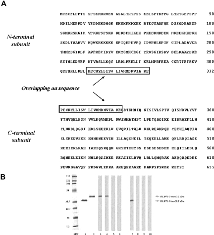
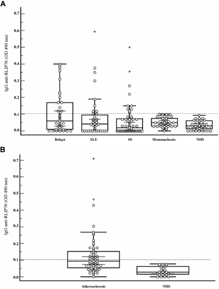
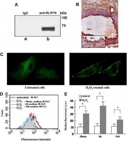
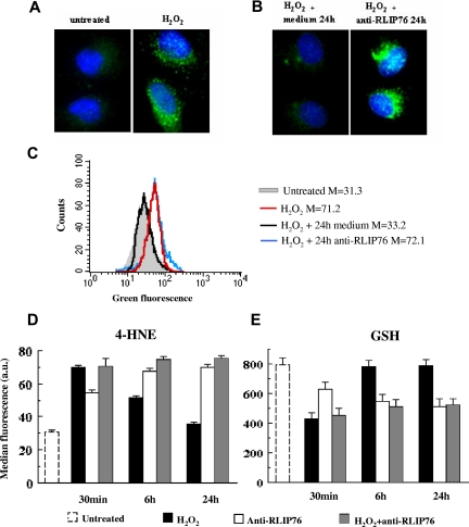
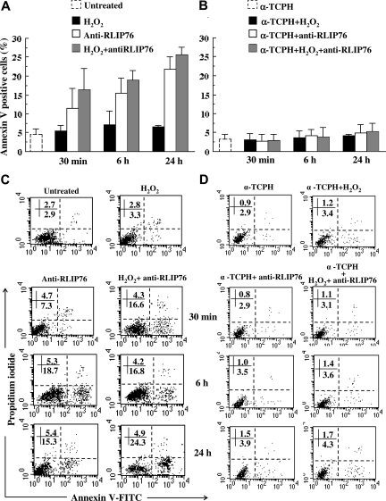
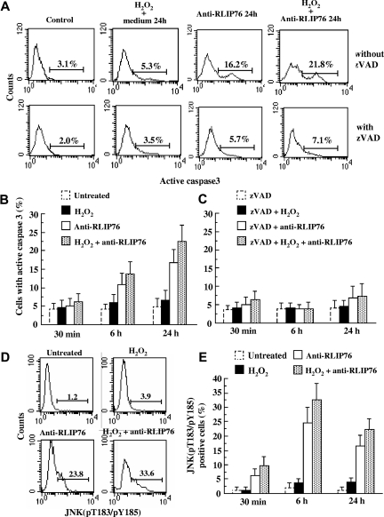
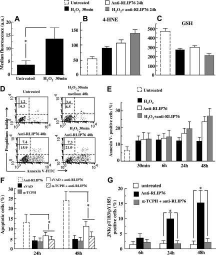
Although detection of autoantibodies in the peripheral blood from patients with immune-mediated endothelial dysfunctions has so far failed to provide tools of diagnostic or pathogenetic value, putative bioindicators include anti-endothelial cell antibodies, a heterogeneous family of antibodies that react with autoantigens expressed by endothelial cells. In this study, to identify endothelial autoantigens involved in the autoimmune processes causing endothelial damage, we screened a human microvascular endothelial cell cDNA library with sera from patients with Behçet's disease. We identified antibodies to the C-terminus of Ral binding protein1 (RLIP76), a protein that catalyzes the ATP-dependent transport of glutathione (GSH) conjugates including GSH-4-hydroxy-t-2,3-nonenal, in the serum of a significant percentage of patients with various diseases characterized by immune-mediated endothelial dysfunction, including Behçet disease, systemic sclerosis, systemic lupus erythematosus and carotid atherosclerosis. These autoantibodies increased intracellular levels of 4-hydroxy-t-2,3-nonenal, decreased levels of GSH and activated C-Jun NH2 Kinase signaling (JNK), thus inducing oxidative stress-mediated endothelial cell apoptosis. The dietary antioxidant alpha-tocopherol counteracted endothelial cell demise. These findings suggest that autoantibodies to RLIP76 play a pathogenetic role in immune-mediated vascular diseases and represent a valuable peripheral blood bioindicator of atherosclerosis and immune-mediated vascular diseases.


Figures

Figure 1
Figure 1
The amino acid sequence and the immunochemical characterization of the N- and C-terminal regions of RLIP76. (A) The nucleotide sequence of the cloned cDNA (GenBank accession number NM 006788) was divided in 2 subunits by PCR with specific primers. The cDNA subunits were cloned in an expression vector and the N- and C-terminal regions of the protein were expressed and purified. The overlapped amino acids were squared. (B) The molecular size and the purity of the expressed proteins were confirmed by 10% SDS-PAGE stained by Coomassie blue (lane 1, N-terminal region; lane 2, C-terminal region) and serum immunoreactivity was analyzed by immunoblotting (lanes 3-6, C-terminal region; lanes 7-10, N-terminal region). Lanes 3,7: monoclonal antibody specific to 6-histidine tail; lanes 4,8: serum pool from the 2 patients with Behçet disease (BD) used in screening the library; lane 5,9: representative serum from a healthy subject; lanes 6,10: control without serum.
Figure 2
Figure 2
Anti-RLIP76 C-ter antibodies in patients and healthy controls. (A) Box-whisker plot of anti-RLIP76 C-ter IgG in patients with BD, SLE, SS, infectious mononucleosis, and from sex- and age-matched healthy donors (NHS). (B) Box-whisker plot of anti-RLIP76 C-ter IgG in patients with carotid atherosclerosis and from sex- and age-matched healthy donors (NHS). Median, quartiles, range, and possibly extreme values are indicated. The broken line represents the cutoff (mean + 2 SD for the healthy controls). Outliers are represented as +.
Figure 3
Figure 3
RLIP76 expression and localization.(A) EAhy926 were immunoprecipitated with mouse polyclonal anti-RLIP76 C-ter antibodies. The immunoprecipitates were analyzed by Western blotting, using anti–human RLIP76 C-ter antibodies. Bound antibodies were visualized with HRP-conjugated anti–human IgG and immunoreactivity was assessed by ECL. Virtually no reactivity was found with immunoprecipitates obtained using non RLIP76-specific IgG (irrelevant). (B) RLIP76 expression in vascular tissue was detected by immunohistochemistry on tissue arrays with histologic sections from normal vascular human tissue incubated with mouse anti-RLIP76 C-ter antibodies. Intense immunoreactivity was observed in vascular endothelium. (C) Immunofluorescence analysis of RLIP76 distribution in EAhy926 untreated cells (left panel) or treated with 30 μM H2O2 30 minutes (right panel). Original magnification 500×, objective 100×, numeric aperture 1.4. (D,E) Flow cytometric analysis after surface staining of EAhy926 cells with antibodies to the RLIP76 C-terminus. (D) Results obtained in a representative experiment. Full light-gray histogram: untreated control cells; red histogram: H2O2-treated cells incubated for 30 minutes with fresh medium; black histogram: H2O2-treated cells incubated for 6 hours with fresh medium; blue histogram: H2O2-treated cells incubated for 24 hours with fresh medium. (E) Time-course evaluation of RLIP76 expression in untreated cells and in cells treated with H2O2 and then incubated for 30 minutes, 6 hours, and 24 hours in fresh medium (mean ± SD of the results obtained from 3 different experiments). *P < .01 by Student t test.
Figure 4
Figure 4
Intracellular 4-HNE and GSH levels in EAhy926 endothelial cells. (A,B) Immunofluorescence analysis of the formation of 4-HNE adducts with histidine in EAhy926 cells stained with a 4-HNE specific antibody and counterstained with Hoechst dye to reveal nuclei. Panel A left, untreated cells; panel A right, H2O2-treated cells; panel B left, H2O2-treated cells incubated for 24 hours with fresh medium; panel B right, H2O2-treated cells incubated for 24 hours with medium containing anti-RLIP76 C-ter antibodies. Magnification 1500×, objective 100×, numeric aperture 1.4. (C) Quantitative analysis of 4-HNE adducts by flow cytometry in a representative experiment. Gray histogram, untreated control cells; red histogram, H2O2-treated cells; black histogram, H2O2-treated cells incubated for 24 hours with fresh medium; blue histogram, H2O2-treated cells incubated for 24 hours with medium containing anti-RLIP76 C-ter antibodies. Numbers represent median values of fluorescence intensity. (D) Quantitative time-course evaluation of 4-HNE intracellular content in untreated control cells; in cells incubated for 30 minutes, 6 hours, and 24 hours with anti-RLIP76 C-ter antibodies; in cells treated with H2O2; and in cells treated with H2O2 and then incubated for an additional 30 minutes, 6 hours, and 24 hours in fresh medium or in medium containing anti-RLIP76 C-ter antibodies. Statistical analysis performed by Student t test indicated P < .01 for: control untreated cells versus cells incubated with anti-RLIP76 C-ter antibodies for 30 minutes, 6 hours, and 24 hours; untreated cells versus H2O2-treated cells incubated for 30 minutes and 6 hours in fresh medium; H2O2-treated cells incubated in fresh medium versus H2O2-treated cells incubated for 6 hours and 24 hours with medium containing anti-RLIP76 C-ter antibodies. (E) Quantitative time-course evaluation of GSH intracellular content. Statistical analysis performed by Student t test indicated P < .01 for: untreated cells versus H2O2-treated cells incubated for 30 minutes in fresh medium; untreated cells versus cells incubated for 30 minutes, 6 hours, and 24 hours with anti-RLIP76 C-ter antibodies; H2O2-treated cells incubated in fresh medium versus H2O2-treated cells incubated for 6 hours and 24 hours with medium containing anti-RLIP76 C-ter antibodies. Data reported in panels D and E are the mean plus or minus SD of the results obtained from 3 different experiments.
Figure 5
Figure 5
Induction of apoptosis by anti-RLIP76 C-ter antibodies in EAhy926 endothelial cells.(A,C) Flow cytometric analysis after double staining with annexin V/propidium iodide of untreated control cells; cells incubated for 30 minutes, 6 hours, and 24 hours with anti-RLIP76 C-ter antibodies; and in cells treated with H2O2 and then incubated additional 30 minutes, 6 hours, and 24 hours in fresh medium or with medium containing anti-RLIP76 C-ter antibodies. (B,D) Cells were also pretreated with α-TCPH. (A,B) Results obtained from 3 independent experiments are reported as mean plus or minus SD. (C,D) Dot plots from a representative experiment. Numbers represent the percentage of annexin V single positive (early apoptosis, bottom right quadrant) or annexin V/PI double positive cells (late apoptosis, bottom right quadrant). Statistical analysis performed by Student t test indicated P < .01 for: control untreated cells versus H2O2-treated cells; control untreated cells versus cells incubated for 30 minutes, 6 hours, and 24 hours with anti-RLIP76 C-ter antibodies; and cells treated with H2O2 and then incubated in fresh medium versus cells treated with H2O2 and then incubated for 6 hours and 24 hours with medium containing anti-RLIP76 C-ter antibodies. P < .01 for any treatment (panel A) versus the same treatment performed after α-TCPH preincubation (panel B).
Figure 6
Figure 6
Apoptotic pathway induced by anti-RLIP76 C-ter in EAhy926 cell line. (A) Flow cytometric data were obtained in the absence (first row) or in the presence (second row) of 30 μM pan-caspase inhibitor zVAD. The numbers in each panel refer to the percentage of cells containing caspase 3 in its active form. Results obtained in a representative experiment are reported. (B,C) Graphs showing the mean plus or minus SD of the percentages of cells with the active form of caspase 3 obtained from 3 different experiments done without (panel B) or with (panel C) zVAD. Statistical analyses indicate a significant (P < .01) decrease in caspase 3 activity in cells pretreated with zVAD before anti-RLIP76 C-ter antibody exposure. (D) Quantitative flow cytometric analysis investigating the JNK activation state 6 hours after the various treatments with a polyclonal antibody able to identify JNK (pT183/pY185). The numbers in each panel refer to the percentage of cells containing JNK in its active form. Results obtained in a representative experiment are reported. (E) Time-course analysis of the activation state of JNK. Statistical analyses of the results obtained from 3 independent experiments (reported as mean ± SD) indicate a significant difference (P < .01) between cells treated with RLIP76 C-ter antibodies, alone or in combination with H2O2, and both untreated and H2O2-treated cells at any time point considered.
Figure 7
Figure 7
Effects of anti-RLIP76 C-ter antibodies on human microvascular primary cells. (A) Flow cytometric analysis of HMVEC-L cells, either untreated or H2O2-treated cells, after surface staining with antibodies to RLIP76. Mean plus or minus SD of the results obtained from 3 different experiments. *P < .01 by Student t test. (B,C) Quantitative flow cytometric analysis of 4-HNE adducts and GSH intracellular content in: untreated control cells, cells treated with H2O2 for 30 minutes, cells incubated with anti-RLIP76 C-ter antibodies for 24 hours, and cells treated with H2O2 for 30 minutes and then incubated for an additional 24 hours in medium containing anti-RLIP76 C-ter antibodies. Data reported in panels B and C are the mean plus or minus SD of the results obtained from 3 different experiments. Student t test indicated P < .01 for: control untreated cells versus H2O2-treated cells; untreated cells versus cells incubated with anti-RLIP76 C-ter antibodies for 24 hours; and untreated cells versus H2O2-treated cells incubated for 24 hours with medium containing anti-RLIP76 C-ter antibodies. (D) Dot plots from a representative experiment performed 48 hours after different treatments. Numbers represent the percentage of annexin V single positive (early apoptosis, bottom right quadrant) or annexin V/PI double positive cells (late apoptosis, bottom right quadrant). (E) Flow cytometric analysis of apoptosis after double staining with annexin V/propidium iodide of untreated control cells; cells treated with H2O2 and then incubated at different time points with fresh medium or with medium containing anti-RLIP76 C-ter antibodies. Cells were also treated for the same times with anti-RLIP76 C-ter antibodies given alone. Results obtained from 3 independent experiments are reported as mean plus or minus SD. Student t test indicated P < .01 for: control untreated cells versus cells incubated for 24 and 48 hours with anti-RLIP76 C-ter antibodies, and cells treated with H2O2 and then incubated in fresh medium versus cells treated with H2O2 and then incubated for 24 and 48 hours with medium containing anti-RLIP76 C-ter antibodies. (F) Quantitative flow cytometric analysis of apoptosis in cells treated with anti-RLIP76 C-ter antibodies for 24 and 48 hours pretreated or not with zVAD or α-TCPH as indicated in “Methods.” As control, cells were also treated at the same time points with zVAD or α-TCPH given alone. Results obtained from 3 independent experiments are reported as mean plus or minus SD. (G) Quantitative flow cytometric analysis of the JNK activation state obtained with a polyclonal antibody specific for active form of JNK (pT183/pY185) in cells treated with anti-RLIP76 C-ter antibodies in the presence or absence of α-TCPH. * represents P < .01 by Student t test.

Similar articles

Cited by

References

    1. Lee SJ, Kavanaugh A. 4. Autoimmunity, vasculitis, and autoantibodies. J Allergy Clin Immunol. 2006;117:S445–S450. - PubMed
    1. Martin F, Chan AC. Pathogenic roles of B cells in human autoimmunity; insights from the clinic. Immunity. 2004;20:517–527. - PubMed
    1. Meroni P, Ronda N, Raschi E, Borghi MO. Humoral autoimmunity against endothelium: theory or reality? Trends Immunol. 2005;26:275–281. - PubMed
    1. Belizna CC, Duijvestijn A, Hamidou M, Cohen Tervaert JW. Antiendothelial cell antibodies in vasculitis and connective tissue disease. Ann Rheum Dis. 2006;65:1545–1550. - PMC - PubMed
    1. Youinou P, Le Dantec C, Bendaoud B, Renaudineau Y, Pers JO, Jamin C. Endothelium, a target for immune-mediated assault in connective tissue disease. Autoimmun Rev. 2006;5:222–228. - PubMed

Publication types