. 2008 Feb 1;111(3):1610-6.
doi: 10.1182/blood-2007-06-093823. Epub 2007 Nov 9.

Antigen activation and impaired Fas-induced death-inducing signaling complex formation in T-large-granular lymphocyte leukemia

Affiliations

Antigen activation and impaired Fas-induced death-inducing signaling complex formation in T-large-granular lymphocyte leukemia

Jun Yang et al. Blood. .

Abstract

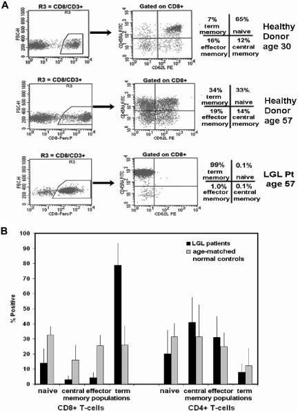
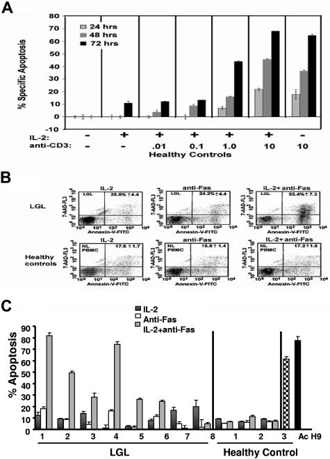
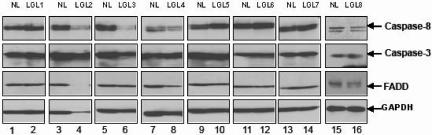
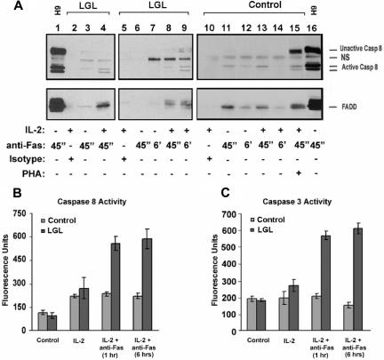
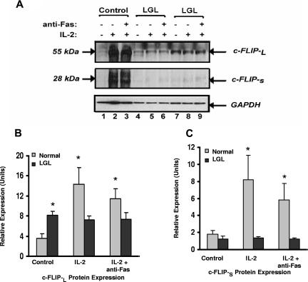
Clonal T-cell expansion in patients with T-large-granular lymphocyte (LGL) leukemia occurs by an undefined mechanism that may be related to Fas apoptosis resistance. Here, we demonstrate polarized expansion of CD8(+) terminal-memory differentiation in such patients, as demonstrated by CD45RA expression and absence of CD62L expression, suggesting repeated stimulation by antigen in vivo. Elimination of antigen-stimulated T cells normally occurs through Fas-mediated apoptosis. We show that cells from LGL leukemia patients express increased levels of c-FLIP and display resistance to Fas-mediated apoptosis and abridged recruitment of proteins that comprise the death-inducing signaling complex (DISC), including the Fas-associated protein with death-domain (FADD) and caspase-8. Exposure to interleukin-2 (IL-2) for only 24 hours sensitized leukemic LGL to Fas-mediated apoptosis with enhanced formation of the DISC, and increased caspase-8 and caspase-3 activities. We observed dysregulation of c-FLIP by IL-2 in leukemic LGL, suggesting a role in Fas resistance. Our results demonstrate that expanded T cells in patients with LGL leukemia display both functional and phenotypic characteristics of prior antigen activation in vivo and display reduced capacity for Fas-mediated DISC formation.


Figures

Figure 1
Figure 1
Naive and memory phenotyp13.6pe distribution in CD8+ and CD4+ T cells from patients with LGL leukemia compared with healthy control subjects using coexpression patterns of CD45RA and CD62L. (A) Flow cytometry was performed on cells stained with CD3, CD8, CD62L, and CD45RA. A gate was set on CD3+ cells and a second gate set on CD8+ (R3) and CD8 (CD4+) T cells. CD62L (x-axis) and CD45RA (y- axis) were used to distinguish specific T-cell subsets that included naive (top right quadrant), central memory (bottom right quadrant), effector memory (bottom left quadrant), and terminal-effector memory (top left quadrant) T cells. Representative results from healthy donors aged 30 and 57 years and a patient with LGL leukemia aged 57 years are shown. (B) Graphic representation of the mean percentage positive of these 4 distinct T-cell subsets in patients with LGL leukemia (n = 6) compared with age-matched healthy control subjects (n = 6). Standard deviation (SD) is indicated, and statistical significance was determined by a Wilcoxon sum rank test.
Figure 2
Figure 2
TCR-independent sensitivity to Fas-mediated apoptosis in PBMCs from LGL leukemia patients. (A) The percentage of apoptotic cells was determined by annexin-V-FITC/7-AAD staining in healthy control subjects treated in the absence of IL-2 and anti-CD3-antibody (dose 0) or in the presence of IL-2 (500 IU/mL) in combination with increasing doses of plate-bound anti-CD3 antibody (0.01, 0.1, 1.0, and 10 μg/mL). (B) Flow cytometric dot plots of PBMCs from a representative control donor (lower row) and from an LGL leukemia patient (upper row). Cells that stained with both annexin-V-FITC (x axis) and 7-AAD (y axis) plus cells stained with annexin-V-FITC alone were considered apoptotic and the values are shown in the upper right hand corner of each dot plot. (C) The percentage of specific apoptosis was determined in cells from 8 patients with LGL leukemia and from 3 healthy donors. Cells were assigned to one of 3 treatment groups: (1) IL-2 (500 IU/mL) for 48 hours, (2) anti-Fas (1.0 μg/mL) for 24 hours, or (3) IL-2 for 24 hours plus anti-Fas for an additional 24 hours. H9 cells and PHA/IL-2-activated normal PBMCs served as positive control. Results shown represent the mean percentage of specific apoptosis in individual samples using the equation described in “Apoptosis assay.” Standard error of the mean (SEM) is indicated for experiments performed in triplicate.
Figure 3
Figure 3
Expression of caspase-8, caspase-3, and FADD protein. The basal protein expression levels of caspase-8, caspase-3, and FADD were determined by Western blot assay using whole-cell lysates from 8 patients with LGL leukemia and from 8 healthy donors. Bands indicate the position of caspase-8, caspase-3, and FADD. Western blot analysis for GAPDH was performed to confirm equal loading of total protein in each lane.
Figure 4
Figure 4
DISC formation in PBMC from LGL leukemia patients and from healthy donors. (A) Fas-mediated DISC formation was determined after 45 minutes and after 6 hours of cross-linking with (500 ng/mL) anti-Fas (APO-1, IgG3) antibody (lanes 1, 3, 4, 6-9, 11-14) in samples from 5 patients with LGL leukemia. Cells were cultured for 24 hours with medium in the absence (−, lanes 1, 3, 6, 7, and 11) and presence of 500 IU/mL IL-2 (+, lanes 2, 4, 5, 8-10, 12, 13) for 24 hours, or PHA (1 μg/mL) plus IL-2 (500 IU/mL) for 5 to 7 days (indicated as PHA in lane 14). IgG3 isotype control antibody (500 ng/mL) was added to demonstrate specificity of protein interactions with the FasR (lanes 2, 5, and 10). After immunoprecipitation and gel electrophoresis, Western blot analysis was performed to detect caspase-8 (top panel) and FADD (bottom panel) in these FasR immunoprecipitates. Immunoprecipitation after anti-Fas antibody cross-linking of H9 cells (T-cell leukemia cell line) was used as a positive control. (B) Whole-cell lysates were prepared from a fraction of cells studied in these immunoprecipitation experiments after 1 and 6 hours of cross-linking with anti-Fas (Apo-1) antibody and isotype control (IgG3). Results shown represent the means (± SEM) of caspase-8, and -3/7 activities that were determined by a fluorometric enzyme activity assay. Protein bands for full-length caspase-8 (casp-8), FADD, and an activated cleaved product of caspase-8 (Ac casp-8) are indicated. NS = nonspecific band observed with the anti-caspase-8 antibody.
Figure 5
Figure 5
Expression of c-FLIP proteins in samples from patients with LGL leukemia and healthy control subjects. (A) c-FLIP Western blot analysis of whole cell lysates from a representative normal donor (control) and from 2 representative patients with LGL leukemia treated with medium alone, IL-2 (500 IU/mL), or IL-2 plus anti-Fas (APO-1) 1.0 μg/mL for 24 hours before lysis. Western blot analysis for GAPDH was performed to confirm equal loading of total protein in each lane. (B) Densitometry was performed on Western blot results from patients 8 LGL leukemia and 6 healthy control subjects to determine the average level of c-FLIP-L and c-FLIP-S protein expression in medium control (control), IL-2, and IL-2 plus anti-Fas (APO-1)-cultured cells. Indicated on the gel are c-FLIP-L and c-FLIP-S of molecular masses 55 and 28 kDa, respectively. *P ≤ .05.

References

    1. Yoon HJ, Sugamura K, Loughran TP., Jr Activation of leukemic large granular lymphocytes by interleukin-2 via the p75 interleukin-2 receptor. Leukemia. 1990;4:848–850. - PubMed
    1. Langerak AW, van Den Beemd R, Wolvers-Tettero IL, et al. Molecular and flow cytometric analysis of the Vbeta repertoire for clonality assessment in mature TCRalphabeta T cell proliferations. Blood. 2001;98:165–173. - PubMed
    1. Melenhorst JJ, Sorbara L, Kirby M, Hensel NF, Barrett AJ. Large granular lymphocyte leukaemia is characterized by a clonal T cell receptor rearrangement in both memory and effector CD8(+) lymphocyte populations. Br J Haematol. 2001;112:189–194. - PubMed
    1. Wlodarski MW, O'Keefe C, Howe EC, et al. Pathologic clonal cytotoxic T cell responses: nonrandom nature of the T cell-receptor restriction in large granular lymphocyte leukemia. Blood. 2005;106:2769–2780. - PubMed
    1. Saunthararajah Y, Molldrem JL, Rivera M, et al. Coincident myelodysplastic syndrome and T cell large granular lymphocytic disease: clinical and pathophysiological features. Br J Haematol. 2001;112:195–200. - PubMed

Publication types

MeSH terms