

Canine model of ischemic stroke with permanent middle cerebral artery occlusion: clinical and histopathological findings
- PMID: 17993751
- PMCID: PMC2868153
- DOI: 10.4142/jvs.2007.8.4.369
Canine model of ischemic stroke with permanent middle cerebral artery occlusion: clinical and histopathological findings
Abstract
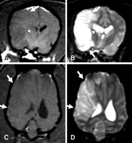
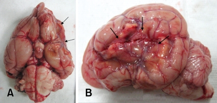
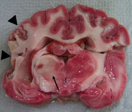
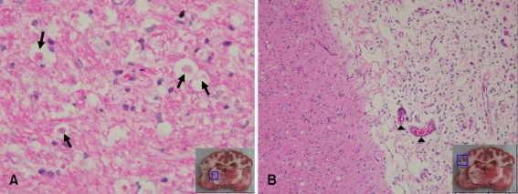
The aim of the present study was to assess the clinical and histopathological findings in a canine model of ischemic stroke. Cerebral ischemic stroke was induced by middle cerebral artery occlusion in four healthy beagle dogs using silicone plugs. They showed neurological signs of forebrain dysfunction such as reduced responsiveness, head turning, circling, postural reaction deficits, perceptual deficits, and hemianopsia. These signs gradually regressed within 4 weeks without therapy. On magnetic resonance imaging, T2 hyperintensity and T1 hypointensity were found in the cerebral cortex and basal ganglia. These lesions were well-defined and sharply demarcated from adjacent brain parenchyma with a homogenous appearance. No abnormalities of the cerebrospinal fluid were observed. At necropsy, atrophic and necrotic lesions were observed in the cerebral cortex. The cerebral cortex, basal ganglia, and thalamus were partially unstained with triphenyl- tetrazolium chloride. Histopathologically, typical features of infarction were identified in cortical and thalamic lesions. This study demonstrates that our canine model resembles the conditions of real stroke patients.
Figures
References
-
- Berg JM, Joseph RJ. Cerebellar infarcts in two dogs diagnosed with magnetic resonance imaging. J Am Anim Hosp Assoc. 2003;39:203–207. - PubMed
-
- D'Arceuil HE, Duggan M, He J, Pryor J, de Crespigny A. Middle cerebral artery occlusion in Macaca fascicularis: acute and chronic stroke evolution. J Med Primatol. 2006;35:78–86. - PubMed
-
- Diaz FG, Mastri AR, Ausman JI, Chou SN. Acute cerebral revascularization: Part I. Cerebral ischemia experimental animal model. Surg Neurol. 1979;12:353–362. - PubMed
-
- Garosi LS, McConnell JF. Ischaemic stroke in dogs and humans: a comparative review. J Small Anim Pract. 2005;46:521–529. - PubMed
Publication types
MeSH terms
LinkOut - more resources
Full Text Sources
