

The determination of dark adaptation time using electroretinography in conscious miniature Schnauzer dogs
- PMID: 17993756
- PMCID: PMC2868158
- DOI: 10.4142/jvs.2007.8.4.409
The determination of dark adaptation time using electroretinography in conscious miniature Schnauzer dogs
Abstract
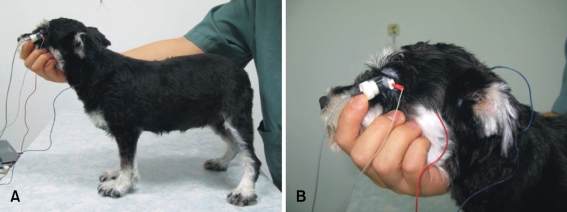
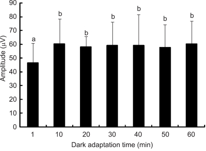
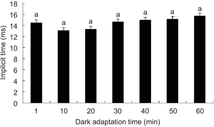
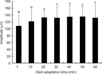
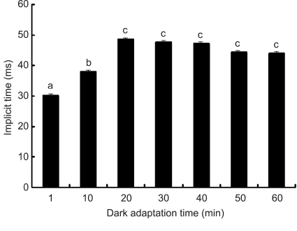
The optimal dark adaptation time of electroretinograms (ERG's) performed on conscious dogs were determined using a commercially available ERG unit with a contact lens electrode and a built-in light source (LED-electrode). The ERG recordings were performed on nine healthy Miniature Schnauzer dogs. The bilateral ERG's at seven different dark adaptation times at an intensity of 2.5 cd.s/m(2) was performed. Signal averaging (4 flashes of light stimuli) was adopted to reduce electrophysiologic noise. As the dark adaptation time increased, a significant increase in the mean a-wave amplitudes was observed in comparison to base-line levels up to 10 min (p < 0.05). Thereafter, no significant differences in amplitude occurred over the dark adaptation time. Moreover, at this time the mean amplitude was 60.30 +/- 18.47 microV. However, no significant changes were observed for the implicit times of the a-wave. The implicit times and amplitude of the b-wave increased significantly up to 20 min of dark adaptation (p < 0.05). Beyond this time, the mean b-wave amplitudes was 132.92 +/- 17.79 microV. The results of the present study demonstrate that, the optimal dark adaptation time when performing ERG's, should be at least 20 min in conscious Miniature Schnauzer dogs.
Figures
Similar articles
-
Dark adaptation time in canine electroretinography using a contact lens electrode with a built-in light source.J Vet Med Sci. 2015 Oct;77(10):1335-8. doi: 10.1292/jvms.14-0647. Epub 2015 Jun 13. J Vet Med Sci. 2015. PMID: 26074341 Free PMC article.
-
Effects of stimulus intensity for electroretinogram in conscious Miniature Schnauzers.J Vet Med Sci. 2008 Aug;70(8):857-9. doi: 10.1292/jvms.70.857. J Vet Med Sci. 2008. PMID: 18772566
-
Changes in oscillatory potentials in the canine electroretinogram during dark adaptation.Am J Vet Res. 1990 Oct;51(10):1580-6. Am J Vet Res. 1990. PMID: 2240780
-
The effects of different anesthetic agents on short electroretinography protocol in dogs.J Vet Med Sci. 2009 Jun;71(6):763-8. doi: 10.1292/jvms.71.763. J Vet Med Sci. 2009. PMID: 19578285
-
Beta wave of the scotopic (rod) electroretinogram as a measure of the activity of human on-bipolar cells.J Opt Soc Am A Opt Image Sci Vis. 1996 Mar;13(3):623-33. doi: 10.1364/josaa.13.000623. J Opt Soc Am A Opt Image Sci Vis. 1996. PMID: 8627419 Review.
Cited by
-
Anti-inflammatory α-Melanocyte-Stimulating Hormone Protects Retina After Ischemia/Reperfusion Injury in Type I Diabetes.Front Neurosci. 2022 Feb 25;16:799739. doi: 10.3389/fnins.2022.799739. eCollection 2022. Front Neurosci. 2022. PMID: 35281489 Free PMC article.
-
Comparison of two sedation protocols for long electroretinography in horses using the Koijman electrode.BMC Vet Res. 2023 Aug 4;19(1):106. doi: 10.1186/s12917-023-03654-9. BMC Vet Res. 2023. PMID: 37537621 Free PMC article.
References
-
- Acland GM. Diagnosis and differentiation of retinal diseases in small animals by electroretinography. Semin Vet Med Surg (Small Anim) 1988;3:15–27. - PubMed
-
- Andréasson S, Tornqvist K, Ehinger B. Full-field electroretinograms during general anesthesia in normal children compared to examination with topical anesthesia. Acta Ophthalmol(Copenh) 1993;71:491–495. - PubMed
-
- Dyer RS, Rigdon GC. Urethane affects the rat visual system at subanesthetic doses. Physiol Behav. 1987;41:327–330. - PubMed
-
- Fortune B, Cull G, Wang L, Van Buskirk EM, Cioffi GA. Factors affecting the use of multifocal electroretinography to monitor function in a primate model of glaucoma. Doc Ophthalmol. 2002;105:151–178. - PubMed
-
- Gallemore RP, Steinberg RH. Light-evoked modulation of basolateral membrane Cl- conductance in chick retinal pigment epithelium: the light peak and fast oscillation. J Neurophysiol. 1993;70:1669–1680. - PubMed
Publication types
MeSH terms
LinkOut - more resources
Full Text Sources
Research Materials
