Review
. 2008;47(4):644-52.
doi: 10.1002/anie.200702450.

Multiplexed detection methods for profiling microRNA expression in biological samples

Affiliations
Review

Multiplexed detection methods for profiling microRNA expression in biological samples

Alastair W Wark et al. Angew Chem Int Ed Engl. 2008.

Abstract

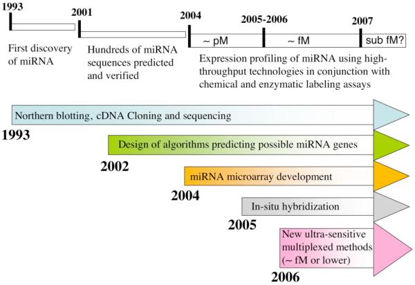
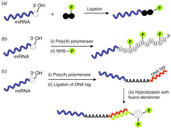
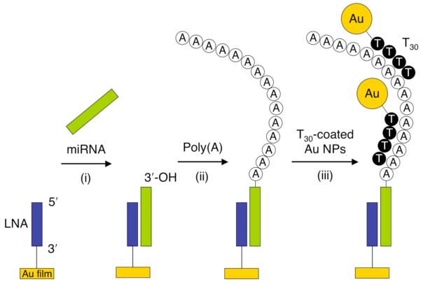
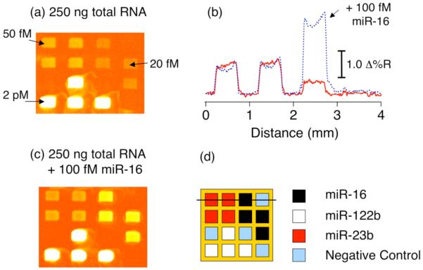
The recent discovery of short, non-protein coding RNA molecules, such as microRNA molecules (miRNAs), that can control gene expression has unveiled a whole new layer of complexity in the regulation of cell function. Since 2001, there has been a surge of interest in understanding the regulatory role of the hundreds to thousands of miRNAs expressed in both plants and animals. Significant progress in this area requires the development of quantitative bioanalytical methods for the rapid, multiplexed detection of all miRNAs that are present in a particular cell or tissue sample. In this Minireview, we discuss some of the latest methods for high-throughput miRNA profiling and the unique technological challenges that must be surmounted in this endeavor.


Figures

Figure 1
Figure 1
Timeline for microRNA discovery and detection. Initially, expression analysis was performed using northern blotting. Cloning and sequencing methods were later used to discover hundreds more miRNAs. The improved understanding of miRNA properties has enabled the development of computer algorithms that search for possible miRNA gene locations and targets. Experimental methods have progressed through several generations of microarray-based strategies with improved sensitivity and accuracy.
Figure 2
Figure 2
Single-molecule direct detection of miRNA. Step 1: Two spectrally distinct fluorescent LNA probes (F1-LNA and F2-LNA) are hybridized in solution to each target miRNA in a sandwich assay format to form the duplex labeled F1-LNA + F2-LNA)/miRNA. Step 2: Complementary DNA probes modified with a quencher molecule (Q-DNA1 and Q-DNA2) are then added which hybridize to the remaining “free” LNA probes forming two different duplexes (Q-DNA2/F2-LNA and Q-DNA1/F1-LNA). Step 3: The sample solution is subsequently flown through a microfluidic capillary within which light from lasers operating at the F1 and F2 exciting wavelengths is focused closely together. A signal corresponding to the target miRNA is observed as two coincident spikes when both probes F1 and F2 are excited simultaneously. Adapted from reference [39].
Figure 3
Figure 3
(a) Schematic of target (T) single-stranded RNA hybridization adsorption onto a probe (P) ssDNA microarray element. (b) A representative plot of relative surface coverage as a function of target RNA concentration. The solid line is a Langmuir isotherm fit to the data, from which a value of KAds = 2 × 10⁷ M-1 was determined. The inset is a representative SPRI difference image obtained by subtracting images acquired before and after sequence-specific RNA hybridization adsorption on a two-component DNA microarray.
Figure 4
Figure 4
Chemical strategies for miRNA labeling. (a) Reaction of a platinum fluorophore complex (Ulysis™ Alexa Fluor reagent) to form a coordinative bond at the N7 position of any guanine (G) base. (b) An alkylation reaction (Label IT™ reagent) which targets any reactive N heteroatom on the miRNA with some preference for the guanine (G), cytosine (C) and adenine (A) bases. (c) A two-step end labeling reaction where the 2′, 3′-diol on the ribose ring at the 3′-terminus of the miRNA is first oxidized to a dialdehyde by sodium periodate followed by a condensation reaction with a hydrazide derivative linked to a labeling moiety.
Figure 5
Figure 5
Enzymatic strategies for miRNA labeling. (a) Ligation of a fluorophore-conjugated dinucleotide to the 3′-OH end of miRNA using T4 RNA ligase. (b) E. Coli poly(A) polymerase is used to catalyze the multiple addition of nucleotides a create a poly(U) tail at the 3′-end of each miRNA. A fraction of the added nucleotides are amine-modified which are then covalently coupled to a fluorophore containing a N-Hydroxysuccinimide (NHS) ester reactive group. (c) The enzyme catalyzed formation of a 3′ poly(A) tail is followed by a second enzymatic reaction where a DNA tag sequence is ligated to the 3′-end of the tail. The tagged miRNA is then hybridized to a microarray and subsequently detected via a DNA-modified dendrimer that is complementary to the tag sequence and which also incorporates up to several hundred fluorophores for amplified detection.
Figure 6
Figure 6
Detection of microRNAs using a combination of surface polyadenylation and nanoparticle-amplified SPRI. Step (i) - hybridization adsorption of miRNA onto a complementary LNA array element. Step (ii) - poly(A) tail addition at the 3′-end of surface bound miRNAs using poly(A) polymerase. Step (iii) - hybridization adsorption of T30-coated Au nanoparticles to poly(A) tails. Reprinted with permission from reference [49].
Figure 7
Figure 7
Quantitative analysis of miRNAs from 250 ng of mouse liver total RNA using polyadenylation-nanoparticle amplified SPRI measurements. (a) SPRI difference image obtained by subtracting images acquired before and after the nanoparticle amplification step. (b) An SPRI difference image obtained from a separate chip using the same total RNA concentration as the top image plus the addition of 100 fM synthetic miR-16. (c) Comparison of line profiles taken from both SPRI difference images with the solid and dashed lines corresponding to top and bottom images respectively. (d) Schematic of the four-component LNA probe microarray and the line profile location. Reprinted with permission from reference [49].

Similar articles

Cited by

References

    1. Ambros V, Bartel B, Bartel DP, Burge CB, Carrington JC, Chen X, Dreyfuss G, Eddy SR, Griffiths-Jones S, Marshall M, Matzke M, Ruvkun G, Tuschl T. RNA. 2003;9:277. - PMC - PubMed
    1. Bartel D. Cell. 2004;116:281. - PubMed
    1. He L, Hannon GJ. Nat. Rev. Genetics. 2004;5:522. - PubMed
    1. Kim VN. Nat. Rev. Mol. Cell Biol. 2005;6:376. - PubMed
    1. Pasquinelli AE, Hunter S, Bracht J. Curr. Opin. Genet. Dev. 2005;15:200. - PubMed

Publication types

MeSH terms

LinkOut - more resources