Comparative Study
. 2008 Feb;104(4):1116-31.
doi: 10.1111/j.1471-4159.2007.05039.x. Epub 2007 Nov 6.

Carnosic acid, a catechol-type electrophilic compound, protects neurons both in vitro and in vivo through activation of the Keap1/Nrf2 pathway via S-alkylation of targeted cysteines on Keap1

Affiliations
Comparative Study

Carnosic acid, a catechol-type electrophilic compound, protects neurons both in vitro and in vivo through activation of the Keap1/Nrf2 pathway via S-alkylation of targeted cysteines on Keap1

Takumi Satoh et al. J Neurochem. 2008 Feb.

Abstract

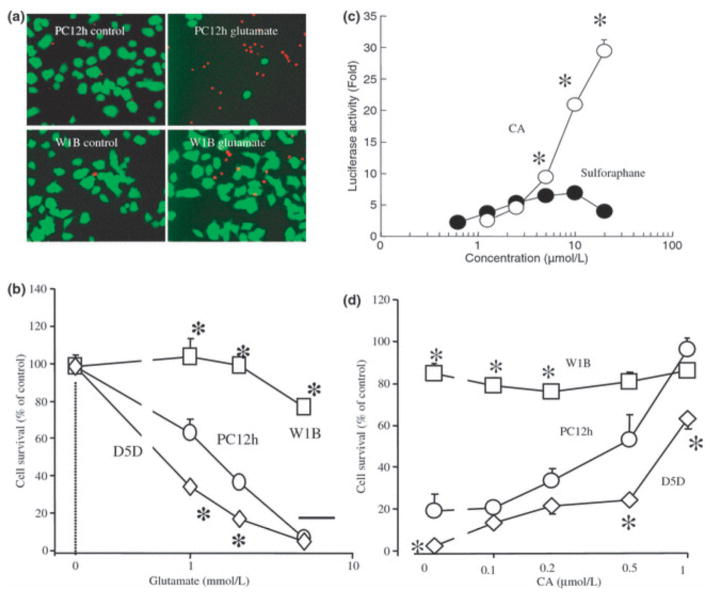
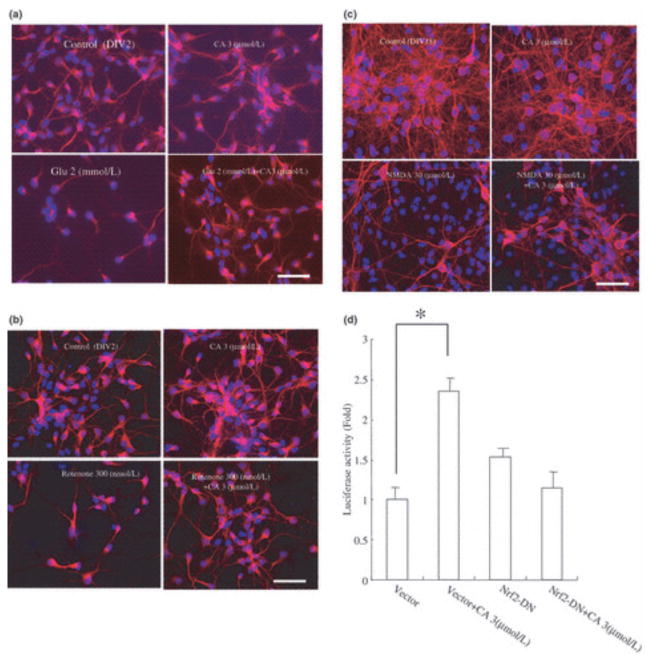
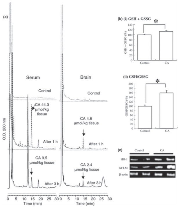
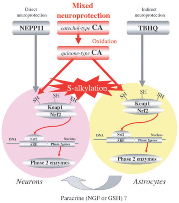
Electrophilic compounds are a newly recognized class of redox-active neuroprotective compounds with electron deficient, electrophilic carbon centers that react with specific cysteine residues on targeted proteins via thiol (S-)alkylation. Although plants produce a variety of physiologically active electrophilic compounds, the detailed mechanism of action of these compounds remains unknown. Catechol ring-containing compounds have attracted attention because they become electrophilic quinones upon oxidation, although they are not themselves electrophilic. In this study, we focused on the neuroprotective effects of one such compound, carnosic acid (CA), found in the herb rosemary obtained from Rosmarinus officinalis. We found that CA activates the Keap1/Nrf2 transcriptional pathway by binding to specific Keap1 cysteine residues, thus protecting neurons from oxidative stress and excitotoxicity. In cerebrocortical cultures, CA-biotin accumulates in non-neuronal cells at low concentrations and in neurons at higher concentrations. We present evidence that both the neuronal and non-neuronal distribution of CA may contribute to its neuroprotective effect. Furthermore, CA translocates into the brain, increases the level of reduced glutathione in vivo, and protects the brain against middle cerebral artery ischemia/reperfusion, suggesting that CA may represent a new type of neuroprotective electrophilic compound.


Figures

Figure 1
Figure 1
Core chemical structures of neuroprotective electrophilic compounds of the catechol and enone types.
Figure 2
Figure 2
Thiols from GSH and BSA as targets for CA binding. (a) Chemical reactions leading to GS-CA adduct formation as proposed from the results of NMR analysis. The catechol form of CA slowly oxidizes to the quinone derivative, an electrophilic compound, which serves as a target for nucleophilic attack by GSH or other protein thiols. The asterisk (*) indicates the electrophilic carbon in the quinone form of CA. The chemical structure of the GS-CA adduct was confirmed by 1H-NMR, 13C-NMR, and nuclear overhauser effect analysis in deuteriumized DMSO. Nuclear overhauser effects, highlighted by red double-headed arrows, yielded evidence that C(14) was subjected to nucleophilic attack by GSH. Note: The quinone-ring of CA returns to a catechol-type ring in response to adduct formation because the thiol of GSH donates an electron pair to the ring. Rectangular box: Time course of GS-CA adduct formation. GSH (18 mmol/L) and CA (6 mmol/L) were incubated and slowly mixed in 50% ethanol-PBS, pH 7.4, at 37°C for 7 h. GS-CA formation was then quantified by HPLC under the following conditions: column, μBondasphere C18; temperature, 40°C; HPLC system, Shimadzu (Kyoto, Japan) LC10Avp; detector, UV 230 nm; running solvent (i) 2% acetic acid and (ii) acetonitrile, 30% B isocratic at 1 mL/min. (b) Chemical structure of CAB. Biotin was conjugated at the carbonic acid site of CA with a chemical linker. (c) Dose-dependent BSA-CA adduct formation. BSA (1 μg/lane) was incubated with various concentrations of CAB (1–100 μmol/L) for 5 h at 23°C. The BSA/CAB samples were subjected to electrophoresis and probed with peroxidase-conjugated streptavidin (upper panel). A duplicate gel was stained with Coomassie brilliant blue as a control to assess loading of BSA. (d) Inhibition of BSA-CA adduct formation by NEM. BSA (1 μg/lane) was incubated with various concentrations of NEM (10–1000 μmol/L) for 30 min at 23°C. Vehicle (lane 1) or CAB (10 μmol/L, lanes 2–7) was then added, and the samples were incubated for 5 h at 23°C prior to blotting and probing with peroxidase-conjugated streptavidin (upper panel). A duplicate gel was stained with Coomassie brilliant blue (lower panel).
Figure 3
Figure 3
Activation of the Keap1/Nrf2 pathway by CA in COS7 and PC12h cells. (a) Schematic representation of Keap1 deletion mutants. Deletion mutants for the major Keap1 domains (BTB, IVR, and DGR) and HA (N-terminal)-tagged wild-type Keap1(HA-WT Keap1) were constructed. Numbers represent the position of cysteine residues in Keap1. Red-colored cysteines (151, 273, and 288) were previously reported to be electrophilic targets (Zhang et al. 2004; Eggler et al. 2005; Hong et al. 2005; Hosoya et al. 2005; Kobayashi et al. 2006). (b) CA binding to BTB or IVR domains of Keap1 protein. COS-7 cells were transfected with WT Keap1 or deletion mutant expression vectors and cultured for 48 h. Then the cells were treated with CAB (10 μmol/L) for 1 h. Cell lysates were blotted with anti-HA antibody (INPUT) or immunoprecipitated with streptavidin–agarose beads and blotted by anti-HA antibody (IP). (c) Nuclear translocation of Nrf2 following treatment with CA. COS7 cells were either not treated (NT) or treated with (CA) for 48 h and then stained with anti-Nrf2 monoclonal antibody (green) and 4′,6-diamino-2-phenylindole (blue). Arrows indicate nuclei. (d) CA binding to Keap1 in PC12h cells. PC12h cells were transfected with pEF6-Keap1, a Keap1 expression vector without an HA tag, and cultured for 48 h. Cell lysates were blotted with anti-Keap1 antibody (INPUT) or immunoprecipitated with streptavidin–agarose beads and probed with anti-Keap1 antibody (IP). (e) CA induces phase 2 gene expression in PC12h cells. PC12h cells were incubated with CA (10 μmol/L) for various times, and total RNA was extracted and subjected to RT-PCR. (f) CA activates the ARE in PC12h cells. PC12h cells were transfected with an ARE(GSTYa)-luciferase reporter gene construct or empty vector plus Nrf2DN- or Keap 1-expression vector. (g) CA activates the ARE in PC12h cells. PC12h cells were transfected with an ARE(GSTYa)-luciferase reporter gene construct plus either vehicle, 10 μmol/L CA, 10 μmol/L CAB, or 20 μmol/L CAB.
Figure 4
Figure 4
Carnosic acid (CA) protects PC12h cells via Nrf2. (a) Nrf2 inhibits cell death. PC12h cells and PC12hW1B cells (expressing Nrf2WT) were exposed to 5 mmol/L glutamate for 20 h and then incubated in a solution of fluorescein diacetate (10 μmol/L) and propidium iodide (1 μg/mL). Viable cells stained with fluorescein (green) and dead cells with propidium iodide (red). (b) Nrf2DN increases cell death. PC12h cells, PC12hW1B cells, and PC12hD5D cells (expressing Nrf2DN) were incubated with various concentrations of glutamate for 20 h. Cell survival was assessed using the 3-(4,5-dimethylthiazol-2-yl)-2,5-diphenyltetrazolium bromide assay. *Significantly different (p < 0.01) from PC12h cells by anova. (c) Dose-dependent activation of the ARE by CA and sulforaphane. PC12h cells were tranfected with an ARE-luciferase reporter gene plasmid, and then incubated for 20 h with various concentrations of CA or sulforaphane. *Significantly different (p < 0.01) between CA and sulforaphane by anova. (d) CA protects PC12h cells in an Nrf2-dependent manner. Various concentrations of CA were added to PC12, PC12hW1B, or PC12hD5D cells 1 h prior to exposure to 5 mmol/L glutamate for 20 h. Viability was then assessed by the 3-(4,5-dimethylthiazol-2-yl)-2,5-diphenyltetrazolium bromide assay. *Significantly different (p < 0.01) from PC12h cells by anova.
Figure 5
Figure 5
Carnosic acid (CA) protects cortical neurons via Nrf2. (a and b) CA inhibits oxidative glutamate toxicity. CA (3 μmol/L) or vehicle was added to cerebrocortical cultures (E17, DIV2) 60 min prior to exposure to glutamate or rotenone. The cultures were then incubated for 20 h and stained with anti-MAP2 and anti-NeuN (red) as well as with Hoechst dye (blue). (c) CA inhibits excitotoxicity. CA (3 μmol/L) or vehicle was added to cerebrocortical cultures (E17, DIV21) 60 min prior to exposure to NMDA (50 μmol/L for 15 min). The cultures were then incubated for 20 h and subsequently stained with anti-MAP2 and anti-NeuN (red) as well as with Hoechst dye (blue). (d) CA activates the ARE in an Nrf2-dependent manner. Cortical cultures (E17, DIV21) were transfected with ARE-luciferase reporter gene DNA (1 μg/well) and co-transfected with pEF6 or pEFNrf2DN. CA (3 μmol/L) or vehicle was then added to the cultures. After a 24-h incubation, cell lysates were used for luciferase reporter gene assays. Values are mean ± SEM; *p < 0.01 by anova.
Figure 6
Figure 6
Carnosic acid (CA) accumulates both in neurons and in non-neuronal cells. (a, c, and e) Cortical cultures (E17, DIV21) treated with various concentrations of CAB for 20 h were stained with anti-MAP2 monoclonal antibody to identify neurons (green) and rhodamine-conjugated streptavidin antibody to detect intracellular CAB (red). Hoechst dye 33,258 was used to label nuclei (blue). (b, d, and f) Anti-S100 monoclonal antibody was used to identify non-neuronal cells (green). Yellow and white arrows indicate accumulation of CAB in non-neuronal cells and neurons, respectively. Scale bar: 25 μm.
Figure 7
Figure 7
Carnosic acid (CA) penetration into brain parenchyma. (a) Translocation of CA into the brain. CA was dissolved in olive oil (10 mg/mL), and 0.3 mL/mouse (representing 3 mg CA) or vehicle was then administered orally. One to 3 h later, serum and brain tissue were subjected to HPLC analysis for CA. (b) CA increased reducing equivalents of GSH in the brain. Mice were fed 0.03% CA for 1 week, and their brains were then removed, lysed, and subjected to GSH and GSSG measurement. (c) Induction of phase 2 enzymes in the brain by CA. CA was again administered orally; 8 h later, total RNA was extracted from brain tissue and subjected to RT-PCR using the indicated primers.
Figure 8
Figure 8
Carnosic acid (CA) protects against cerebral ischemia induced by 2-h MCAO/24-h reperfusion. (a) Representative TTC staining of brain sections after stroke in CA- versus vehicle-treated mice. CA (1 mg/kg body weight) was administrated intraperitoneally in vehicle solution (10% DMSO in PBS) 1 h prior to MCAO. After 24-h reperfusion, coronal sections, 1 mm in thickness, were prepared and stained with TTC. (b) Quantification of infarct volume by TTC staining. CA decreased infarct volume compared with vehicle-treated mice. Data represent mean ± SEM (vehicle-treated, n = 9; CA-treated, n = 9; *p < 0.05 by anova).
Figure 9
Figure 9
Proposed mechanism of neuroprotective action of CA, NEPP11, and TBHQ. NEPP11 appears to protect neurons directly, TBHQ via effects on astrocytes, which in turn may release survival or neurotrophic factors (Ahlgren-Beckendorf et al. 1999; Kosaka and Yokoi 2003), and CA by a ‘mixed’ type of protection mediated by actions on both neurons and astrocyt

Similar articles

Cited by

References

    1. Ahlgren-Beckendorf JA, Reising AM, Schander MA, Herdler JW, Johnson JA. Coordinate regulation of NAD(P)H: quinone oxidoreductase and glutathione-S-transferase in primary cultures of rat neurons and glias: role of the antioxidant/electrophile responsive element. Glia. 1999;15:131–142. - PubMed
    1. Alam J, Stewart D, Touchard C, Boinapally S, Choi AM, Cook JL. Nrf2, a cap’n’collar transcription factor, regulates induction of the heme oxygenase-1 gene. J Biol Chem. 1999;274:26071–26078. - PubMed
    1. Ankacrona M, Dypbukt JM, Bonfoco E, Zhivotovsky B, Orrenius S, Lipton SA, Nicotera P. Glutamate-induced neuronal death: a succession of necrosis or apoptosis depending on mitochondrial function. Neuron. 1995;15:961–973. - PubMed
    1. Aruoma OI, Halliwell B, Aeschbach R, Loligers J. Antioxidant and pro oxidant properties of active rosemary constituents: carnosol and carnosic acid. Xenobiotica. 1992;22:257–268. - PubMed
    1. Barco A, Bailey CH, Kandel ER. Common molecular mechanisms in explicit and implicit memory. J Neurochem. 2006;97:1520–1533. - PubMed

Publication types

MeSH terms