. 2008 Jan;104(1):74-88.
doi: 10.1111/j.1471-4159.2007.05018.x. Epub 2007 Nov 6.

PACAP-38 induces neuronal differentiation of human SH-SY5Y neuroblastoma cells via cAMP-mediated activation of ERK and p38 MAP kinases

Affiliations

PACAP-38 induces neuronal differentiation of human SH-SY5Y neuroblastoma cells via cAMP-mediated activation of ERK and p38 MAP kinases

T K Monaghan et al. J Neurochem. 2008 Jan.

Abstract

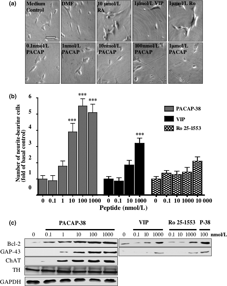
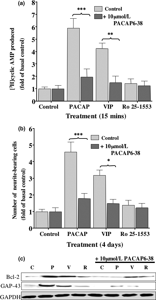
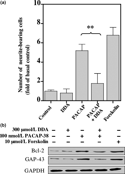
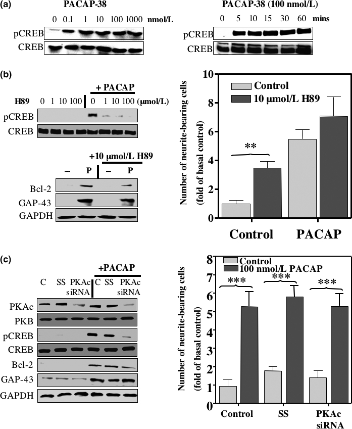
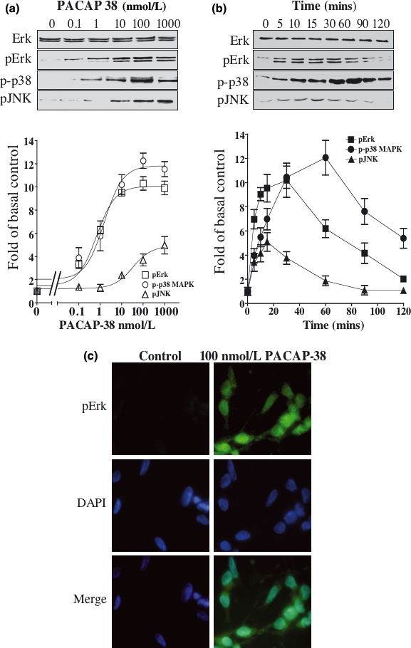
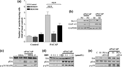
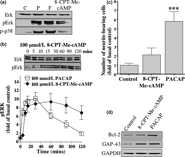
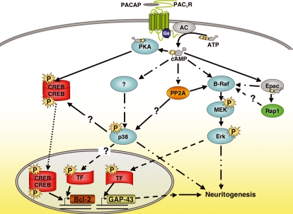
The intracellular signaling pathways mediating the neurotrophic actions of pituitary adenylate cyclase-activating polypeptide (PACAP) were investigated in human neuroblastoma SH-SY5Y cells. Previously, we showed that SH-SY5Y cells express the PAC(1) and VIP/PACAP receptor type 2 (VPAC(2)) receptors, and that the robust cAMP production in response to PACAP and vasoactive intestinal peptide (VIP) was mediated by PAC(1) receptors (Lutz et al. 2006). Here, we investigated the ability of PACAP-38 to differentiate SH-SY5Y cells by measuring morphological changes and the expression of neuronal markers. PACAP-38 caused a concentration-dependent increase in the number of neurite-bearing cells and an up-regulation in the expression of the neuronal proteins Bcl-2, growth-associated protein-43 (GAP-43) and choline acetyltransferase: VIP was less effective than PACAP-38 and the VPAC(2) receptor-specific agonist, Ro 25-1553, had no effect. The effects of PACAP-38 and VIP were blocked by the PAC(1) receptor antagonist, PACAP6-38. As observed with PACAP-38, the adenylyl cyclase activator, forskolin, also induced an increase in the number of neurite-bearing cells and an up-regulation in the expression of Bcl-2 and GAP-43. PACAP-induced differentiation was prevented by the adenylyl cyclase inhibitor, 2',5'-dideoxyadenosine (DDA), but not the protein kinase A (PKA) inhibitor, H89, or by siRNA-mediated knock-down of the PKA catalytic subunit. PACAP-38 and forskolin stimulated the activation of extracellular signal-regulated kinase (ERK), mitogen-activated protein kinase (MAP; p38 MAP kinase) and c-Jun N-terminal kinase (JNK). PACAP-induced neuritogenesis was blocked by the MEK1 inhibitor PD98059 and partially by the p38 MAP kinase inhibitor SB203580. Activation of exchange protein directly activated by cAMP (Epac) partially mimicked the effects of PACAP-38, and led to the phosphorylation of ERK but not p38 MAP kinase. These results provide evidence that the neurotrophic effects of PACAP-38 on human SH-SY5Y neuroblastoma cells are mediated by the PAC(1) receptor through a cAMP-dependent but PKA-independent mechanism, and furthermore suggest that this involves Epac-dependent activation of ERK as well as activation of the p38 MAP kinase signaling pathway.


Figures

Fig. 1
Fig. 1
The effect of PACAP on differentiation of SH-SY5Y cells. SH-SY5Y cells were cultured in low serum medium for 24 h before being incubated with increasing concentrations of PACAP-38 (PACAP), retinoic acid (RA), VIP, Ro 25-1553 or dimethylformamide, as indicated. Medium and treatments were replaced after 2 days and micrographs captured after 4 days (a). Micrographs are set to the same scale; the scale bar represents 50 μm. (b) The total number of cells, and the number of cells bearing neurites greater than the length of the cell body were counted and the data expressed as fold of basal control. Experiments were performed in triplicate and one- and two-way ANOVA carried out to determine differences between treatments (***p < 0.001). (c) After capture of micrographs, whole cell extracts were taken and the proteins separated and blotted as detailed in Materials and methods. Antibodies specific for Bcl-2, GAP-43, ChAT, TH and GAPDH were used as indicated. Cell extracts from cells treated with 100 nmol/L PACAP-38 (P-38) were run alongside those from cells treated with VIP and Ro 25-1553 at the indicated concentrations for comparison. Each blot is representative of three independent experiments.
Fig. 2
Fig. 2
The effects of the PAC1 receptor antagonist PACAP6-38 on SH-SY5Y cells. SH-SY5Y cells were maintained in low serum medium as described in Materials and methods. (a) Histogram showing fold change in cAMP levels for cells treated for 15 min with 100 nmol/L PACAP-38, 1 μM VIP or 1 μM Ro 25-1553, in the presence (dark gray bars), or absence (light gray bars) of 10 μM PACAP6-38. The basal level was determined for cells maintained in low serum medium alone. (b) Histogram showing numbers of neurite-bearing cells (fold of basal control) following treatment for 4 days with 100 nmol/L PACAP-38, 1 μM VIP or 1 μM Ro 25-1553, in the presence (dark gray bars) or absence (light gray bars) of 10 μM PACAP6-38. Standard error bars are shown (n = 3) and one-way ANOVA performed to identify significant differences between treatments (***p < 0.001, **p < 0.01, *p < 0.05). (c) Western blot analysis of cell extracts using antibodies specific for Bcl-2 and GAP-43 was carried out following the different treatments for 4 days. Protein loading was determined using an antibody specific for GAPDH. Each blot represents three independent experiments. (C = control, p = 100 nmol/L PACAP-38, V = 1 μM VIP, R = 1 μmol/L Ro 25-1553).
Fig. 3
Fig. 3
The effects of DDA and Forskolin on differentiation of SH-SY5Y cells. SH-SY5Y cells were maintained in low serum medium as previously described. (a) Histogram showing for the numbers of neurite-bearing cells following treatment with 100 nmol/L PACAP-38 in the presence or absence of 300 μmol/L DDA, or for cells treated with 10 μmol/L forskolin for 4 days. Standard error bars are shown (n = 3), one-way ANOVA analysis was carried out to identify significant differences between treatments (**p < 0.01). (b) Western blot analysis of cell extracts using antibodies specific for Bcl-2 and GAP-43 was carried out following the different treatments for 4 days. Protein loading was determined using an antibody specific for GAPDH. Each blot represents three independent experiments.
Fig. 4
Fig. 4
The effects of PKA inhibition on differentiation of SH-SY5Y cells. SH-SY5Y cells were maintained in low serum medium as previously described. (a) Western blots showing changes in CREB phosphorylation following a 15 min. treatment with increasing concentrations of PACAP-38 (left panel) and a time course of 100 nmol/L PACAP-38-induced CREB phosphorylation (right panel). The total CREB protein in each sample is shown in the lower panels. (b) The effects of treating cells with H89 were assessed by Western blot analysis of pCREB levels in whole cell extracts from cells treated with 1, 10 and 100 μmol/L H89 in the presence or absence of 100 nmol/L PACAP-38 for 15 min (upper panel). The levels of total CREB protein were also assessed for each sample. The effect of 10 μmol/L H89 on levels of Bcl-2 and GAP-43 protein expression in cells in the presence or absence of 100 nmol/L PACAP-38 for 4 days are shown in the lower panels. The right panel is a histogram showing numbers of neurite-bearing cells (fold of basal control) following treatment with 10 μmol/L H-89, in the presence (dark gray bars) or absence (light gray bars) of 100 nmol/L PACAP-38. (c) Western blots showing changes in expression of PKAc and PKB 2 days after siRNA transfection to knock-down PKAc expression, and its affect on 100 nmol/L PACAP-38-induced CREB (C = control, SS = scrambled siRNA sequence, PKAc siRNA = specific siRNA sequence targeting the catalytic subunit of PKA). The right panel is a histogram showing numbers of neurite-bearing cells (fold of basal control) following siRNA transfection to knock-down PKAc expression, in the presence (dark gray bars) or absence (light gray bars) of 100 nmol/L PACAP-38. ANOVA analysis was performed to identify significant differences between treatments (***p < 0.001, **p < 0.01). Protein loading in (b) and (c) was determined using an antibody specific for GAPDH. Each blot is representative of three independent experiments.
Fig. 5
Fig. 5
PACAP-38-mediated activation of ERK, p38 MAP kinase and JNK. SH-SY5Y cells were cultured in low serum medium for 24 h and incubated with increasing concentrations of PACAP-38 for 15 min (a) or with 100 nmol/L PACAP-38 for up to 2 h (b). Western blot analysis of whole cell extracts was carried out using antibodies specific for ERK, pERK, p-p38 MAP kinase and pJNK. The increase in phosphorylation levels was determined by densitometry as described in Materials and methods and is graphically represented in the lower section of the figure. Standard error bars are shown (n = 5). (c) The cells were incubated with a pERK-specific antibody and its respective fluorescent dye-conjugated secondary antibody (green) following treatment with 100 nmol/L PACAP-38 for 30 min. Cells were also stained with DAPI, the DNA-binding fluorescent stain (blue). Each micrograph is representative of three independent experiments.
Fig. 6
Fig. 6
The effect of ERK and p38 MAP kinase on PACAP-induced differentiation of SH-SY5Y cells. (a–b) SH-SY5Y cells were cultured in low serum medium for 24 h and incubated with 10 μmol/L PD98059 (PD) or 10 μmol/L SB203580 (SB) for 45 min before treatment with 100 nmol/L PACAP-38 or low serum medium as control for 4 days. Treatments were replaced after the second day. (a) The fold increase in neurite-bearing cells was determined as described in Materials and methods. Standard error bars are shown (n = 3) and ANOVA was carried out to identify significant differences between treatments (***p < 0.001). (b) Western blot analysis of whole cell extracts using antibodies specific for Bcl-2 and GAP-43 was carried out on day 4. Protein loading was determined using an antibody specific for GAPDH. (c-e) Western blot analysis of whole cell extracts was carried out using antibodies specific for ERK, pERK and p-p38 MAP kinase. (c) Cells cultured in low serum medium for 24 h were incubated with 300 μmol/L DDA in the presence, or absence, of 100 nmol/L PACAP-38 or 10 μmol/L forskolin for 15 min. (d) Cells maintained in low serum medium for 24 h were treated with 0.1, 1 and 10 μmol/L H89 for 45 min, then with or without 100 nmol/L PACAP-38 for 15 min. (e) Expression of PKAc was inhibited by siRNA knock-down as described in Materials and methods (C = control, SS = scrambled siRNA sequence, PKAc siRNA = specific siRNA sequence targeting the catalytic subunit of PKA). Cells were incubated in the presence, or absence, of 100 nmol/L PACAP-38 for 15 minutes. Each blot is representative of three independent experiments.
Fig. 7
Fig. 7
Involvement of Epac. (a) Western blot analysis of whole cell extracts was carried out using antibodies specific for ERK, pERK and p-p38 MAP kinase. The cells were maintained in low serum medium for 24 h and stimulated with the indicated factors for 15 min C = control, p = 100 nmol/L PACAP-38, F = 10 μmol/L forskolin and CPT-cAMP = 100 μmol/L 8-CPT-Me-cAMP. (b) Western blot analysis of whole cell extracts was carried out using antibodies specific for ERK and pERK. Cells were stimulated for up to 2 h with 100 μmol/L 8-CPT-Me-cAMP. The increase in phosphorylation levels was determined by densitometry as described in Materials and methods and is graphically represented in the lower section of the figure. Standard error bars are shown. (c) Cells were maintained in low serum medium for 24 h then stimulated with 100 μmol/L 8-CPT-Me-cAMP or with 100 nmol/L PACAP-38 for 4 days. Control cells were kept in low serum medium alone. The fold increase in neurite-bearing cells was determined as described in the Materials and methods. Standard error bars are shown (n = 3) and ANOVA analysis was carried out to identify significant differences between treatments (***p < 0.001). (e) Western blot analysis of whole cell extracts using antibodies specific for Bcl-2 and GAP-43 was carried out. Protein loading was determined using an antibody specific for GAPDH. Cells were treated for 4 days in low serum medium alone (Control) or treated with 100 μmol/L 8-CPT-Me-cAMP (CPT-cAMP) or with 100 nmol/L PACAP-38 (PACAP). Each blot is representative of three independent experiments.
Fig. 8
Fig. 8
Proposed model for the cAMP-dependent signaling events involved in the neurotrophic actions of PACAP-38 on SH-SY5Y cells. PACAP-38 binding to the PAC1 receptor, which couples to the activation of AC, evokes an increase in the intracellular levels of cAMP (Lutz et al. 2006). This in turn leads to the activation of PKA-dependent and PKA-independent mechanisms that are involved in the induction of expression of neuronal proteins and in neuritogenesis, respectively. The PKA-dependent mechanism involves PKA phosphorylation of the transcription factor CREB, initiating its activation and translocation to the nucleus where it may be involved in the up-regulated expression of proteins involved in neuronal differentiation, including Bcl-2. The PKA-independent mechanisms involve activation of MEK and ERK as well as p38 MAP kinase that are required for neuritogenesis. The MEK/ERK activation possibly occurs through one or a combination of different mechanisms. The neuronal MAP kinase signaling cascade involves B-Raf as the first component (Dugan et al. 1999) and it has been shown that the 95 kDa B-Raf kinase is expressed in SH-SY5Y cells (Stephens et al. 1992). However, it is still unclear how cAMP activates B-Raf (Dumaz and Marais 2005). This may occur through activation of the cAMP-dependent Rap 1 GEF, Epac, or through another mechanism that may be potentiated by Epac (Lin et al. 2003) but may or may not involve Rap1 (Dumaz and Marais 2005). An additional possibility is the cAMP-mediated activation of PP2A (Feschenko et al. 2002) may directly induce B-Raf activation (Strack 2002). Activated ERK is required for neuritogenesis as well as being involved in the up-regulated expression of GAP-43, possibly through translocation to the nucleus. cAMP mediates the activation of p38 MAP kinase through a non-canonical pathway that may be similar to that observed in Th2 cells (Chen et al. 2000) and in cardiac fibroblasts (Yin et al. 2006), and which also may involve PP2A (Boudreau et al. 2004). Activated p38 MAP kinase in turn is involved in the increased expression of Bcl-2, possibly through phosphorylating CREB (Yin et al. 2006) and/or other factors, as well as in neuritogenesis. The arrows indicate direct interactions, the dotted arrows translocation, and the dot dash arrows the possibility of one or a number of intermediary steps that have not been worked out.

Similar articles

Cited by

References

    1. Alleaume C, Eychène A, Harnois T, Bourmeyster N, Constantin B, Caigneaux E, Muller J-M, Philippe M. Vasoactive intestinal peptide-induced neurite remodeling in human neuroblastoma SH-SY5Y cells implicates the cdc42 GTPase and is independent of Ras-ERK pathway. Exp. Cell Res. 2004;299:511–524. - PubMed
    1. Boeshore KL, Schreiber RC, Vaccariello SA, Sachs HH, Salazar R, Lee J, Ratan RR, Leahy P, Zigmond RE. Novel changes in gene expression following axotomy of a sympathetic ganglion: a microarray analysis. J. Neurobiol. 2004;59:216–235. - PubMed
    1. Borba JC, Henze IP, Silveira MS, Kubrusly RC, Gardino PF, de Mello MC, Hokoc JN, de Mello FG. Pituitary adenylate cyclase-activating polypeptide (PACAP) can act as determinant of the tyrosine hydroxylase phenotype of dopaminergic cells during retina development. Brain Res. Dev. Brain Res. 2005;156:193–201. - PubMed
    1. Boudreau RTM, Hoskin DW, Lin T-J. Phosphatase inhibition potentiates IL-6 production by mast cells in response to FcεRI-mediated activation: involvement of p38 MAPK. J. Leukoc. Biol. 2004;76:1075–1081. - PubMed
    1. Braas KM, May V. Pituitary adenylate cyclase-activating polypeptides, PACAP-38 and PACAP-27, regulation of sympathetic neuron catecholamine, and neuropeptide Y expression through activation of type I PACAP/VIP receptor isoforms. Ann. NY Acad. Sci. 1996;805:204–216. - PubMed

Publication types

MeSH terms

Substances