

Studies on the actin-binding protein HS1 in platelets
- PMID: 17996076
- PMCID: PMC2203996
- DOI: 10.1186/1471-2121-8-46
Studies on the actin-binding protein HS1 in platelets
Abstract
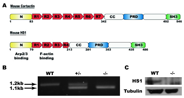
Background: The platelet cytoskeleton mediates the dramatic change in platelet morphology that takes place upon activation and stabilizes thrombus formation. The Arp2/3 complex plays a vital role in these processes, providing the protrusive force for lamellipodia formation. The Arp2/3 complex is highly regulated by a number of actin-binding proteins including the haematopoietic-specific protein HS1 and its homologue cortactin. The present study investigates the role of HS1 in platelets using HS1-/- mice.
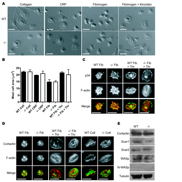
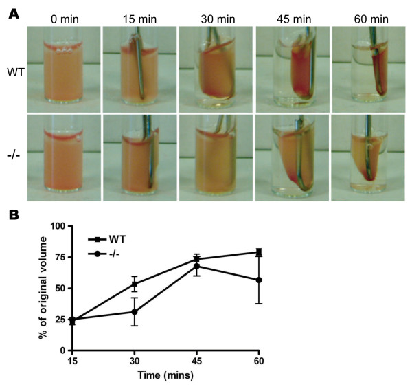
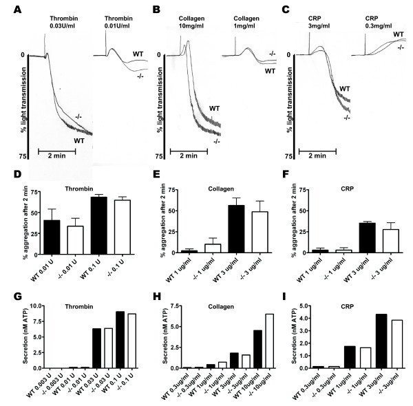
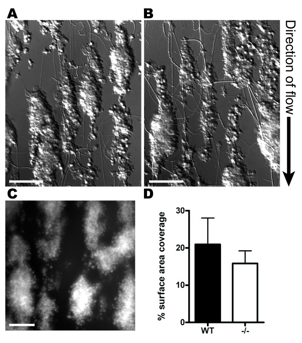
Results: The present results demonstrate that HS1 is not required for platelet activation, shape change or aggregation. Platelets from HS1-/- mice spread normally on a variety of adhesion proteins and have normal F-actin and Arp2/3 complex distributions. Clot retraction, an actin-dependent process, is also normal in these mice. Platelet aggregation and secretion is indistinguishable between knock out and littermates and there is no increase in bleeding using the tail bleeding assay.
Conclusion: This study concludes that HS1 does not play a major role in platelet function. It is possible that a role for HS1 is masked by the presence of cortactin.
Figures

Similar articles
-
The actin binding proteins cortactin and HS1 are dispensable for platelet actin nodule and megakaryocyte podosome formation.Platelets. 2017 Jun;28(4):372-379. doi: 10.1080/09537104.2016.1235688. Epub 2016 Oct 25. Platelets. 2017. PMID: 27778524 Free PMC article.
-
The cytoskeletal crosslinking protein MACF1 is dispensable for thrombus formation and hemostasis.Sci Rep. 2019 May 22;9(1):7726. doi: 10.1038/s41598-019-44183-6. Sci Rep. 2019. PMID: 31118482 Free PMC article.
-
Wdr-1 is essential for F-actin interaction with focal adhesions in platelets.Blood Coagul Fibrinolysis. 2018 Sep;29(6):540-545. doi: 10.1097/MBC.0000000000000756. Blood Coagul Fibrinolysis. 2018. PMID: 29995657 Free PMC article.
-
Cortactin: Cell Functions of A Multifaceted Actin-Binding Protein.Trends Cell Biol. 2018 Feb;28(2):79-98. doi: 10.1016/j.tcb.2017.10.009. Epub 2017 Nov 20. Trends Cell Biol. 2018. PMID: 29162307 Review.
-
Platelet Shape Changes during Thrombus Formation: Role of Actin-Based Protrusions.Hamostaseologie. 2021 Feb;41(1):14-21. doi: 10.1055/a-1325-0993. Epub 2021 Feb 15. Hamostaseologie. 2021. PMID: 33588449 Review.
Cited by
-
Sex differences in the acute in vivo effects of different human SP-A variants on the mouse alveolar macrophage proteome.J Proteomics. 2014 Aug 28;108:427-44. doi: 10.1016/j.jprot.2014.06.007. Epub 2014 Jun 18. J Proteomics. 2014. PMID: 24954098 Free PMC article.
-
Termination of bleeding by a specific, anticatalytic antibody against plasmin.J Thromb Haemost. 2019 Sep;17(9):1461-1469. doi: 10.1111/jth.14522. Epub 2019 Jun 23. J Thromb Haemost. 2019. PMID: 31136076 Free PMC article.
-
Thrombin mutant W215A/E217A treatment improves neurological outcome and reduces cerebral infarct size in a mouse model of ischemic stroke.Stroke. 2011 Jun;42(6):1736-41. doi: 10.1161/STROKEAHA.110.603811. Epub 2011 Apr 21. Stroke. 2011. PMID: 21512172 Free PMC article.
-
The actin binding proteins cortactin and HS1 are dispensable for platelet actin nodule and megakaryocyte podosome formation.Platelets. 2017 Jun;28(4):372-379. doi: 10.1080/09537104.2016.1235688. Epub 2016 Oct 25. Platelets. 2017. PMID: 27778524 Free PMC article.
-
Immune pathology associated with altered actin cytoskeleton regulation.Autoimmunity. 2010 Feb;43(1):64-75. doi: 10.3109/08916930903374634. Autoimmunity. 2010. PMID: 20001423 Free PMC article. Review.
References
-
- Shattil SJ, Kashiwagi H, Pampori N. Integrin Signaling: The Platelet Paradigm. Blood. 1998;91:2645–2657. - PubMed
Publication types
MeSH terms
Substances
Grants and funding
LinkOut - more resources
Full Text Sources
Other Literature Sources
Molecular Biology Databases
Research Materials
Miscellaneous
