. 2007;9(6):R117.
doi: 10.1186/ar2325.

Chondroitin and glucosamine sulfate in combination decrease the pro-resorptive properties of human osteoarthritis subchondral bone osteoblasts: a basic science study

Affiliations

Chondroitin and glucosamine sulfate in combination decrease the pro-resorptive properties of human osteoarthritis subchondral bone osteoblasts: a basic science study

Steeve Kwan Tat et al. Arthritis Res Ther. 2007.

Abstract

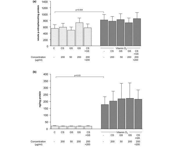
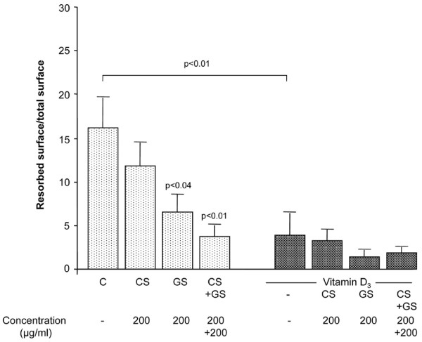
Early in the pathological process of osteoarthritis (OA), subchondral bone remodelling, which is related to altered osteoblast metabolism, takes place. In the present study, we explored in human OA subchondral bone whether chondroitin sulfate (CS), glucosamine sulfate (GS), or both together affect the major bone biomarkers, osteoprotegerin (OPG), receptor activator of nuclear factor-kappa B ligand (RANKL), and the pro-resorptive activity of OA osteoblasts. The effect of CS (200 mug/mL), GS (50 and 200 mug/mL), or both together on human OA subchondral bone osteoblasts, in the presence or absence of 1,25(OH)2D3 (vitamin D3) (50 nM), was determined on the bone biomarkers alkaline phosphatase and osteocalcin, on the expression (mRNA) and production (enzyme-linked immunosorbent assay) of bone remodelling factors OPG and RANKL, and on the pro-resorptive activity of these cells. For the latter experiments, human OA osteoblasts were incubated with differentiated peripheral blood mononuclear cells on a sub-micron synthetic calcium phosphate thin film. Data showed that CS and GS affected neither basal nor vitamin D3-induced alkaline phosphatase or osteocalcin release. Interestingly, OPG expression and production under basal conditions or vitamin D3 treatment were upregulated by CS and by both CS and GS incubated together. Under basal conditions, RANKL expression was significantly reduced by CS and by both drugs incubated together. Under vitamin D3, these drugs also showed a decrease in RANKL level, which, however, did not reach statistical significance. Importantly, under basal conditions, CS and both compounds combined significantly upregulated the expression ratio of OPG/RANKL. Vitamin D3 decreased this ratio, and GS further decreased it. Both drugs reduced the resorption activity, and statistical significance was reached for GS and when CS and GS were incubated together. Our data indicate that CS and GS do not overly affect cell integrity or bone biomarkers. Yet CS and both compounds together increase the expression ratio of OPG/RANKL, suggesting a positive effect on OA subchondral bone structural changes. This was confirmed by the decreased resorptive activity for the combination of CS and GS. These data are of major significance and may help to explain how these two drugs exert a positive effect on OA pathophysiology.


Figures

Figure 1
Figure 1
Levels of alkaline phosphatase and osteocalcin in human osteoarthritis subchondral bone osteoblasts. Alkaline phosphatase activity (a) and osteocalcin level (b) were determined after treatment with chondroitin sulfate (CS) (200 μg/mL), glucosamine sulfate (GS) (50 or 200 μg/mL), or both (200 μg/mL each) in the absence or presence of vitamin D3 at 50 nM. Alkaline phosphatase activity (a) was determined in the cell lysate by substrate hydrolysis using p-nitrophenylphosphate, whereas osteocalcin level (b) was determined in the culture media by using a specific enzyme immunoassay. Data are from eight independent experiments. Statistical significance was assessed by paired Student t test. P value indicates the statistical difference between control (C, basal conditions) and vitamin D3-treated specimens.
Figure 2
Figure 2
Levels of osteoprotegerin (OPG), receptor activator of nuclear factor-kappa B ligand (RANKL), and OPG/RANKL ratio in human osteoarthritis subchondral bone osteoblasts. Expression and production of OPG (a), expression of RANKL (b), and expression ratio of OPG/RANKL (c) of cells incubated in the absence or presence of chondroitin sulfate (CS) (200 μg/mL), glucosamine sulfate (GS) (50 or 200 μg/mL), or both (200 μg/mL each) in the absence or presence of vitamin D3 at 50 nM. Total RNA was extracted and processed for quantitative polymerase chain reaction (qPCR), and the data are expressed as the mean ± standard error of the mean of arbitrary unit. The release of OPG was determined in the culture medium by a specific enzyme-linked immunosorbent assay. Data are from eight independent experiments. Statistical significance was assessed by paired Student t test versus autologous control. Underlined p value indicates the statistical difference between control (C, basal conditions) and vitamin D3-treated specimens.
Figure 3
Figure 3
Pro-resorptive activity of human osteoarthritis subchondral bone osteoblasts. Resorption activity of osteoblasts co-incubated with differentiated peripheral blood mononuclear cells in the presence of macrophage colony-stimulating factor and in the absence or presence of chondroitin sulfate (CS) (200 μg/mL), glucosamine sulfate (GS) (200 μg/mL), or both (200 μg/mL each) in the absence or presence of vitamin D3 at 50 nM. Data are in the absence or presence of vitamin D3 from nine or five independent experiments, respectively. They are expressed as the mean resorbed surface per total surface upon treatment with the factors. Statistical significance was assessed by paired Student t test versus autologous control. Underlined p value indicates the statistical difference between control (C, basal conditions) and vitamin D3-treated specimens.

Similar articles

Cited by

References

    1. Martel-Pelletier J, Lajeunesse D, Pelletier JP. Etiopathogenesis of osteoarthritis. In: Koopman WJ, Moreland LW, editor. Arthritis and Allied Conditions: A Textbook of Rheumatology. Baltimore: Lippincott, Williams Wilkins; 2005. pp. 2199–2226.
    1. Lajeunesse D, Massicotte F, Pelletier JP, Martel-Pelletier J. Subchondral bone sclerosis in osteoarthritis: not just an innocent bystander. Modern Rheumatology. 2003;13:7–14. doi: 10.1007/s101650300001. - DOI - PubMed
    1. Lippiello L. Glucosamine and chondroitin sulfate: biological response modifiers of chondrocytes under simulated conditions of joint stress. Osteoarthritis Cartilage. 2003;11:335–342. doi: 10.1016/S1063-4584(03)00026-8. - DOI - PubMed
    1. McAlindon TE, LaValley MP, Gulin JP, Felson DT. Glucosamine and chondroitin for treatment of osteoarthritis: a systematic quality assessment and meta-analysis. JAMA. 2000;283:1469–1475. doi: 10.1001/jama.283.11.1469. - DOI - PubMed
    1. Richy F, Bruyere O, Ethgen O, Cucherat M, Henrotin Y, Reginster JY. Structural and symptomatic efficacy of glucosamine and chondroitin in knee osteoarthritis: a comprehensive meta-analysis. Arch Intern Med. 2003;163:1514–1522. doi: 10.1001/archinte.163.13.1514. - DOI - PubMed

Publication types

MeSH terms