. 2007 Nov 9:8:47.
doi: 10.1186/1471-2121-8-47.

Immobility, inheritance and plasticity of shape of the yeast nucleus

Affiliations

Immobility, inheritance and plasticity of shape of the yeast nucleus

Thomas Hattier et al. BMC Cell Biol. .

Abstract

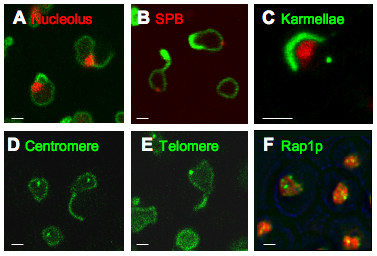
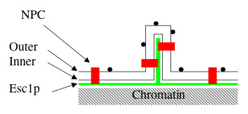
Background: Since S. cerevisiae undergoes closed mitosis, the nuclear envelope of the daughter nucleus is continuous with that of the maternal nucleus at anaphase. Nevertheless, several constitutents of the maternal nucleus are not present in the daughter nucleus. The present study aims to identify proteins which impact the shape of the yeast nucleus and to learn whether modifications of shape are passed on to the next mitotic generation. The Esc1p protein of S. cerevisiae localizes to the periphery of the nucleoplasm, can anchor chromatin, and has been implicated in targeted silencing both at telomeres and at HMR.

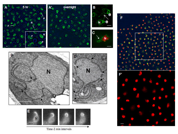
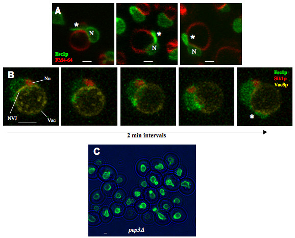
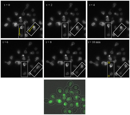
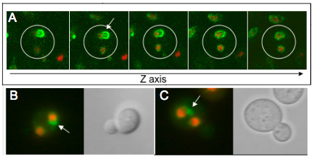
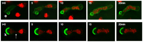
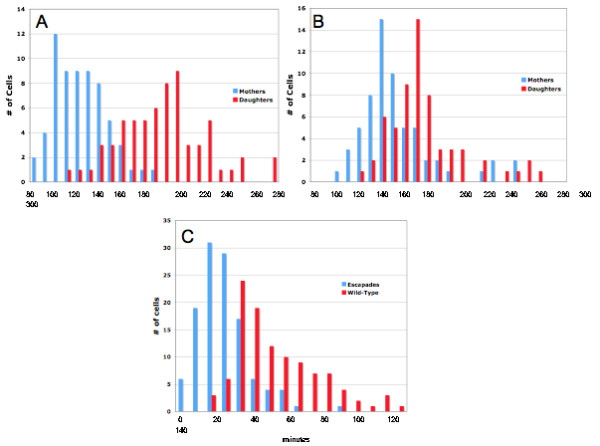
Results: Upon increased Esc1p expression, cell division continues and dramatic elaborations of the nuclear envelope extend into the cytoplasm. These "escapades" include nuclear pores and associate with the nucleolus, but exclude chromatin. Escapades are not inherited by daughter nuclei. This exclusion reflects their relative immobility, which we document in studies of prezygotes. Moreover, excess Esc1p affects the levels of multiple transcripts, not all of which originate at telomere-proximal loci. Unlike Esc1p and the colocalizing protein, Mlp1p, overexpression of selected proteins of the inner nuclear membrane is toxic.

Conclusion: Esc1p is the first non-membrane protein of the nuclear periphery which - like proteins of the nuclear lamina of higher eukaryotes - can modify the shape of the yeast nucleus. The elaborations of the nuclear envelope ("escapades") which appear upon induction of excess Esc1p are not inherited during mitotic growth. The lack of inheritance of such components could help sustain cell growth when parental nuclei have acquired potentially deleterious characteristics.


Figures

Figure 1
Figure 1
Structure of Escapades. (A) (A') Haploid cells expressing GFP-Esc1p from a galactose-inducible promoter (ATY3258) were induced for 5 hrs (left) or overnight (right) with 2% galactose. Escapades are seen as "fins" (F), "rings" (R) or "patches" (P). Phase images are in blue. Induction with 10–100-fold lower concentrations of galactose reduced the number of cells which were fluorescent but did not affect the appearance of their escapades. Note that fewer fins are seen upon overnight induction. The scale bar in this and all figures designates 1 micron. (B) Strain ATY2957 expressing both untagged Esc1p from a galactose-inducible promoter and a GFP-tagged ER marker (a truncated form of HMG-CoA reductase 1-GFP [59]), after overnight induction. Two cells are illustrated – one toward the upper right and one in the lower middle portion of the field. Note the ring-like escapades (e) in both cells. Fin-like escapades are seen after shorter periods of induction and no such structures are seen in the absence of galactose. The peripheral ER is indicated by the arrow. N: The nucleoplasmic volume. (C) Cells which express mRFP-tagged Htb2p and Nup49p-GFP as well as galactose-inducible untagged Esc1p (ATY3156) were induced overnight. Note the presence of the Nup49p-GFP signal (arrow) outside the margin of the red chromatin mass. By contrast, controls in glucose medium show a conventional circumferential distribution of GFP signal (not shown). (D) (D') GFP-Esc1p-expressing cells (ATY1483) were induced overnight, fixed and processed for transmission EM. Note the constant width of the double membranes that extend from the surface of the nucleus, and the presence of nuclear pores in these extensions (arrows). (E) Time-lapse sequence of a GFP-Esc1 expressing strain (ATY2102) after overnight induction. Note the progressive conversion of the fin to a ring-like escapade. Such conversions are seen only infrequently. N: The spherical portion of the nucleus. (F) (F') Htb2p-mRFP-expressing cells induced for 5 hr to express GFP-Esc1p (ATY3281). The square in the upper field is enlarged and illustrated without the green signal below to clarify the distribution of Htb2p-mRFP. Note the absence of tagged chromatin from the escapades.
Figure 2
Figure 2
Relation to Nuclear Structures. (A) GFP-Esc1p was induced for 5 hr to compare the localization of GFP-tagged escapades to the nucleolus, in a strain (ATY2101) which expresses Sik1p-mRFP. Systematic examination of through-focal series shows that > 90% of escapades contact the perimeter of the nucleus at or immediately adjacent to the mRFP-positive nucleolus. See Fig. S5. (B) Comparison of the localization of GFP-tagged escapades with the spindle pole body, in a strain (ATY3276) which expresses Spc42p-mRFP and has been induced for 5 hr. An overview is given in Fig S6. (C) Comparison of the localization of GFP-tagged karmellae with the nucleolus and the spindle pole body, in a strain (ATY1577) which expresses Sik1p-mRFP and Spc42p-GFP and carries plasmid [pGAL-HMG-CoA Reductase I-GFP]. Karmellae are ER membrane stacks associated with the outer nuclear membrane which result from overexpression of HMG-CoA reductase type1. (D) Comparison of the localization of GFP-tagged escapades with a centromere, in a strain (ATY2098) which expresses a GFP-lac repressor fusion and an insertion of lac operator arrays near CENIV and has been induced for 5 hr. An overeview is given in Fig. S7. (E) Comparison of the localization of GFP-tagged escapades with a telomere, in a strain (ATY2097) which expresses a GFP-lac repressor fusion and carries an insertion of lac operator arrays near telomere XIVL and has been induced for 5 hr. An overview is given in Fig. S8. (F) Comparison of the localization of the GFP-tagged telomere-associated protein, Rap1p, and Htb2p-mRFP-tagged chromatin in ATY3275. Induction was for 5 hr. An overview is given in Fig. S9.
Figure 3
Figure 3
Escapades Associate with the Vacuole and Originate at the NVJ. (A) Association with the vacuole, as detected by confocal examination of strain ATY2102 stained with FM4-64 (red) after galactose induction for 3 hrs. In each case, the chromatin-containing portion of the nucleus is designated (N) and the escapade is indicated by (*). (B) Time-lapse confocal sequence of strain ATY2513 that expresses Sik1p-mRFP (nucleolus: Nu) and Vac8p-YFP, which marks the vacuole membrane (Vac) and concentrates at the nucleus-vacuole junction (NVJ). GFP-Esc1p expression was induced with galactose 30 minutes prior to and maintained throughout time-lapse imaging, to follow the progression of GFP-Esc1p accumulation. Note the GFP-Esc1p at the periphery of the nucleus (N) and its progressive extension along the surface of the vacuole, generating an escapade (*). The MET25p-VAC8-EYFP plasmid was from D. Goldfarb. (C)pep3Δ cells which allow induction of GFPEsc1p were induced for 5 hr and examined (ATY2103).
Figure 4
Figure 4
Escapades are Asymmetrically Inherited. GAL-GFP-Esc1p strain ATY2102 was induced for 3 hours at 23°C prior to imaging at 2 min intervals in glucose medium. Boxed regions indicate cells undergoing mitosis and the arrows indicate the direction of elongation of the nucleus during anaphase. Mother (M) and daughter (D) nuclei are indicated in the last panel. Note the retention of escapades by the mother. The color image at the bottom includes a phase image to illustrate the cell outlines just prior to t = 0.
Figure 5
Figure 5
Distribution of Escapades in Binucleates. Cells expressing GFP-Esc1p and Htb2p-mRFP (ATY2509) were pre-induced, washed and recultured for 8 hrs at 23°C in glucose medium supplemented with latrunculin A to produce binucleate cells, resulting from occasional incorrect orientation of the spindle. A diploid strain was used to facilitate spatial resolution. (A) Confocal z-sections of a binucleate cell show retention of escapades (arrow) in a single nucleus. (B, C) Epifluorescent imaging of binucleate cells. Phase images indicate that the nuclei are contained within the maternal cytoplasm.
Figure 6
Figure 6
Distribution of Escapades and Karmellae upon Karyogamy. Escapades and karmellae remain with the nucleus of their origin upon fusion with nuclei which express Htb2p-mRFP. Top: A strain which allows induction of GFP-Esc1p (ATY1550) was crossed with a strain expressing mRFP-tagged histone Htb2p (ATY2835). Bottom: ATY1650, which carries a galactose-inducible plasmid allowing induction of Hmg1p-Co A Reductase-GFP and expresses a mRFP-tagged form of the SPB-protein, Spc42p (arrow), was crossed with ATY2835. Upon nuclear fusion, the red histone signal gradually invades the trans nucleus, but neither the escapades (e) nor karmellae (k) changes its location over at least 20 min. ATY1550 and ATY1650 were pre-grown overnight in galactose medium. For both time series, note that mRFP-tagged chromatin is absent from the regions immediately underlying the escapades or karmellae. These volumes are occupied by the nucleoli. Also, note the infusion of GFP-Esc1p into the trans nucleus upon karyogamy, indicative of facile diffusion.
Figure 7
Figure 7
Kinetics of Bud Formation. GAL-GFP-Esc1p strain ATY2102 and an isogenic wild-type strain (ATY2500) were pre-induced in galactose media for 3 hrs, washed and examined on agarose pads in glucose media over 5 hr, during which time escapades remained visible (ATY2500). DIC images captured every 10 min allowed measurement of the timing of bud initiation for each cell. Time measurements were all relative to the emergence of the first bud by each mother cell, M1. For example, the mother's bud interval, M2-M1 measures the delay prior to the appearance of the second bud on the mother. Correspondingly, D-M1 measures the interval prior to the appearance of the first bud on the daughter. (A) Histogram showing the bud intervals for a single experiment for the wild type strain. Note that there is minimal overlap between mothers and daughters and that mothers re-bud considerably faster than daughter cells. (B) Histogram showing the bud intervals for an isogenic escapade strain. Note, in contrast to (A), that there is considerable overlap between mothers and daughters. The bud interval is slowed in mothers and accelerated in daughters. (C) Histogram illustrating the mother-daughter bud differential calculated as: (D-M2 = bud differential) where D is the time at which a bud appears in the daughter cell and M2 is the time at which a second bud appears in the mother cell. Note that the bud differential is decreased by 50% by the presence of escapades (average bud differential is 65 min. in wild-type strain and 28 min. in escapade strain). Results are cumulative over four separate experiments for each strain.
Figure 8
Figure 8
Model of Escapade Structure. Nuclear pore complexes (NPC), outer nuclear membrane (Outer), inner nuclear membrane (Inner), and Esc1p are indicated. The small circles at the surface of the membranes are ribosomes. The model has been drawn to illustrate a tight apposition of Esc1p layers within the escapade, which may account for the constant width that is observed with EM and the exclusion of chromatin.

References

    1. Jaspersen SL, Winey M. The budding yeast spindle pole body: structure, duplication, and function. Annu Rev Cell Dev Biol. 2004;20:1–28. doi: 10.1146/annurev.cellbio.20.022003.114106. - DOI - PubMed
    1. Loidl J. Chromosomes of the budding yeast Saccharomyces cerevisiae. Int Rev Cytol. 2003;222:141–196. - PubMed
    1. Gruenbaum Y, Margalit A, Goldman RD, Shumaker DK, Wilson KL. The nuclear lamina comes of age. Nat Rev Mol Cell Biol. 2005;6:21–31. doi: 10.1038/nrm1550. - DOI - PubMed
    1. Best S, Salvati F, Kallo J, Garner C, Height S, Thein SL, Rees DC. Lamin B-receptor mutations in Pelger-Huet anomaly. Br J Haematol. 2003;123:542–544. doi: 10.1046/j.1365-2141.2003.04621.x. - DOI - PubMed
    1. Davido T, Getzenberg RH. Nuclear matrix proteins as cancer markers. J Cell Biochem Suppl. 2000:136–141. doi: 10.1002/1097-4644(2000)79:35+<136::AID-JCB1137>3.0.CO;2-E. - DOI - PubMed

Publication types

MeSH terms

Substances