Review
. 2008 Jan;1784(1):16-26.
doi: 10.1016/j.bbapap.2007.10.002. Epub 2007 Oct 12.

Signaling through cAMP and cAMP-dependent protein kinase: diverse strategies for drug design

Affiliations
Review

Signaling through cAMP and cAMP-dependent protein kinase: diverse strategies for drug design

Susan S Taylor et al. Biochim Biophys Acta. 2008 Jan.

Abstract

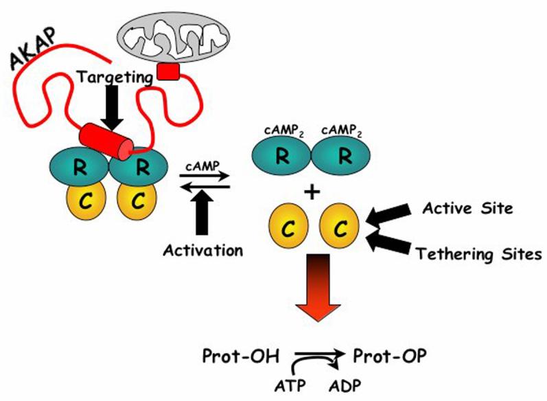
The catalytic subunit of cAMP-dependent protein kinase has served as a prototype for the protein kinase superfamily for many years while structures of the cAMP-bound regulatory subunits have defined the conserved cyclic nucleotide binding (CNB) motif. It is only structures of the holoenzymes, however, that enable us to appreciate the molecular features of inhibition by the regulatory subunits as well as activation by cAMP. These structures reveal for the first time the remarkable malleability of the regulatory subunits and the CNB domains. At the same time, they allow us to appreciate that the catalytic subunit is not only a catalyst but also a scaffold that mediates a wide variety of protein:protein interactions. The holoenzyme structures also provide a new paradigm for designing isoform-specific activators and inhibitors of PKA. In addition to binding to the catalytic subunits, the regulatory subunits also use their N-terminal dimerization/docking domain to bind with high affinity to A Kinase Anchoring Proteins using an amphipathic helical motif. This targeting mechanism, which localizes PKA near to its protein substrates, is also a target for therapeutic intervention of PKA signaling.


Figures

Figure 1
Figure 1. Conformational states of PKA and candidate sites for engineering inhibitors of PKA signaling
The free catalytic (C) subunit can be inactivated with inhibitors to the active site and by inhibitors that disrupt tethering of protein substrates and inhibitors. Therapeutic intervention can also disrupt the activation of the PKA holoenzyme complex and the targeting of PKA through the AKAPs. C-subunit is yellow, R-subunit is teal and AKAP is red. Potential sites for therapeutic intervention are shown by black arrows.
Figure 2
Figure 2. N- and C-terminal tails of AGC kinases are cis-regulatory elements
The upper panels show the N- and C-terminal tails of the PKA catalytic subunit. On the left is the N-tail showing its different sites of covalent modification. On the right is the C-tail showing the Active Site Tether (AST) in red and the N- and C-lobe tethers in white. In the middle panel one can see how the αC Helix and the αC-β4 Loop are anchored by the NLT and CLT. AGC conserved residues are also highlighted. The lower panels show how the features of the C-tail are conserved in the AGC subfamily of protein kinases. On the lower right are the segments of the C-tail: N-lobe Tether (NLT), Active Site Tether (AST), C-lobe Tether (CLT). The C-lobe Anchor (CLA) contains the AGC Insert between the αH and αI Helices. The panels on the left show the C-terminal tail conformation in three AGC kinase structures (PKA – PDB code:1ATP, AKT – PDB code:1GZN, and PKC – PBDcode:1XJD). The disordered regions are shown in dotted representation.
Figure 3
Figure 3. Domain organization of the R-subunit isoforms and the structure of the cAMP binding domains of RIα
The domain organization of RIα, RIIα, and RIIβ is shown on the left. On the right is the structure of RIα(91-379) where the cAMP Binding Domain A is shown in dark teal and Domain B is in turquoise.
Figure 4
Figure 4. Models of the catalytic subunit bound to different inhibitors
On the left is shown the complex with the PKI(5-24) Inhibitor Peptide. In the middle is the RIα(91-244):C complex (PDB code:1U7E). On the right is the RIα(91-379):C complex (PDB code:2QCS). All three structures have bound Mg2ATP and are in a closed conformation. The catalytic subunits are rendered as space filling models while the inhibitors are shown as ribbons. CBD A is in dark teal while CBD B is in turquoise. The αB/αC Helix in CBD A is in red.
Figure 5
Figure 5. The active site cleft of the catalytic subunit is filled by the pseudosubstrate inhibitor segment of RIα
The left panel shows how the P−5 to P+1 peptide occupies the active site cleft. On the right is a close up of the site that is nucleated by the P+1 Val. Key residues that nucleate this site are Tyr205 from the PBC of Domain A and Tyr247 from the αG Helix of the C-subunit.
Figure 6
Figure 6. Dynamic malleability of the RIα subunit
On the top is shown the conformation of RIα (91-244) bound to cAMP (left) and bound to Mn2ATP and the C-subunit. The αB/αC Helix, shown in red, undergoes dramatic conformational rearrangement. On the bottom are the conformational changes associates with the RIIα(91-379) construct that contains both the Domain A and B. The conformational changes in domain A are comparable to what was seen in the smaller construct, but the B-domain which immediately follows the Domain A is dramatically changed by the snapping open of the αB/αC Helix, again shown in red.
Figure 7
Figure 7. The catalytic subunit is a scaffold as well as a catalyst
Panel A shows the conserved kinase core of the catalytic subunit (residues 40-300) while Panel B shows how the N-and C-tails wrap around the surface of the kinase core. The large lobe is in tan and the small lobe in cream. Panel C shows the surfaces that are masked by the inhibitor peptide (red), the PKI amphipathic helix (blue), the A Domain of RIα (green), and the P+5 Arg of the RIα Inhibitor Site.
Figure 8
Figure 8. Ordered pathway for activation of the RI holoenzyme
A cartoon of the ordered activation mechanism is shown at the top. Close up of the cAMP binding site in Domain A in the presence of cAMP is on the bottom left while the site in the absence of cAMP is shown on the bottom right. The PBC is shown as a ribbon through the transparent space filling model.
Figure 9
Figure 9. Fluorescence anisotropy assay for inhibitors and activators of PKA
The strategy for the assay is indicated at the top while the results of two different assays are indicated in the middle.
Figure 10
Figure 10. The AKAP binding motif docked to the dimerization/docking domain of the RIIα subunit
On the left is shown the peptide arrays where isoform specific properties of the AKAP peptide were identified. By overlaying the array with the RIIα or RIα D/D domain, it was possible to identify isoform differences [37]. The hydrophobic surface of the AKAP peptides is shown between the two arrays. On the right is a model of the D-AKAP2 peptide bound to the RIIα D/D domain, based on the recently solved crystal structure[10]. The peptide is shown in red while the two protomers of the D/D domain are shown in tan and grey. The density of the side chains is shown in green. There also are no water molecules in the interface.

References

    1. Walsh DA, Perkins JP, Krebs EG. An adenosine 3′,5′-monophosphate-dependant protein kinase from rabbit skeletal muscle. J Biol Chem. 1968;243:3763–5. - PubMed
    1. Shoji S, Parmelee DC, Wade RD, Kumar S, Ericsson LH, Walsh KA, Neurath H, Long GL, Demaille JG, Fischer EH, Titani K. Complete amino acid sequence of the catalytic subunit of bovine cardiac muscle cyclic AMP-dependent protein kinase. Proc Natl Acad Sci U S A. 1981;78:848–51. - PMC - PubMed
    1. Uhler MD, Carmichael DF, Lee DC, Chrivia JC, Krebs EG, McKnight GS. Isolation of cDNA clones coding for the catalytic subunit of mouse cAMP-dependent protein kinase. Proc Natl Acad Sci U S A. 1986;83:1300–4. - PMC - PubMed
    1. Knighton DR, Zheng JH, Ten Eyck LF, Ashford VA, Xuong NH, Taylor SS, Sowadski JM. Crystal structure of the catalytic subunit of cyclic adenosine monophosphate-dependent protein kinase. Science. 1991;253:407–14. - PubMed
    1. Madhusudan, Akamine P, Xuong NH, Taylor SS. Crystal structure of a transition state mimic of the catalytic subunit of cAMP-dependent protein kinase. Nat Struct Biol. 2002;9:273–7. - PubMed

Publication types

MeSH terms

LinkOut - more resources