

Corneal crystallins and the development of cellular transparency
- PMID: 17997336
- PMCID: PMC2275913
- DOI: 10.1016/j.semcdb.2007.09.015
Corneal crystallins and the development of cellular transparency
Abstract
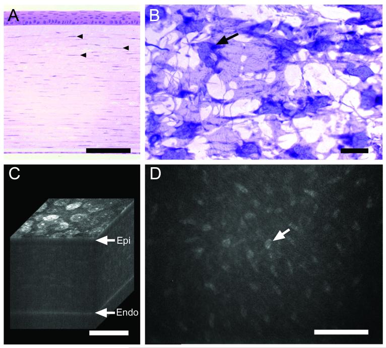
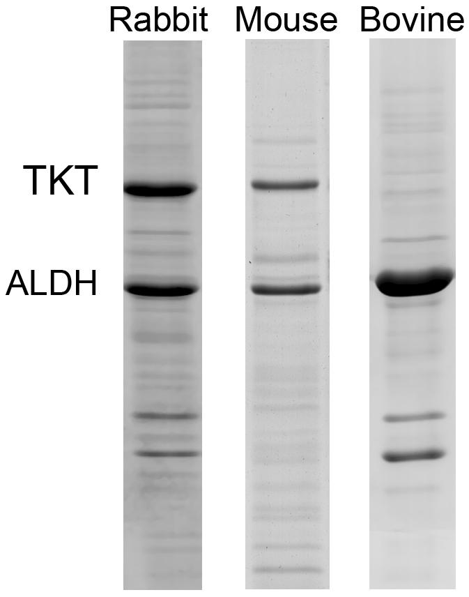
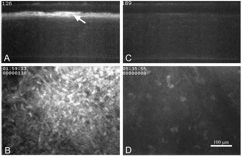
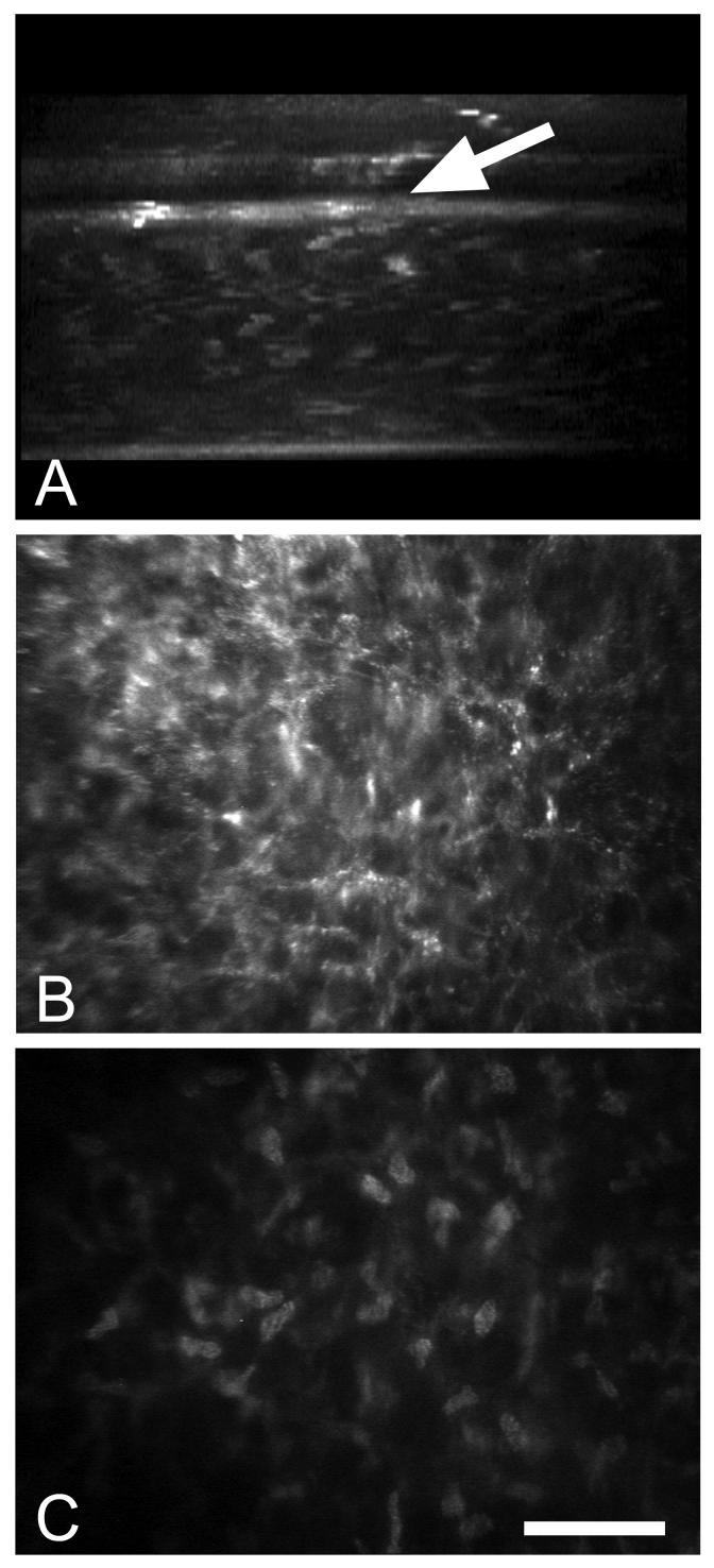
Past studies have established that the cornea like the lens abundantly expresses a few water-soluble enzyme/proteins in a taxon specific fashion. Based on these similarities it has been proposed that the lens and the cornea form a structural unit, the 'refracton', that has co-evolved through gene sharing to maximize light transmission and refraction to the retina. Thus far, the analogy between corneal crystallins and lens crystallins has been limited to similarities in the abundant expression, with few reports concerning their structural function. This review covers recent studies that establish a clear relationship between expression of corneal crystallins and light scattering from corneal stromal cells, i.e. keratocytes, that support a structural role for corneal crystallins in the development of transparency similar to that of lens crystallins that would be consistent with the 'refracton' hypothesis.
Figures
Similar articles
-
Review: A case for corneal crystallins.J Ocul Pharmacol Ther. 2000 Apr;16(2):173-80. doi: 10.1089/jop.2000.16.173. J Ocul Pharmacol Ther. 2000. PMID: 10803428 Review.
-
The role of corneal crystallins in the cellular defense mechanisms against oxidative stress.Semin Cell Dev Biol. 2008 Apr;19(2):100-12. doi: 10.1016/j.semcdb.2007.10.004. Epub 2007 Oct 10. Semin Cell Dev Biol. 2008. PMID: 18077195 Review.
-
Ubiquitous lens alpha-, beta-, and gamma-crystallins accumulate in anuran cornea as corneal crystallins.J Biol Chem. 2007 Jun 29;282(26):18953-9. doi: 10.1074/jbc.M609275200. Epub 2007 Apr 23. J Biol Chem. 2007. PMID: 17452334
-
The cellular basis of corneal transparency: evidence for 'corneal crystallins'.J Cell Sci. 1999 Mar;112 ( Pt 5):613-22. doi: 10.1242/jcs.112.5.613. J Cell Sci. 1999. PMID: 9973596
-
Abundant corneal gelsolin in Zebrafish and the 'four-eyed' fish, Anableps anableps: possible analogy with multifunctional lens crystallins.Exp Eye Res. 2004 Dec;79(6):949-56. doi: 10.1016/j.exer.2004.04.002. Exp Eye Res. 2004. PMID: 15642334 Free PMC article.
Cited by
-
C-MAF Expression in Adult Human Ocular Surface and its Implication in Pterygium Pathogenesis.Rep Biochem Mol Biol. 2020 Jan;8(4):419-423. Rep Biochem Mol Biol. 2020. PMID: 32582801 Free PMC article.
-
Localization of αA-Crystallin in Rat Retinal Müller Glial Cells and Photoreceptors.Microsc Microanal. 2018 Oct;24(5):545-552. doi: 10.1017/S1431927618015118. Epub 2018 Sep 26. Microsc Microanal. 2018. PMID: 30253817 Free PMC article.
-
Identification of a peptide ligand for human ALDH3A1 through peptide phage display: Prediction and characterization of protein interaction sites and inhibition of ALDH3A1 enzymatic activity.Front Mol Biosci. 2023 Mar 20;10:1161111. doi: 10.3389/fmolb.2023.1161111. eCollection 2023. Front Mol Biosci. 2023. PMID: 37021113 Free PMC article.
-
TGFβ1-driven SMAD2/3 phosphorylation and myofibroblast emergence are fully dependent on the TGFβ1 pre-activation of MAPKs and controlled by maternal leucine zipper kinase.Cell Signal. 2024 Jan;113:110963. doi: 10.1016/j.cellsig.2023.110963. Epub 2023 Nov 4. Cell Signal. 2024. PMID: 37931692 Free PMC article.
-
Collagen fibril diameter and alignment promote the quiescent keratocyte phenotype.J Biomed Mater Res A. 2012 Mar;100(3):613-21. doi: 10.1002/jbm.a.33284. Epub 2011 Dec 30. J Biomed Mater Res A. 2012. PMID: 22213336 Free PMC article.
References
-
- Holt WS, Kinoshita JH. The soluble proteins of the bovine cornea. Invest Ophthalmol Vis Sci. 1973;12:114. - PubMed
-
- Alexander RJ, Silverman B, Henley WL. Isolation and characterization of BCP 54, the major water soluble protein of bovine cornea. Exp Eye Res. 1981;32:205. - PubMed
-
- Silverman B, Alexander RJ, Henley WL. Tissue and species specificity of BCP 54, the major soluble protein of bovine cornea. Exp Eye Res. 1981;33:19. - PubMed
-
- Abedinia M, Pain T, Algar EM, Holmes RS. Bovine corneal aldehyde dehydrogenase: the major soluble corneal protein with a possible dual protective role for the eye. Exp Eye Res. 1990;51:419. - PubMed
-
- Cooper DL, Baptist EW, Enghild JJ, Isola NR, Klintworth GK. Bovine corneal protein 54 K (BCP540 is a homologue of the tumor-associated (class 3) rat aldehyde dehydrogenase (TAT_ALD) Gene. 1991;98:201. - PubMed
Publication types
MeSH terms
Substances
Grants and funding
LinkOut - more resources
Full Text Sources
Miscellaneous
