. 2007 Nov;3(11):e171.
doi: 10.1371/journal.ppat.0030171.

Release of hepatic Plasmodium yoelii merozoites into the pulmonary microvasculature

Affiliations

Release of hepatic Plasmodium yoelii merozoites into the pulmonary microvasculature

Kerstin Baer et al. PLoS Pathog. 2007 Nov.

Abstract

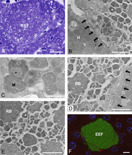
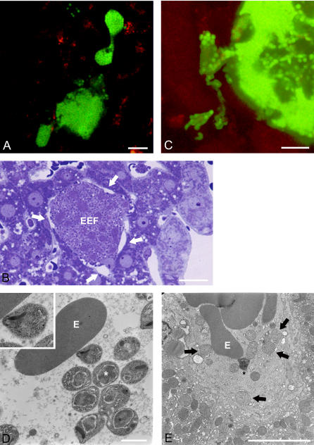
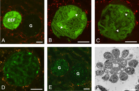
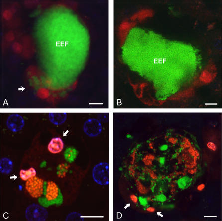
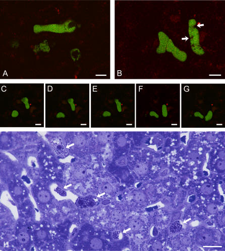
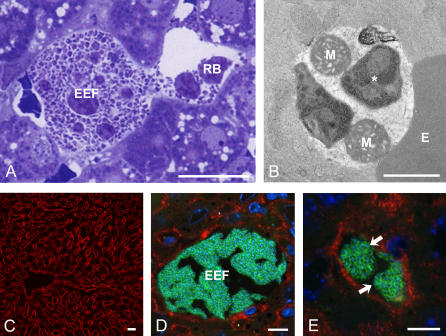
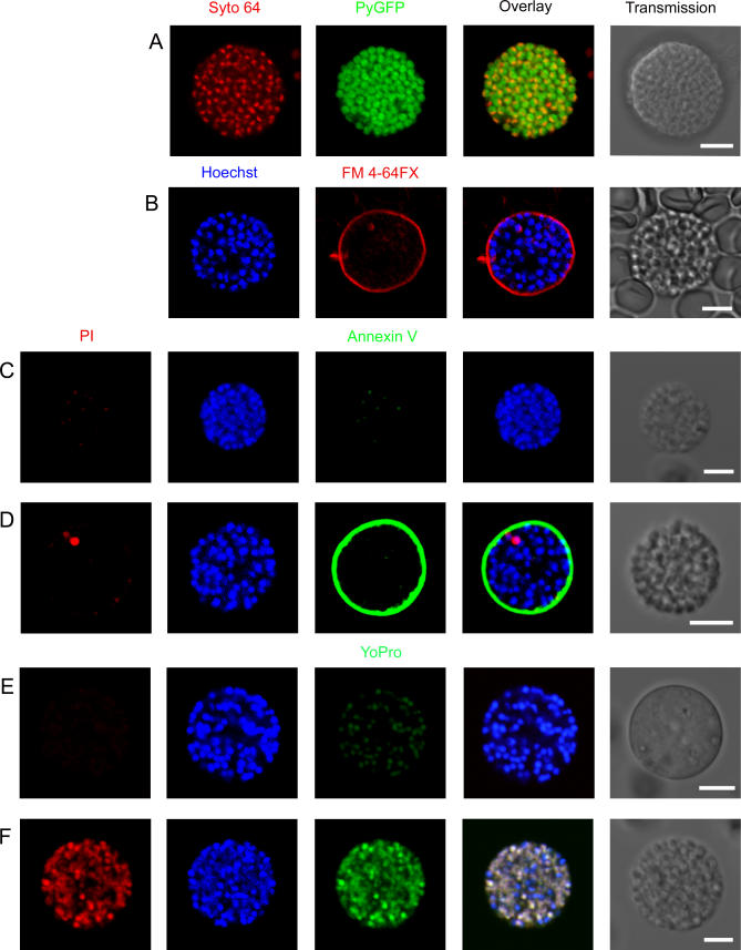
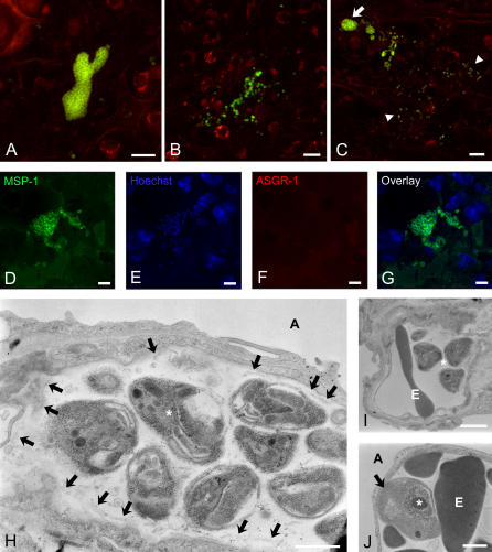
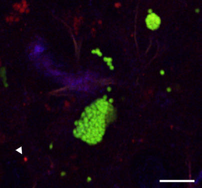
Plasmodium undergoes one round of multiplication in the liver prior to invading erythrocytes and initiating the symptomatic blood phase of the malaria infection. Productive hepatocyte infection by sporozoites leads to the generation of thousands of merozoites capable of erythrocyte invasion. Merozoites are released from infected hepatocytes as merosomes, packets of hundreds of parasites surrounded by host cell membrane. Intravital microscopy of green fluorescent protein-expressing P. yoelii parasites showed that the majority of merosomes exit the liver intact, adapt a relatively uniform size of 12-18 microm, and contain 100-200 merozoites. Merosomes survived the subsequent passage through the right heart undamaged and accumulated in the lungs. Merosomes were absent from blood harvested from the left ventricle and from tail vein blood, indicating that the lungs effectively cleared the blood from all large parasite aggregates. Accordingly, merosomes were not detectable in major organs such as brain, kidney, and spleen. The failure of annexin V to label merosomes collected from hepatic effluent indicates that phosphatidylserine is not exposed on the surface of the merosome membrane suggesting the infected hepatocyte did not undergo apoptosis prior to merosome release. Merosomal merozoites continued to express green fluorescent protein and did not incorporate propidium iodide or YO-PRO-1 indicating parasite viability and an intact merosome membrane. Evidence of merosomal merozoite infectivity was provided by hepatic effluent containing merosomes being significantly more infective than blood with an identical low-level parasitemia. Ex vivo analysis showed that merosomes eventually disintegrate inside pulmonary capillaries, thus liberating merozoites into the bloodstream. We conclude that merosome packaging protects hepatic merozoites from phagocytic attack by sinusoidal Kupffer cells, and that release into the lung microvasculature enhances the chance of successful erythrocyte invasion. We believe this previously unknown part of the plasmodial life cycle ensures an effective transition from the liver to the blood phase of the malaria infection.


Conflict of interest statement

Competing interests. The authors have declared that no competing interests exist.

Figures

Figure 1
Figure 1. Characterization of Late P. yoelii EEFs
(A) Semithin Epon section showing a mature EEF containing thousands of merozoites at 52 h after P. yoelii sporozoite infection. Note that few sinusoids are visible surrounding the EEF. Bar = 10 μm. (B) An infected hepatocyte containing a mixture of merozoites and host cell organelles indicating that the PVM has ruptured. The membrane of the infected cell is in close contact (arrows) with a neighboring hepatocyte. H, hepatocyte. *, merozoites. Bar = 5 μm. (C) Merozoites lie in close apposition to ultrastructurally well-preserved host cell mitochondria. M, mitochondria; *, merozoites. Bar = 1 μm. (D) EEF containing numerous merozoites and a few host cell organelles and remnant bodies. Note that one adjacent sinusoid has collapsed (arrows), while another contains an erythrocyte in its lumen indicating preserved blood flow. RB, remnant bodies. Bar = 5 μm. (E) Merozoites and remnant bodies of various shapes and sizes are embedded in a loose matrix. RB, remnant bodies; *, merozoites. Bar = 5 μm. (F) Intravital micrograph showing a mature PyGFP EEF at the onset of merosome formation. The nuclei of the host cell and the neighboring hepatocytes are visualized by Hoechst staining. Bar = 10 μm.
Figure 2
Figure 2. Merozoite Release by Merosome Formation
(A) Intravital microscopy of merosomes budding from an EEF. Note that a few individual merozoites are located in the vicinity of the EEF (see Videos S1–S3). Bar = 10 μm. (B) Semithin Epon section showing an EEF that has lost contact with the surrounding parenchyma (arrows). Bar = 10 μm. (C) Projection of an EEF in the process of disintegration; note that the cell releases individual merozoites into the environment (see Video S4). Bar = 10 μm. (D) Electron micrograph showing merozoites located free in a liver sinusoid. The apical pole of one of the merozoites is in close contact with an erythrocyte (insert). E, erythrocyte; *, merozoites. Bar = 1 μm. (E) Liver sinusoid containing free hepatocyte mitochondria (arrows) and a free merozoite. E, erythrocyte; *, merozoite. Bar = 5 μm.
Figure 3
Figure 3. Rapid EEF Decay
(A–D) Individual frames from an intravital video showing the process of liver stage transformation into an EEF ghost (see Video S5). (A) The still image shows an intact EEF adjacent to a ghost. G, ghost. (B) Individual frame showing rapid decay of the EEF shown on the left of (A). Note that loss of cytosolic GFP reveals the presence and arrangement of the merozoites (arrow). (C–D) Only a few of the thousands of merozoites remain visible in the faintly fluorescent EEF ghost. (E) Eventually, the infected hepatocyte has transformed into an EEF ghost. Note that both ghosts retain close contact with the surrounding tissue (A–E). Bars = 20 μm. (F) Electron micrograph showing a liver sinusoid with merozoites that are incompletely separated from a remnant body. RB, remnant body; G, ghost; *, merozoite. Bar = 1 μm.
Figure 4
Figure 4. Hepatocyte and Merozoite Viability during Merosome Formation
At 52–70 h after infection with PyGFP, mice were intravenously injected with a mixture of the membrane-permeable DNA stain Hoechst 33342 and the dead cell dye PI prior to intravital microscopic examination. (A) Confocal scan showing an intact EEF that has not incorporated PI (red) and is therefore considered viable. Note that merozoites have been discharged into surrounding cells (arrow) some of which have taken up the red dead cell stain. (B) Due to merosome formation, this EEF has decreased in size and detached from the surrounding tissue. The neighboring cells appear to be compressed and are stained with PI (red). (C) A few merozoites are left behind in an EEF that has disintegrated after repeated merosome budding. The two nuclei of the host cell (arrows) have incorporated predominantly PI (red) and some Hoechst stain (blue) and appear pink. In addition, some of the merozoites have lost their green fluorescence and appear red due to PI staining, while others have retained GFP and excluded PI and are therefore viable. The nuclei of the neighboring hepatocytes are visualized by Hoechst staining (blue). (D) EEF at a late stage of disintegration. Some remaining merozoites are viable (green), while other merozoites are dead (red). The nuclei of some surrounding cells have also incorporated PI (arrows). Bars = 10 μm.
Figure 5
Figure 5. Dynamics of Merosome Transport in the Liver
(A) Multiple merosomes separate from an EEF and are rapidly transported towards the central vein (see Video S6). Bar = 10μm. (B) Large merosomes move slowly within the sinusoidal lumen (see Video S7). Remnant bodies can be differentiated from merozoites by their larger size and lack of GFP (arrows). Bar = 10 μm. (C–G) Individual frames of an intravital confocal video showing two smaller merosomes gliding along a sinusoid. Bars = 10 μm. (H) Semithin liver section 52 h after infection with P. yoelii. Multiple merosomes, presumably released from a single EEF, can be found inside the sinusoids (arrows). Note the presence of remnant bodies in merosomes. Bar = 10 μm.
Figure 6
Figure 6. Merosomes Contain Viable Merozoites and Host Cell Organelles
(A) Semithin section showing an EEF releasing a merosome into a sinusoid. Note that remnant bodies are a normal component of the merosomal cytoplasm. RB, remnant bodies. Bar = 10 μm. (B) Electron microscopy documents that merosomes contain well-preserved merozoites and hepatocyte mitochondria. E, erythrocyte; M, hepatocyte mitochondria; *, merozoite. Bar = 1 μm. (C) Immunolabeling of frozen liver sections for ASGR-1 reveals the presence of the receptor on the basolateral portion of the membrane of all hepatocytes (red). Bar = 20 μm. Neither EEFs (D) nor merosomes (E, arrows) express ASGR-1 on their surface. The merozoites were visualized with Hoechst (blue) and an antibody against MSP-1 in combination with PA-FITC (green). Bars = 10 μm.
Figure 7
Figure 7. In Vitro Characterization of Hepatic Merosomes
(A) Merozoite nuclei in a PyGFP merosome were visualized with the membrane-permeable DNA stain SYTO-64 (red). Differential interference contrast was used to capture the transmission image on the right of the panel. (B) The phospholipid marker FM 4–64FX was used to visualize the membrane surrounding the merosome (red). Merozoite nuclei were stained with Hoechst (blue). (C) Immediately after harvesting from the hepatic effluent, the majority of merosomes were negative for annexin V (green) and PI (red). (D) Prolonged in vitro cultivation led to PS exposure (green) in the outer leaflet of the merosomal membrane and to gradual incorporation of PI (red) into individual merozoite nuclei. Parasite nuclei were visualized with Hoechst (blue). (E and F) Freshly harvested merosomes excluded YO-PRO-1 (green), a DNA stain that selectively passes across apoptotic membranes, and the dead cell stain PI (red). In vitro incubation of the merosomes led to the simultaneous uptake of both nucleic acid stains suggesting that merozoite death occurred by necrosis rather than programmed cell death. Note that colocalization of YO-PRO-1, PI, and Hoechst causes nuclei to appear white. Bars = 5 μm.
Figure 8
Figure 8. Merosomes Accumulate in the Lungs and Release Merozoites into the Pulmonary Microcirculation
(A) Ex vivo confocal microscopy of a mouse lung 52 h after infection with PyGFP shows a large merosome located inside an alveolar capillary. (B) A group of individual merozoites inside pulmonary capillaries. (C) Small parasite aggregates and individual merozoites (arrowheads) fanning out in one direction from a merosome (arrow). This asymmetric parasite distribution suggests that merosomes release merozoites into the pulmonary microcirculation. (A–C) Bars = 10 μm. (D–G) Mouse lung fixed 52 h after infection with P. yoelii. The frozen section was immunolabeled with MSP-1 followed by protein A-FITC (D, green) indicating that the merozoites are intact. Labeling with goat-anti-ASGR-1 followed by rabbit anti-goat IgG-TX (E, red) revealed the absence of the receptor on the merosomal membrane. Nuclei were visualized by Hoechst staining (F, blue). (H) Electron micrograph showing well-preserved merozoites (∗) in the lumen of an alveolar capillary. Only fragments of the merosomal membrane are detectable (arrows). *, merozoite; A, alveolar lumen. Bar = 1 μm. (I) Free merozoites released into the lumen of a pulmonary capillary. *, merozoite; E, erythrocyte. Bar = 1 μm. (J) Alveolar capillary showing an erythrocyte (arrow) harboring a recently invaded merozoite in addition to several uninfected erythrocytes. *, merozoite; E, erythrocyte. Bar = 1 μm.
Figure 9
Figure 9. Pulmonary Merosomes Are Viable
Mouse lung analyzed ex vivo 48 h after infection with PyGFP. Generally, neither merosomes nor free merozoites incorporated intravenously injected PI indicating that the parasites were viable. Note that one of the merozoites is PI-positive (arrowhead). Nuclei were visualized by Hoechst staining (blue). Bar = 10 μm.
Figure 10
Figure 10. Model of Merosome Dissemination and Merozoite Liberation
(A) After malaria sporozoite entry into a hepatocyte, the parasite begins to grow inside a PV to a size larger than its original host cell. Schizogonic division results in the formation of thousands of erythrocyte-infective merozoites. During the final stage of differentiation, the PVM dissolves and allows the parasites to mix with the remaining host cell organelles. Eventually, the plasma membrane of the infected hepatocyte bulges out and forms merosomes; thus releasing merozoites, remnant bodies, and host cell mitochondria into the sinusoidal lumen. Camouflaged by host cell membrane, merosomes are not recognized by Kupffer cells and are shuttled out of the liver. Infiltration of the remains of the infected host cell by mononuclear phagocytes and neutrophil granulocytes gives rise to the formation of a small granuloma. Me, merosomes; RB, remnant bodies; Mi, host cell mitochondria; KC, Kupffer cells; M∅, mononuclear phagocytes; S, sporozoite. (B) Shear forces inside the hepatic and other larger veins cause the merosomes to break down into smaller units. After passing through the right heart, these small merosomes are arrested inside lung capillaries where (1) they eventually release infectious merozoites into the pulmonary microvasculature; (2) local stagnation of the blood flow due to capillary occlusion by merosomes may result in dense erythrocyte packing; and thus (3) facilitate merozoite infection. ∗, alveolar space; H, hepatocyte; N, nucleus; E, endothelium; P-I, type I pneumocyte; P-II, type II pneumocyte; F, fibroblast; aM∅, alveolar macrophage; PMN, polymorphnuclear macrophage.

Similar articles

Cited by

References

    1. Nussenzweig RS, Vanderberg J, Most H, Orton C. Protective immunity produced by the injection of x-irradiated sporozoites of Plasmodium berghei . Nature. 1967;216:160–162. - PubMed
    1. Epstein JE, Giersing B, Mullen G, Moorthy V, Richie TL. Malaria vaccines: are we getting closer? Curr Opin Mol Ther. 2007;9:12–24. - PubMed
    1. Vanderberg JP, Frevert U. Intravital microscopy demonstrating antibody-mediated immobilization of Plasmodium berghei sporozoites injected into skin by mosquitoes. Int J Parasitol. 2004;34:991–996. - PubMed
    1. Amino R, Thiberge S, Martin B, Celli S, Shorte S, et al. Quantitative imaging of Plasmodium transmission from mosquito to mammal. Nat Med. 2006;12:220–224. - PubMed
    1. Meis JFGM, Verhave JP. Exoerythrocytic development of malaria parasites. Adv Parasitol. 1988;27:1–61. - PubMed

Publication types