. 2007;8(11):R239.
doi: 10.1186/gb-2007-8-11-r239.

The distributions, mechanisms, and structures of metabolite-binding riboswitches

Affiliations

The distributions, mechanisms, and structures of metabolite-binding riboswitches

Jeffrey E Barrick et al. Genome Biol. 2007.

Abstract

Background: Riboswitches are noncoding RNA structures that appropriately regulate genes in response to changing cellular conditions. The expression of many proteins involved in fundamental metabolic processes is controlled by riboswitches that sense relevant small molecule ligands. Metabolite-binding riboswitches that recognize adenosylcobalamin (AdoCbl), thiamin pyrophosphate (TPP), lysine, glycine, flavin mononucleotide (FMN), guanine, adenine, glucosamine-6-phosphate (GlcN6P), 7-aminoethyl 7-deazaguanine (preQ1), and S-adenosylmethionine (SAM) have been reported.

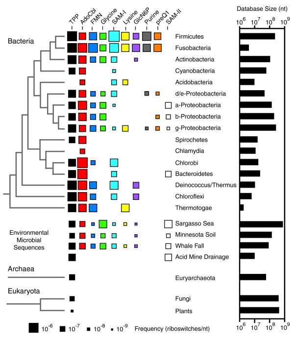
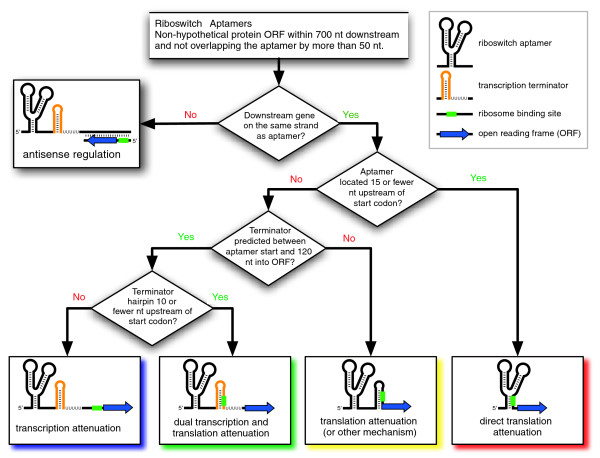
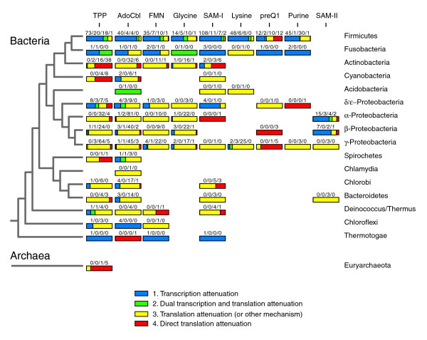
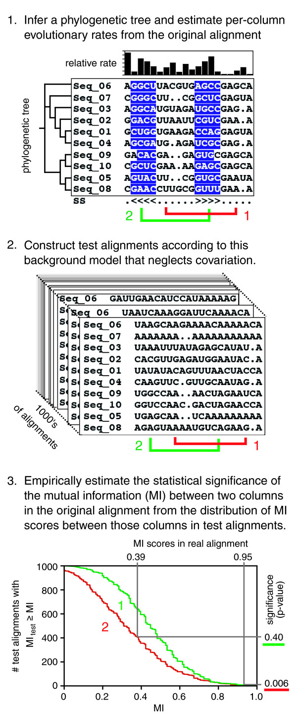
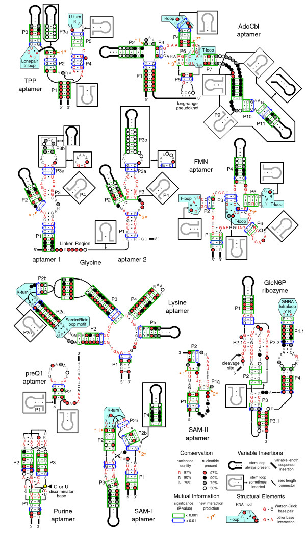
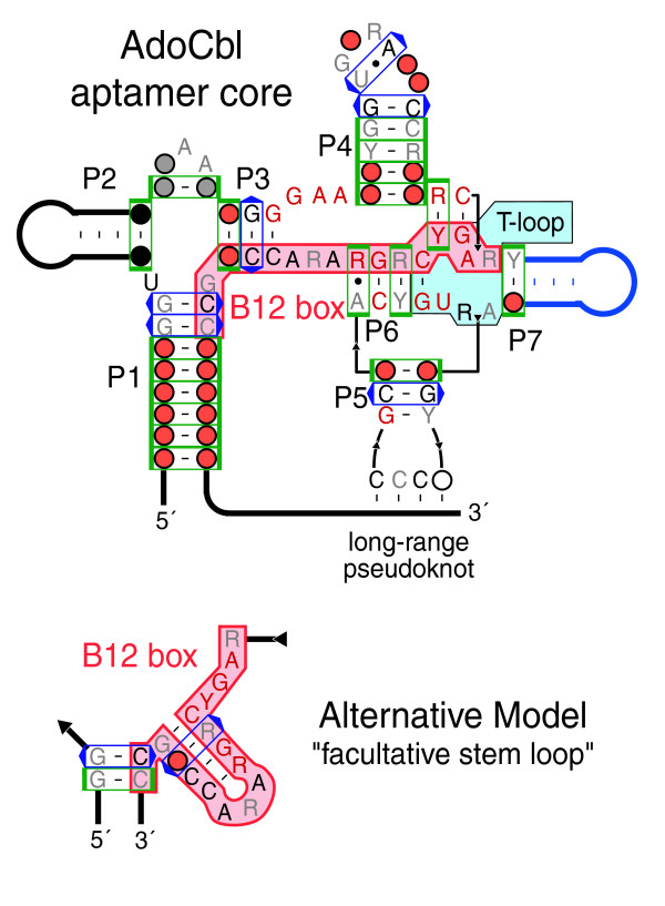
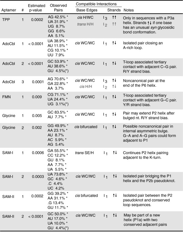
Results: We have used covariance model searches to identify examples of ten widespread riboswitch classes in the genomes of organisms from all three domains of life. This data set rigorously defines the phylogenetic distributions of these riboswitch classes and reveals how their gene control mechanisms vary across different microbial groups. By examining the expanded aptamer sequence alignments resulting from these searches, we have also re-evaluated and refined their consensus secondary structures. Updated riboswitch structure models highlight additional RNA structure motifs, including an unusual double T-loop arrangement common to AdoCbl and FMN riboswitch aptamers, and incorporate new, sometimes noncanonical, base-base interactions predicted by a mutual information analysis.

Conclusion: Riboswitches are vital components of many genomes. The additional riboswitch variants and updated aptamer structure models reported here will improve future efforts to annotate these widespread regulatory RNAs in genomic sequences and inform ongoing structural biology efforts. There remain significant questions about what physiological and evolutionary forces influence the distributions and mechanisms of riboswitches and about what forms of regulation substitute for riboswitches that appear to be missing in certain lineages.


Figures

Figure 1
Figure 1
Riboswitch distributions. The dimensions of each square are proportional to the frequency with which a given riboswitch occurs in the corresponding taxonomic group. A phylogenetic tree with the standard accepted branching order for each group of organisms is shown on the left. For bacteria, this tree is adapted from [92] with the addition of Fusobacteria [93]. On the right is a graph depicting the total number of nucleotides from each taxonomic division in the sequence databases that were searched.
Figure 2
Figure 2
Riboswitch mechanism prediction scheme. The decision tree used to classify riboswitch mechanisms into five categories is shown. Depicted are OFF switches in their ligand-bound state where a P1 switching helix has formed. See the main text and Materials and methods for additional details.
Figure 3
Figure 3
Riboswitch mechanisms. The mechanisms that riboswitches from different taxonomic groups use to regulate gene expression were classified on the basis of expression platform features (Figure 2). The fractions of riboswitch expression platforms in each category are displayed visually as shaded bars with the actual numbers observed written above in the order given in the legend. The phylogenetic tree on the left is described in the legend to Figure 1.
Figure 4
Figure 4
Procedure for estimating MI significance between alignment columns. See the main text and Materials and methods for a complete description of the procedure used to estimate the statistical significance of MI scores between columns in a multiple sequence alignment in order to evaluate riboswitch secondary structures and predict new base-base interactions.
Figure 5
Figure 5
Riboswitch aptamer structures. The consensus secondary structure models based on expanded riboswitch sequence alignments are depicted according to the symbols defined in the inset. Each structure is further annotated with RNA structure motifs and the statistical significances (p values) of the mutual information scores between base-paired alignment columns. New predictions of interacting bases from the MI analysis are numbered and indicated by asterisks. More detailed descriptions of these predictions are provided in Figure 7.
Figure 6
Figure 6
Comparison of B12 box structure models. In addition to the model of the AdoCbl riboswitch aptamer structure presented here [39], an alternative model that folds the highly conserved B12 box sequence (highlighted in red) into a 'facultative stem-loop' has been proposed [20]. The core of the AdoCbl riboswitch aptamer is shown with abbreviated peripheral helices and without the optional P8-P10-P11 domain for comparison with the alternative secondary structure model. The upper model is supported by multiple base pairs with significant MI scores between B12 box bases and remote positions. In it, a portion of the B12 box also forms part of an internal T-loop motif between P6 and P7. Each diagram uses the symbols described in the legend to Figure 5.
Figure 7
Figure 7
New base-base interaction predictions. For each numbered and asterisked prediction in Figure 5 the statistical significance (p value) of the mutual information between the two alignment columns is shown, followed by the relative frequencies with which specific combinations of bases are observed in those columns. Base pair geometries and isostericity groups compatible with the asterisked pairs are described in more detail elsewhere [75]. These descriptions include the relative orientations of the glycosidic bonds across the pair (cis or trans), the edges of each base that interact (WC, Watson-Crick; H, Hoogsteen; SE, sugar edge; bifurcated, intermediate between two edges), and the relative backbone strand geometry (parallel or anti-parallel) assuming both glycosidic bonds are in default anti conformations.

Similar articles

Cited by

References

    1. Mandal M, Breaker RR. Gene regulation by riboswitches. Nat Rev Mol Cell Biol. 2004;5:451–463. - PubMed
    1. Winkler WC, Breaker RR. Regulation of bacterial gene expression by riboswitches. Annu Rev Microbiol. 2005;59:487–517. - PubMed
    1. Winkler WC. Metabolic monitoring by bacterial mRNAs. Arch Microbiol. 2005;183:151–159. - PubMed
    1. Tucker BJ, Breaker RR. Riboswitches as versatile gene control elements. Curr Opin Struct Biol. 2005;15:342–348. - PubMed
    1. Winkler WC, Nahvi A, Roth A, Collins JA, Breaker RR. Control of gene expression by a natural metabolite-responsive ribozyme. Nature. 2004;428:281–286. - PubMed

Publication types

LinkOut - more resources