. 2008 Jan;28(2):609-18.
doi: 10.1128/MCB.01356-07. Epub 2007 Nov 12.

Wdr82 is a C-terminal domain-binding protein that recruits the Setd1A Histone H3-Lys4 methyltransferase complex to transcription start sites of transcribed human genes

Affiliations

Wdr82 is a C-terminal domain-binding protein that recruits the Setd1A Histone H3-Lys4 methyltransferase complex to transcription start sites of transcribed human genes

Jeong-Heon Lee et al. Mol Cell Biol. 2008 Jan.

Abstract

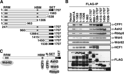
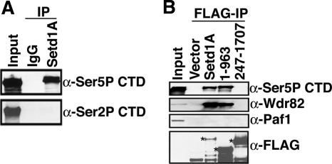
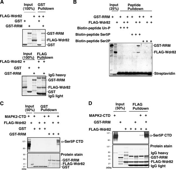
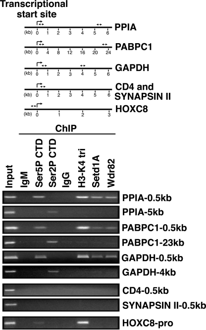
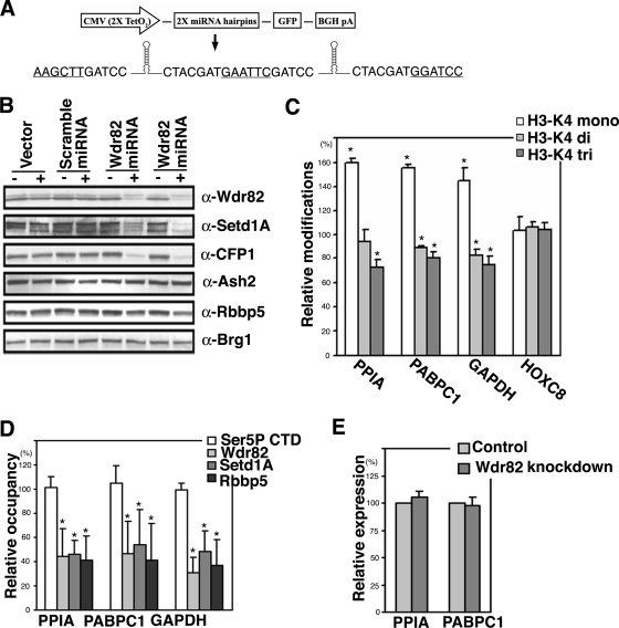
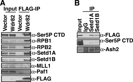
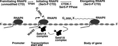
Histone H3-Lys4 trimethylation is associated with the transcription start site of transcribed genes, but the molecular mechanisms that control this distribution in mammals are unclear. The human Setd1A histone H3-Lys4 methyltransferase complex was found to physically associate with the RNA polymerase II large subunit. The Wdr82 component of the Setd1A complex interacts with the RNA recognition motif of Setd1A and additionally binds to the Ser5-phosphorylated C-terminal domain of RNA polymerase II, which is involved in initiation of transcription, but does not bind to an unphosphorylated or Ser2-phosphorylated C-terminal domain. Chromatin immunoprecipitation analysis revealed that Setd1A is localized near the transcription start site of expressed genes. Small interfering RNA-mediated depletion of Wdr82 leads to decreased Setd1A expression and occupancy at transcription start sites and reduced histone H3-Lys4 trimethylation at these sites. However, neither RNA polymerase II (RNAP II) occupancy nor target gene expression levels are altered following Wdr82 depletion. Hence, Wdr82 is required for the targeting of Setd1A-mediated histone H3-Lys4 trimethylation near transcription start sites via tethering to RNA polymerase II, an event that is a consequence of transcription initiation. These results suggest a model for how the mammalian RNAP II machinery is linked with histone H3-Lys4 histone methyltransferase complexes at transcriptionally active genes.


Figures

FIG. 1.
FIG. 1.
Protein-protein interactions within the human Setd1A histone methyltransferase complex. A. Diagram of Setd1A expression constructs. N-SET, N-terminal region of the SET domain; SET, catalytic SET histone methyltransferase domain. Numbers indicate amino acid residues of the human Setd1A protein. All constructs contain an amino-terminal FLAG epitope. B. Inducible T-REx HEK293 cell lines that express various FLAG-tagged human Setd1A fragments were induced for 3 days with 0.5 μg/ml doxycycline. Nuclear extracts were prepared and immunoprecipitated by FLAG-IgG agarose beads, and immunoprecipitates were analyzed by Western blotting using the indicated antisera. Asterisks in the FLAG Western blot indicate the expected size of each FLAG-tagged protein. C. Summary of protein-protein interactions within the human Setd1A complex.
FIG. 2.
FIG. 2.
Setd1A interacts with RNAP II through its RRM domain. A. Nuclear extracts from HEK293 cells were immunoprecipitated as described in Materials and Methods, and immunoprecipitates were analyzed by Western blotting using the indicated phosphorylation-specific CTD antibodies. B. Inducible cell lines that express various human FLAG-tagged Setd1A fragments were induced with doxycycline. Nuclear extracts were prepared, immunoprecipitated, and analyzed by Western blotting using the indicated antisera.
FIG. 3.
FIG. 3.
The Setd1A interaction with Ser5-P CTD is mediated by Wdr82 in vitro. A. The GST-Setd1A RRM domain and FLAG-Wdr82 were purified from E. coli and insect cells, respectively. Purified proteins were used for in vitro GST (top panel) and FLAG (bottom panel) pull-down assays. Proteins were separated by SDS-PAGE and detected by Coomassie staining. Arrows indicate identities of protein bands. B. Biotin-labeled CTD peptides containing various phosphorylation states were synthesized and used for in vitro pull-down assays. Peptides were incubated with purified FLAG-Wdr82 and GST-RRM and captured using streptavidin-agarose. Bound proteins were denatured by SDS sample buffer, separated by SDS-PAGE, and visualized by Coomassie staining. C. Purified GST-RRM, FLAG-Wdr82, and MAPK2-treated full-length CTD were used for in vitro GST pull-down assays. Purified GST was used as a negative control. Proteins were separated by SDS-PAGE and analyzed by Western blotting (top) and Coomassie staining (bottom). D. Purified GST-RRM, FLAG-Wdr82, and MAPK2-treated CTD were used for in vitro FLAG pull-down assays. Proteins were separated by SDS-PAGE and analyzed by Western blotting (top) and Coomassie staining (bottom).
FIG. 4.
FIG. 4.
The Setd1A complex is associated with the transcription start site of active genes. HEK293 cells were fixed and sonicated, and soluble chromatin was immunoprecipitated by the indicated antibodies as described in Materials and Methods. ChIP products were analyzed by PCR and agarose gel electrophoresis. The housekeeping genes PABPC, PPIA, and GAPDH were chosen as genes that are actively transcribed by RNAP II. In contrast, the CD4 and Synapsin II genes were chosen as inactive genes that are not expressed in HEK293 cells. The HOXC8 gene is a target of the MLL1 histone methyltransferase complex. PCR primers were designed to amplify 500-bp regions immediately downstream of each transcription start site (and within the HOXC8 gene promoter) and additionally within the 3′ coding regions of the PPIA, PABPC, and GAPDH genes. Locations of primers within each gene locus are shown in the diagram as double-sided arrows.
FIG. 5.
FIG. 5.
Depletion of Wdr82 decreases Setd1A occupancy and histone H3-Lys4 trimethylation near the transcription start site. A. Diagram of the inducible Wdr82 miRNA construct. Two 118-nt modified miR-30 hairpins for Wdr82 were tandemly subcloned into the pcDNA5/TO vector, which contains a C-terminal green fluorescent protein cDNA and is expressed under control of a tetracycline operator. Restriction enzyme sites used for subcloning are underlined. B. Inducible T-REx HEK293 cell lines that express tandem miRNA of human Wdr82, tandem scramble miRNA, or vector only were induced for 5 days with 1 μg/ml doxycycline. Nuclear extracts were prepared and analyzed by Western blotting using the indicated antisera. The results are shown for two independent clones that express the tandem miRNA of human Wdr82. C. Cell lines that express the tandem miRNA of Wdr82 were induced for 5 days with 1 μg/ml doxycycline. Histone H3-Lys4 methylation near the transcription start site of the PPIA, PABPC1, and GAPDH genes and the promoter region of the HOXC8 gene were analyzed by ChIP and real-time PCR. The level of histone H3-Lys4 methylation in uninduced cells was set at 100%. The data represent a summary of three independent experiments. Error bars indicate the mean standard deviations, and P values were determined by a standard t test. Asterisks indicate P values of <0.05. D. Cell lines that express the tandem miRNA for Wdr82 were induced for 5 days with 1 μg/ml doxycycline. Occupancy by RNAP II containing Ser5-P CTD, Wdr82, Setd1A, and Rbbp5 near the transcription start sites of the indicated genes was analyzed by ChIP and real-time PCR. Occupancy in uninduced cells was set at 100%. The data represent a summary of three independent experiments. Error bars indicate the mean standard deviations, and P values were determined by a standard t test. Asterisks indicate P values of <0.05. E. Cell lines that express tandem miRNA for Wdr82 were induced for 5 days with 1 μg/ml doxycycline. Transcripts were analyzed by reverse transcription and real-time PCR. PPIA and PABPC1 transcripts were normalized to 18S transcripts that are produced by RNA polymerase I. The level of transcripts present in uninduced cells was set at 100%. The data represent a summary of three independent experiments. Error bars indicate the mean standard deviations, and P values were determined by a standard t test.
FIG. 6.
FIG. 6.
Wdr82 associates with a subset of mammalian Set1-like proteins. A. Inducible cell lines that express FLAG-Wdr82 or empty vector were induced for 3 days with 0.5 μg/ml doxycycline. Nuclear extracts were prepared and immunoprecipitated with FLAG-IgG agarose beads, and immunoprecipitates were analyzed by Western blotting using the indicated antisera. B. Nuclear extracts from HEK293 cells carrying FLAG-Wdr82 were immunoprecipitated as described in Materials and Methods. The immunoprecipitates were analyzed by Western blotting using the indicated antisera.
FIG. 7.
FIG. 7.
Role of Wdr82 on histone H3-Lys4 methylation during the transcription cycle. After the formation of the preinitiation complex, CTD kinase (TFIIH) phosphorylates Ser5 residues of CTD repeats within the initiating RNAP II large subunit. The Ser5-P mark of CTD is directly recognized by the Wdr82 subunit of the Setd1A complex, and the Setd1A complex introduces histone H3-Lys4 trimethylation marks on neighboring nucleosomes. As the nascent RNA grows, the Setd1A complex dissociates from RNAP II by an unknown mechanism. CTDK1 phosphorylates Ser2 residues of CTD repeats during the elongation phase of transcription.

References

    1. Bernstein, B. E., M. Kamal, K. Lindblad-Toh, S. Bekiranov, D. K. Bailey, D. J. Huebert, S. McMahan, E. K. Karlsson, E. J. Kulbokas, T. R. Gingeras, S. L. Schreiber, and E. S. Lander. 2005. Genomic maps and comparative analysis of histone modifications in human and mouse. Cell 120169-181. - PubMed
    1. Bernstein, B. E., T. S. Mikkelsen, X. Xie, M. Kamal, D. J. Huebert, J. Cuff, B. Fry, A. Meissner, M. Wernig, K. Plath, R. Jaenisch, A. Wagschal, R. Reil, S. L. Schreiber, and E. S. Lander. 2006. A bivalent chromatin structure marks key developmental genes in embryonic stem cells. Cell 125315-326. - PubMed
    1. Briggs, S. D., M. Bryk, B. D. Strahl, W. L. Cheung, J. K. Davie, S. Y. R. Dent., F. Winston, and C. D. Allis. 2001. Histone H3 lysine 4 methylation is mediated by Set1 and required for cell growth and rDNA silencing in Saccharomyces cerevisiae. Genes Dev. 153286-3295. - PMC - PubMed
    1. Cheng, H., X. He, and C. Moore. 2004. The essential WD repeat protein Swd2 has dual functions in RNA polymerase II transcription termination and lysine 4 methylation of histone H3. Mol. Cell. Biol. 242932-2943. - PMC - PubMed
    1. Couture, J.-F., E. Collazo, and R. C. Trievel. 2006. Molecular recognition of histone H3 by the WD40 protein WDR5. Nat. Struct. Mol. Biol. 13698-703. - PubMed

Publication types

MeSH terms