. 2007 Nov 26;204(12):2963-76.
doi: 10.1084/jem.20071132. Epub 2007 Nov 6.

A protein associated with Toll-like receptor (TLR) 4 (PRAT4A) is required for TLR-dependent immune responses

Affiliations

A protein associated with Toll-like receptor (TLR) 4 (PRAT4A) is required for TLR-dependent immune responses

Koichiro Takahashi et al. J Exp Med. .

Abstract

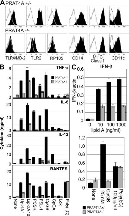
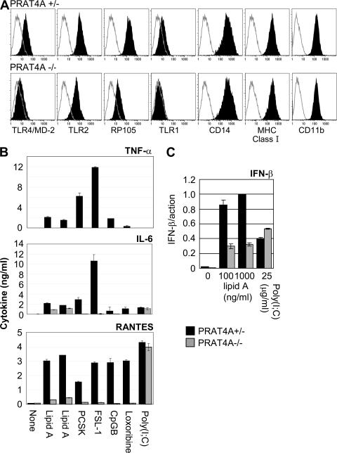
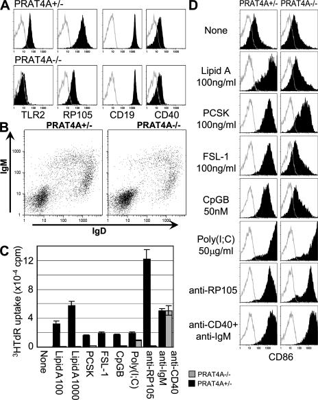
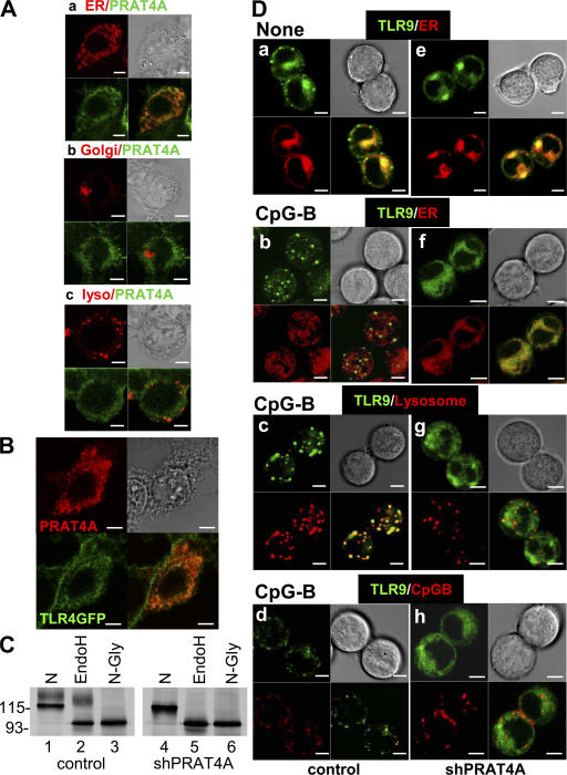
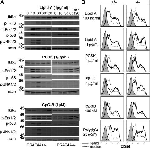
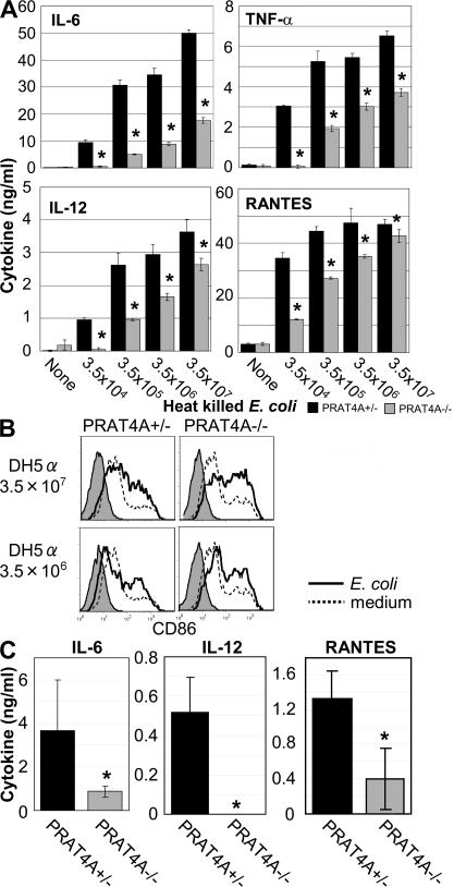
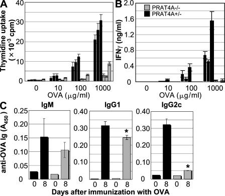
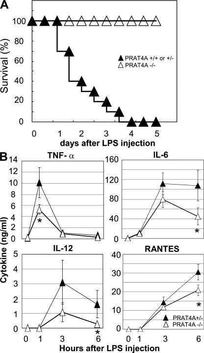
Immune cells express multiple Toll-like receptors (TLRs) that are concomitantly activated by a variety of pathogen products. Although there is presumably a need to coordinate the expression and function of TLRs in individual cells, little is known about the mechanisms governing this process. We show that a protein associated with TLR4 (PRAT4A) is required for multiple TLR responses. PRAT4A resides in the endoplasmic reticulum, and PRAT4A knockdown inhibited trafficking of TLR1 and TLR4 to the cell surface and ligand-induced trafficking of TLR9 to lysosomes. Other cell-surface molecules were expressed normally on immunocytes from PRAT4A-/- mice. There was impaired cytokine production to TLR ligands, except to the TLR3 ligand poly(I:C), and to whole bacteria. Activation of antigen-specific T helper type 1 responses were also defective. Moreover, PRAT4A-/- bone marrow chimeric mice were resistant to lipopolysaccharide-induced sepsis. These results suggest that PRAT4A regulates the subcellular distribution and response of multiple TLRs and is required for both innate and adaptive immune responses.


Figures

Figure 1.
Figure 1.
BM-DCs lacking PRAT4A are impaired in TLR-dependent responses. (A) Shaded histograms show the expression of TLRs and cell-surface markers on BM-DCs from PRAT4A+/− or PRAT4A−/− mice. Open histograms show control staining with the second reagent alone. (B) Bar graphs show cytokine production by BM-DCs in response to a variety of TLR ligands (1 and 10 ng/ml lipid A, 100 ng/ml PCSK, 100 ng/ml FSL-1, 100 nM CpG-B, 1.5 μM CpG-A, 150 μM loxoribine, and 100 μg/ml poly(I:C)). (C) Bar graphs show real-time RT-PCR analyses for mRNA encoding IFN-β, the amount of which was normalized by that of mRNA for β-actin. BM-DCs from BM chimeric mice were stimulated with the indicated TLR ligands, and RNA was collected 2 (lipid A) or 5 (CpG-B and poly(I:C)) h after stimulation, respectively. Data in B and C represent the SD.
Figure 2.
Figure 2.
PRAT4A is required for TLR responses in macrophages. (A) Shaded histograms show cell-surface expression of the indicated markers on BM-macrophages from PRAT4A+/− or PRAT4A−/− mice. Open histograms show control staining with the second reagent alone. (B) Bar graphs show cytokine production by BM-macrophages in response to a variety of TLR ligands (1 and 10 ng/ml lipid A, 100 ng/ml PCSK, 100 ng/ml FSL-1, 100 nM CpG-B, 150 μM loxoribine, and 100 μg/ml poly(I:C)). (C) Bar graphs show real-time RT-PCR analyses for mRNA encoding IFN-β, the amount of which was normalized by that of mRNA for β-actin. BM-macrophages were stimulated with the indicated TLR ligands and RNA was collected 2 (lipid A) or 5 (poly(I:C)) h after stimulation, respectively. Data in B and C represent the SD.
Figure 3.
Figure 3.
PRAT4A is required for TLR responses in B lymphocytes. (A) Shaded histograms show cell-surface expression of the indicated markers on B220-positive splenic B cells. Open histograms show control staining with the second reagent alone. (B) Dot plots show cell-surface expression of IgM and IgD on splenocytes. (C) Proliferation of enriched B cells stimulated with a variety of stimulation (100 and 1,000 ng/ml lipid A, 1,000 ng/ml PCSK, 1,000 ng/ml FSL-1, 500 nM CpG-B, 100 μg/ml poly(I:C), 1 μg/ml anti-RP105 antibody, 1 μg/ml anti-IgM antibody plus 2.25 μg/ml anti-CD40 antibody). Splenic B cells were enriched from BM chimeric mice and stimulated for 3 d. 1 μCi [3H]thymidine was pulsed for the last 6 h, and the incorporated thymidine was counted by a scintillation counter. (D) Shaded histograms show CD86 up-regulation on B cells stimulated with the indicated reagents. The concentration used was as described in C. Open histograms show control staining with the second reagent alone.
Figure 4.
Figure 4.
PRAT4A resides in the ER and is required for TLR4 protein maturation and TLR9 relocation. (A) PRAT4A-GFP was expressed in HEK293 cells together with a marker locating ER (a), the Golgi apparatus (b), or lysosome (c). PRAT4A-GFP (green) and the markers (red) were visualized by confocal microscopy. (B) TLR4-GFP and PRAT4A-FlagHis were expressed in HEK293 cells, and PRAT4A was stained with anti-Flag mAb. TLR4-GFP (green) and PRAT4A-FlagHis (red) were visualized by confocal microscopy. (C) Ba/F3 cells expressing TLR4–Flag/MD-2 were transduced with a control vector (lanes 1–3) or with a vector encoding shPRAT4A (lanes 4–6). Cells were subjected to immunoprecipitation with anti-Flag antibody, followed by treatment with buffer (N), endoH, or N-glycanase (N-Gly), as indicated in the figure. Samples were further subjected to SDS-PAGE and immunoprobing with anti-TLR4 mAb. Molecular masses are shown in kilodaltons. (D) B cell lymphoma M12 cells expressing TLR9-GFP plus a control vector (left) or TLR9-GFP plus shPRAT4A (right) were stimulated with medium (a and e) or 1 μM CpG-B for 2 h (b–d and f–h). Cells were counterstained with markers for ER (a, b, e, and f) or lysosome (c and g) and visualized by confocal microscopy. (bottom) CpG-B conjugated with 1 μM rhodamine was used for stimulation, and its distribution was determined in d and h. Bars, 5 μm.
Figure 5.
Figure 5.
PRAT4A-independent responses in BM-DCs. (A) BM-DCs from BM chimeric mice were stimulated with lipid A, PCSK, or CpG-B, as indicated in the figure. After the indicated periods of time, cells were lysed and subjected to SDS-PAGE, Western blotting, and immunoprobing with the indicated antibodies. (B) CD86 on BM-DCs from BM chimeric mice was stained 48 h after stimulation with the indicated TLR ligands. Shaded histograms show staining with the second reagent alone. Histograms with continuous or dashed lines show CD86 staining of BM-DCs cultured with TLR ligands or medium, respectively.
Figure 6.
Figure 6.
PRAT4A is required for cytokine production in response to heat-killed E. coli. (A) BM-DCs from BM chimeric mice were stimulated with heat-killed E. coli, as indicated in the figure. After 24 h of culture, concentrations of cytokines in the supernatant were determined by ELISA. Data represent the SD. *, P < 0.05. (B) CD86 on BM-DCs from BM chimeric mice was stained 48 h after stimulation with heat-killed E. coli. Shaded histograms show staining with the second reagent alone. Histograms with solid or dashed lines show CD86 staining of BM-DCs cultured with heat-killed E. coli or medium, respectively. (C) BM chimeric mice (n = 5) were injected with heat-killed E. coli (3.5 × 10⁸ cells per mouse) and bled 3 h after injection. Serum cytokines were measured by ELISA. Data represent the SD. *, P < 0.05.
Figure 7.
Figure 7.
Th1 responses are impaired in PRAT4A−/− mice. Cells from draining popliteal lymph nodes were collected from mice 8 d after immunization (hind legs) with OVA in CFA. (A) The proliferation of antigen-specific lymph node cells from PRAT4A+/− or PRAPT4A−/− BM chimeric mice was measured by culturing these cells with OVA for 72 h. (B) IFN-γ production was determined by ELISA from supernatants of antigen-stimulated lymph node cells from PRAT4A+/− or PRAT4A−/− BM chimeric mice. Data are means + SD from each mouse, and the results of three independent mice per group are shown. Similar results were obtained in two independent experiments. (C) Serum was collected before (day 0) and after (day 8) immunization with OVA plus CFA, and the titers of OVA-specific IgM, IgG2a/c, and IgG2b were determined by ELISA. Data are means + SD from three mice per group. Similar results were obtained in two independent experiments. *, P < 0.01.
Figure 8.
Figure 8.
PRAT4A−/− BM chimeric mice are resistant to LPS-induced sepsis. (A) PRAT4A−/− (n = 10), PRAT4A+/− (n = 5), and PRAT4A+/+ (n = 5) BM chimeric mice were administered with 400–500 μg LPS intraperitoneally, and the mortality was shown as the percentage of survival. (B) Blood samples from PRAT4A+/− (n = 6) and PRAT4A−/− (n = 6) mice were collected from the vena cava at 0 (no treatment), 1, 3, and 6 h after injection of a lethal dose of LPS (500 μg per mouse). The concentrations of TNF-α, IL-6, IL-12, and RANTES were determined by ELISA. Data represent the SD. *, P < 0.01.

References

    1. Beutler, B., Z. Jiang, P. Georgel, K. Crozat, B. Croker, S. Rutschmann, X. Du, and K. Hoebe. 2006. Genetic analysis of host resistance: Toll-like receptor signaling and immunity at large. Annu. Rev. Immunol. 24:353–389. - PubMed
    1. Kaisho, T., and S. Akira. 2006. Toll-like receptor function and signaling. J. Allergy Clin. Immunol. 117:979–987. - PubMed
    1. Gautier, G., M. Humbert, F. Deauvieau, M. Scuiller, J. Hiscott, E.E. Bates, G. Trinchieri, C. Caux, and P. Garrone. 2005. A type I interferon autocrine–paracrine loop is involved in Toll-like receptor–induced interleukin-12p70 secretion by dendritic cells. J. Exp. Med. 201:1435–1446. - PMC - PubMed
    1. Napolitani, G., A. Rinaldi, F. Bertoni, F. Sallusto, and A. Lanzavecchia. 2005. Selected Toll-like receptor agonist combinations synergistically trigger a T helper type 1-polarizing program in dendritic cells. Nat. Immunol. 6:769–776. - PMC - PubMed
    1. Bafica, A., C.A. Scanga, C.G. Feng, C. Leifer, A. Cheever, and A. Sher. 2005. TLR9 regulates Th1 responses and cooperates with TLR2 in mediating optimal resistance to Mycobacterium tuberculosis. J. Exp. Med. 202:1715–1724. - PMC - PubMed

Publication types