

Differential expression patterns of NDRG family proteins in the central nervous system
- PMID: 17998568
- PMCID: PMC2324175
- DOI: 10.1369/jhc.7A7323.2007
Differential expression patterns of NDRG family proteins in the central nervous system
Abstract
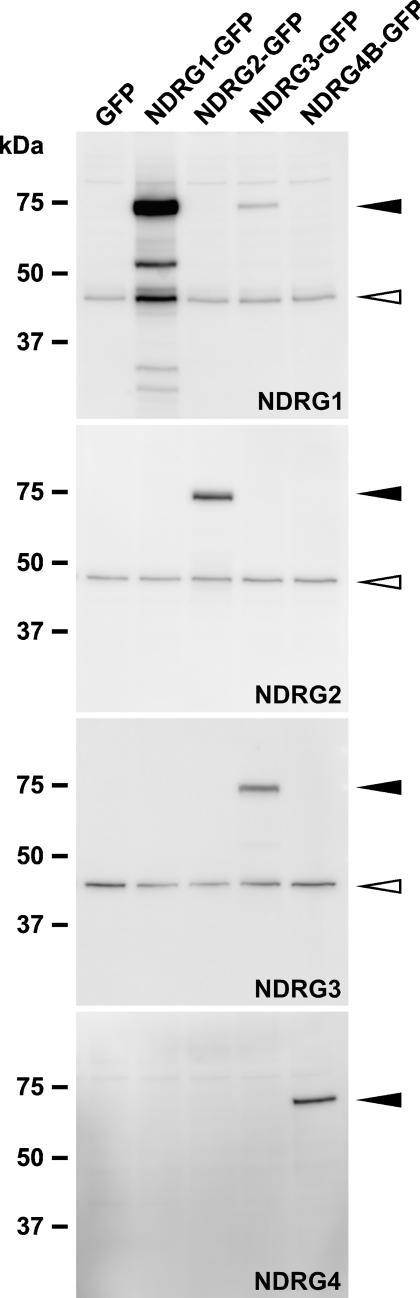
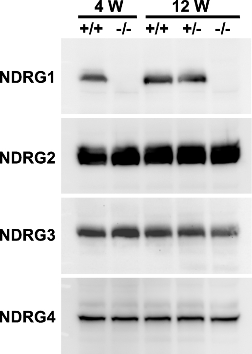
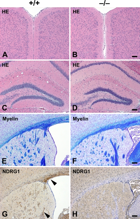
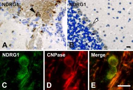
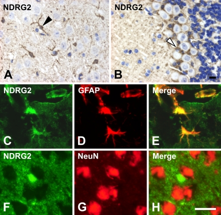
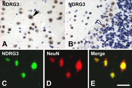
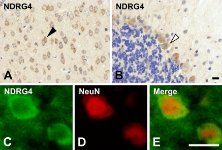
The N-myc downstream-regulated gene (NDRG) family consists of four proteins: NDRG1, NDRG2, NDRG3, and NDRG4 in mammals. NDRG1 has been thoroughly studied as an intracellular protein associated with stress response, cell growth, and differentiation. A nonsense mutation in the NDRG1 gene causes hereditary motor and sensory neuropathy, Charcot-Marie-Tooth disease type 4D. We previously generated Ndrg1-deficient mice and found that they exhibited peripheral nerve degeneration caused by severe demyelination, but that the complicated motor abilities were retained. These results implied that other NDRG family proteins may compensate for the NDRG1 deficiency in the central nervous system. In this study we raised specific antibodies against each member of the NDRG protein family and examined their cellular expression patterns in the mouse brain. In the cerebrum, NDRG1 and NDRG2 were localized in oligodendrocytes and astrocytes, respectively, whereas NDRG3 and NDRG4 were ubiquitous. In the cerebellum, NDRG1 and NDRG4 were localized in Purkinje cells and NDRG2 in Bergmann glial cells. NDRG3 was detected in the nuclei in most cells. These expression patterns demonstrated the cell type-specific and ubiquitous localization of the NDRG family proteins. Each NDRG may play a partially redundant role in specific cells in the brain.
Figures
Similar articles
-
Nervous NDRGs: the N-myc downstream-regulated gene family in the central and peripheral nervous system.Neurogenetics. 2019 Oct;20(4):173-186. doi: 10.1007/s10048-019-00587-0. Epub 2019 Sep 4. Neurogenetics. 2019. PMID: 31485792 Free PMC article. Review.
-
Characterization and expression of three novel differentiation-related genes belong to the human NDRG gene family.Mol Cell Biochem. 2002 Jan;229(1-2):35-44. doi: 10.1023/a:1017934810825. Mol Cell Biochem. 2002. PMID: 11936845
-
Characterization of the human NDRG gene family: a newly identified member, NDRG4, is specifically expressed in brain and heart.Genomics. 2001 Apr 1;73(1):86-97. doi: 10.1006/geno.2000.6496. Genomics. 2001. PMID: 11352569
-
NDRG4 protein-deficient mice exhibit spatial learning deficits and vulnerabilities to cerebral ischemia.J Biol Chem. 2011 Jul 22;286(29):26158-65. doi: 10.1074/jbc.M111.256446. Epub 2011 Jun 2. J Biol Chem. 2011. PMID: 21636852 Free PMC article.
-
A combined literature and in silico analysis enlightens the role of the NDRG family in the gut.Biochim Biophys Acta Gen Subj. 2018 Oct;1862(10):2140-2151. doi: 10.1016/j.bbagen.2018.07.004. Epub 2018 Jul 7. Biochim Biophys Acta Gen Subj. 2018. PMID: 30033230 Review.
Cited by
-
NDRG4 is required for cell cycle progression and survival in glioblastoma cells.J Biol Chem. 2009 Sep 11;284(37):25160-9. doi: 10.1074/jbc.M109.012484. Epub 2009 Jul 10. J Biol Chem. 2009. PMID: 19592488 Free PMC article.
-
Roles of N-myc downstream-regulated gene 2 in the central nervous system: molecular basis and relevance to pathophysiology.Anat Sci Int. 2021 Jan;96(1):1-12. doi: 10.1007/s12565-020-00587-3. Epub 2020 Nov 11. Anat Sci Int. 2021. PMID: 33174183 Review.
-
NDRG4, a novel candidate tumor suppressor, is a predictor of overall survival of colorectal cancer patients.Oncotarget. 2015 Apr 10;6(10):7584-96. doi: 10.18632/oncotarget.3170. Oncotarget. 2015. PMID: 25749388 Free PMC article.
-
A comparative transcriptomic analysis of astrocytes differentiation from human neural progenitor cells.Eur J Neurosci. 2016 Nov;44(10):2858-2870. doi: 10.1111/ejn.13382. Epub 2016 Sep 25. Eur J Neurosci. 2016. PMID: 27564458 Free PMC article.
-
Nervous NDRGs: the N-myc downstream-regulated gene family in the central and peripheral nervous system.Neurogenetics. 2019 Oct;20(4):173-186. doi: 10.1007/s10048-019-00587-0. Epub 2019 Sep 4. Neurogenetics. 2019. PMID: 31485792 Free PMC article. Review.
References
-
- Agarwala KL, Kokame K, Kato H, Miyata T (2000) Phosphorylation of RTP, an ER stress-responsive cytoplasmic protein. Biochem Biophys Res Commun 272:641–647 - PubMed
-
- Bandyopadhyay S, Pai SK, Gross SC, Hirota S, Hosobe S, Miura K, Saito K, et al. (2003) The Drg-1 gene suppresses tumor metastasis in prostate cancer. Cancer Res 63:1731–1736 - PubMed
-
- Berger P, Sirkowski EE, Scherer SS, Suter U (2004) Expression analysis of the N-Myc downstream-regulated gene 1 indicates that myelinating Schwann cells are the primary disease target in hereditary motor and sensory neuropathy-Lom. Neurobiol Dis 17:290–299 - PubMed
-
- Guan RJ, Ford HL, Fu Y, Li Y, Shaw LM, Pardee AB (2000) Drg-1 as a differentiation-related, putative metastatic suppressor gene in human colon cancer. Cancer Res 60:749–755 - PubMed
Publication types
MeSH terms
Substances
LinkOut - more resources
Full Text Sources
Molecular Biology Databases
