. 2008 Mar;56(3):223-31.
doi: 10.1369/jhc.7A7290.2007. Epub 2007 Nov 12.

Aberrant expression of histo-blood group A type 3 antigens in vascular endothelial cells in inflammatory sites

Affiliations

Aberrant expression of histo-blood group A type 3 antigens in vascular endothelial cells in inflammatory sites

Mizuho Nosaka et al. J Histochem Cytochem. 2008 Mar.

Abstract

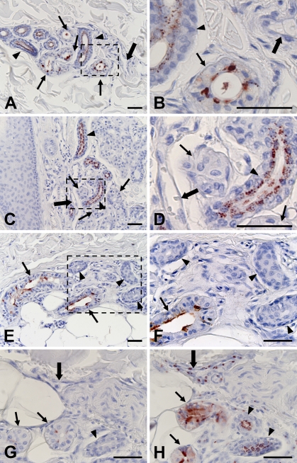
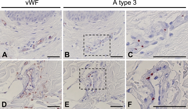
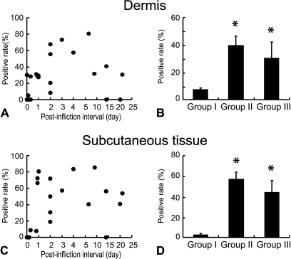
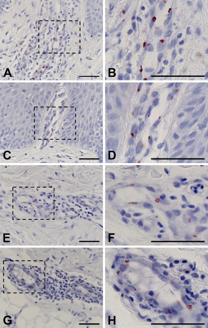
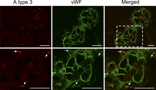
Histo-blood group ABH antigens are widely distributed in human tissues. The epitopes of ABH antigens are carried by at least four different peripheral core isotypes of internal carbohydrate backbones (type 1-4). Each type of ABH antigen is expressed tissue specifically, and aberrant expression of ABH antigens is often observed during oncogenesis. We immunohistochemically examined the expression of A type 3 antigens in wounded and diseased skin tissues (A and AB blood groups). In uninjured skin, the expression of A type 3 antigens was restricted to the eccrine sweat gland. In addition to the sweat glands, A type 3 antigens were found in vascular endothelial cells of the wound sites. The extent of A type 3 antigens expression related to postinfliction intervals. A significantly higher expression rate of A type 3 antigens in endothelial cells was also observed in diseased skin, suggesting that inflammation might induce A type 3 antigen expression in endothelial cells. Double-color immunofluorescence staining of the specimens showed that von Willebrand factor (vWF) was a core-protein of A type 3 determinants aberrantly expressed in endothelial cells in inflamed tissues, suggesting that aberrant expression of A type 3 antigens is involved in stabilization of vWF in inflammation.


Figures

Figure 1
Figure 1
Localization of histo-blood group A type 3 antigens reactive to AR-1, and H type 3/4 antigens reactive to MBr1 in normal skin. A type 3 antigens are localized in dark cells (arrows) and duct epithelial cells (arrowheads) in eccrine sweat glands in specimen from A blood group secretor (A,B). Only duct epithelial cells of eccrine sweat glands were reactive to AR-1 in specimens from non-secretors (C,D). Histo-blood group H type 3/4 antigens were detected in dark cells but not duct cells of eccrine sweat glands in specimens from O blood group secretors (E,F). Most vascular endothelial cells did not express type 3 antigens (large arrows). Areas indicated by boxes in A, C, and E are depicted at higher magnification in B, D, and F, respectively. Two sections from a wound specimen (Group II) were stained with AR-1 absorbed with blood group A (G) and O red cells (H), respectively. Absorption with blood group A red cells abolished reactivity of AR-1 (G). Bar = 50 μm.
Figure 2
Figure 2
Immunohistochemical staining of the skin of a 1-day-old wound (Group II). Vascular endothelial cells in the dermis (A) and subcutaneous region (D) of 1-day-old wound specimens were identified by immunostaining with anti-human von Willebrand factor (vWF) antibody. A type 3 antigens were strongly detected in vascular endothelial cells in the adjacent sections (B,E). Areas indicated by boxes in B and E are depicted at higher magnification in C and F, respectively. Bar = 50 μm.
Figure 3
Figure 3
Ratio of A type 3–positive vascular endothelial cells in the dermis (A) and subcutaneous tissue (C) in relation to postinfliction intervals. Vascular endothelial cells were identified by immunostaining of adjacent sections with anti-human vWF antibody as shown in Figure 2. Mean value and SE of A type 3–positive vascular endothelial cells in the dermis (B) and subcutaneous tissues (D) in each wound group. *Significant difference from Group I was observed statistically (p<0.05).
Figure 4
Figure 4
Immunohistochemical staining of diseased skin tissues. A type 3 antigens were detected in the vascular endothelial cells in the dermis of atopic dermatitis (A), eczema (C), psoriasis (E), and erythema nodosum (G) specimens.Areas indicated by boxes in A, C, E, and G are depicted at higher magnification in B, D, F, and H, respectively. Bar = 50 μm.
Figure 5
Figure 5
Double-color immunofluorescence analysis of wounded skin tissues using a combination of AR-1 and antibody to vWF. A type 3 antigens (red) located in cells stained with anti-vWF (green) in 4-day-old wounded skin. Area indicated by box in merged panel is depicted at higher magnification in bottom panels. All AR-1–reactive spots were found in the regions reactive to anti-human vWF antibody. As shown in bottom panels, the discrete spots (white arrows) reactive to AR1 were completely merged to anti-human vWF-reactive spots. Bar = 20 μm.

Similar articles

Cited by

References

    1. Adobati E, Panza L, Russo G, Colnaghi MI, Canevari S (1997) In vitro mimicry of CaMBr1 tumor-associated antigen by synthetic oligosaccharides. Glycobiology 7:173–178 - PubMed
    1. Bernardo A, Ball C, Nolasco L, Moake JF, Dong JF (2004) Effects of inflammatory cytokines on the release and cleavage of the endothelial cell-derived ultralarge von Willebrand factor multimers under flow. Blood 104:100–106 - PubMed
    1. Bonifati C, Carducci M, Mussi A, D'auria L, Ameglio F (1997) IL-1 α, IL-1β and psoriasis: conflicting results in the literature. Opposite behaviour of the two cytokines in lesional or non-lesional extracts of whole skin. J Biol Regul Homeost Agents 11:133–136 - PubMed
    1. Bowen DJ (2003) An influence of ABO blood group on the rate of proteolysis of von Willebrand factor by ADAMTS13. J Thromb Haemost 1:33–40 - PubMed
    1. Bremer EG, Levery SB, Sonnino S, Ghidoni R, Canevari S, Kannagi R, Hakomori S (1984) Characterization of a glycosphingolipid antigen defined by the monoclonal antibody MBr1 expressed in normal and neoplastic epithelial cells of human mammary gland. J Biol Chem 259:14773–14777 - PubMed

Substances