

Oestrogen supplementation following castration promotes stromal remodelling and histopathological alterations in the Mongolian gerbil ventral prostate
- PMID: 17999680
- PMCID: PMC2525754
- DOI: 10.1111/j.1365-2613.2007.00559.x
Oestrogen supplementation following castration promotes stromal remodelling and histopathological alterations in the Mongolian gerbil ventral prostate
Abstract
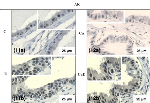
The effect of oestradiol on the intact and castrated adult gerbil prostate was evaluated by focussing on stromal and epithelial disorders, and hormonal receptor immunoreactivity. The experimental animals were studied by histological, histochemical and immunohistochemical techniques, morphometric-stereological analysis and transmission electron microscopy. Epithelial alterations in the oestradiol-treated animals were frequent, with an increase in epithelial cell height, areas of intense dysplasia and hyperplasia and formation of prostatic intraepithelial neoplasia (PIN). Another aspect that did not depend on the presence of testosterone was the arrangement of the fibrillar and non-fibrillar elements of the extracellular matrix among smooth muscle cells (SMC), suggesting a possible role of these cells in rearrangement and synthesis of these components, after oestrogenic treatment. In the castrated animals, an accumulation of extracellular matrix elements under the epithelium was evident, while in the intact animals the same compounds were dispersed and scarce. In the groups of intact and castrated animals, SMC and fibroblasts exhibited a secretory phenotype, which was accentuated after oestradiol administration. There was an increase of the immunoreactivity to alpha-oestrogen and androgen receptors in hyperplastic areas compared to normal epithelium, revealing the involvement of these steroid receptors in the hyperplasia and PIN development.
Figures


Similar articles
-
Tissue evidence of the testosterone role on the abnormal growth and aging effects reversion in the gerbil (Meriones unguiculatus) prostate.Anat Rec A Discov Mol Cell Evol Biol. 2006 Nov;288(11):1190-200. doi: 10.1002/ar.a.20391. Anat Rec A Discov Mol Cell Evol Biol. 2006. PMID: 17031809
-
Progesterone as a morphological regulatory factor of the male and female gerbil prostate.Int J Exp Pathol. 2013 Dec;94(6):373-86. doi: 10.1111/iep.12050. Int J Exp Pathol. 2013. PMID: 24205795 Free PMC article.
-
Estrogen-induced morphological and immunohistochemical changes in stroma and epithelium of rat ventral prostate.Prostate. 1992;21(3):183-99. doi: 10.1002/pros.2990210303. Prostate. 1992. PMID: 1437855
-
Prolactin promotes a partial recovery from the atrophy of both male and female gerbil prostates caused by castration.Reprod Biol Endocrinol. 2021 Jun 22;19(1):94. doi: 10.1186/s12958-021-00777-2. Reprod Biol Endocrinol. 2021. PMID: 34158080 Free PMC article.
-
Histochemical studies of epithelial cell glycoconjugates in atrophic, metaplastic, hyperplastic, and neoplastic canine prostate.Lab Invest. 1984 Mar;50(3):294-302. Lab Invest. 1984. PMID: 6199584 Review.
Cited by
-
Androgen signaling disruption during fetal and postnatal development affects androgen receptor and connexin 43 expression and distribution in adult boar prostate.Biomed Res Int. 2013;2013:407678. doi: 10.1155/2013/407678. Epub 2013 Sep 17. Biomed Res Int. 2013. PMID: 24151599 Free PMC article.
-
Neonatal exposure to ethinylestradiol increases ventral prostate growth and promotes epithelial hyperplasia and inflammation in adult male gerbils.Int J Exp Pathol. 2016 Oct;97(5):380-388. doi: 10.1111/iep.12208. Epub 2016 Dec 5. Int J Exp Pathol. 2016. PMID: 27917613 Free PMC article.
-
Harmful effects of carbamazepine on the postnatal development of the rat ventral prostate.Reprod Biol Endocrinol. 2012 Mar 25;10:22. doi: 10.1186/1477-7827-10-22. Reprod Biol Endocrinol. 2012. PMID: 22443633 Free PMC article.
-
Corticosterone influences gerbil (Meriones unguiculatus) prostatic morphophysiology and alters its proliferation and apoptosis rates.Int J Exp Pathol. 2017 Jun;98(3):134-146. doi: 10.1111/iep.12232. Epub 2017 Jun 29. Int J Exp Pathol. 2017. PMID: 28664583 Free PMC article.
-
The Mongolian Gerbil as a Useful Experimental Model in Reproductive Biology.Reprod Sci. 2023 Jul;30(7):2092-2106. doi: 10.1007/s43032-023-01171-6. Epub 2023 Jan 25. Reprod Sci. 2023. PMID: 36696041 Review.
References
-
- Antonioli E, Della-Colleta HHM, Carvalho HF. Smooth muscle cells behavior in the ventral prostate of catrated rats. J. Androl. 2004;25:50–56. - PubMed
-
- Arai Y, Suzuki Y, Nishizuka Y. Hyperplastic and metaplastic lesions in the reproductive tract of male rats induced by neonatal treatment with diethylstilbestrol. Virchows Arch. 1977;376:21–28. - PubMed
-
- Bosland MC. The role of steroid hormones in prostate carcinogenesis. J. Natl. Cancer Inst. Monogr. 2000;27:39–66. - PubMed
-
- Campos SGP, Zanetoni C, Scarano WR, Vilamaior PSL, Taboga SR. Age-related histopathological lesions in the Mongolian gerbil ventral prostate as a good model for studies of spontaneous hormone-related disorders. Int. J. Exp. Path. 2007 EPub ahead of print; doi 10.1111/j.1365-2613.2007.00550.x. - DOI - PMC - PubMed
-
- Cardoso LEM, Falcao PG, Sampaio FJB. Increased and localized accumulation of chondroitin sulfate proteoglycans in the hyperplastic human prostate. BJU Int. 2004;93:532–538. - PubMed
Publication types
MeSH terms
Substances
LinkOut - more resources
Full Text Sources
