. 2007 Nov 13:2:25.
doi: 10.1186/1749-8104-2-25.

Tissue interactions in the developing chick diencephalon

Affiliations

Tissue interactions in the developing chick diencephalon

Maria Flavia Guinazu et al. Neural Dev. .

Abstract

Background: The developing vertebrate brain is patterned first by global signalling gradients that define crude anteroposterior and dorsoventral coordinates, and subsequently by local signalling centres (organisers) that refine cell fate assignment within pre-patterned regions. The interface between the prethalamus and the thalamus, the zona limitans intrathalamica (ZLI), is one such local signalling centre that is essential for the establishment of these major diencephalic subdivisions by secreting the signalling factor Sonic hedgehog. Various models for ZLI formation have been proposed, but a thorough understanding of how this important local organiser is established is lacking.

Results: Here, we describe tissue explant experiments in chick embryos aimed at characterising the roles of different forebrain areas in ZLI formation. We found that: the ZLI becomes specified unexpectedly early; flanking regions are required for its characteristic morphogenesis; ZLI induction can occur independently from ventral tissues; interaction between any prechordal and epichordal neuroepithelial tissue anterior to the midbrain-hindbrain boundary is able to generate a ZLI; and signals from the dorsal diencephalon antagonise ZLI formation. We further show that a localised source of retinoic acid in the dorsal diencephalon is a likely candidate to mediate this inhibitory signal.

Conclusion: Our results are consistent with a model where planar, rather than vertical, signals position the ZLI at early stages of neural development and they implicate retinoic acid as a novel molecular cue that determines its dorsoventral extent.


Figures

Figure 1
Figure 1
Gene expression in the diencephalon during ZLI formation. (A-F) In situ hybridisation was used to analyse the expression of Shh (blue in (A, D); red in (C, E)), Lfng (blue in (B, E; red in (F)) and Wnt8b to stage HH14 (A, B), HH15 (C), HH18.5 (D) and HH22 (E, F) chick embryos. Whole heads are shown in (A-D) and hemisected brains in (E, F) (anterior to the left, dorsal to the top). Note expression of Shh and Wnt8b in the ZLI (yellow arrowheads in (B, D-F)).
Figure 2
Figure 2
The ZLI is specified before stage HH10. (A) Explanted brain regions are schematically shown in a stage HH14 chick brain stained by in situ hybridisation for the expression of Lfng (anterior to the left, dorsal to the top). (B) Pro-Pth, pro-ZLI and pro-Th explants from stage HH14 brains were analysed by in situ hybridisation for the expression of Shh (left) and Lfng (right) at the time of dissection (pre-cult.) or after culture for 24 hours (post-cult.). Note induction of Shh in cultured pro-ZLI explants and absence of Lfng expression in pro-ZLI explants at the time of dissection and after culture. (C) Pro-Pth, pro-ZLI and pro-Th explants from stage HH10 brains. Note induction of Shh and absence of Lfng expression in cultured pro-ZLI explants. Lfng is expressed in pro-ZLI explants at the time of dissection.
Figure 3
Figure 3
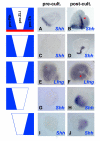
ZLI formation occurs independently from ventral tissues after stage HH10 and ZLI morphogenesis depends on the integrity of its flanking regions. Diencephalic explants were taken from (A-F) stage HH10 and (G-J) stage HH14 embryos and were analysed by in situ hybridisation for the expression of Shh (a-d, g-j) and Lfng (E, F) at the time of dissection (pre-cult.) or after culture for 48 (B, D, J) or 24 hours (F) (post-cult.). (A, B) Explants comprising pro-Pth, pro-ZLI, pro-Th and basal tissue (red). Note basal expression of Shh at the time of dissection and formation of a ZLI after culture (red arrowhead). (C, D) Pro-Pth + pro-ZLI + pro-Th explants excluding basal tissue. Note absence of Shh expression at the time of dissection and induction of a ZLI after culture. (E, F) Explants as in (C, D). Note formation of a Lfng-free stripe of cells after culture (red arrowhead). (G, H) Pro-Pth + pro-ZLI explants; (I, J) pro-ZLI + pro-Th explants. Note induction of Shh in a patch, rather than a line, after culture (compare to (B, D)).
Figure 4
Figure 4
Co-culture of prechordal and epichordal neural explants results in ZLI induction. (A) Explanted brain regions are schematically shown in a stage HH14 chick brain stained by in situ hybridisation for the expression of Lfng (anterior to the left, dorsal to the top): 1, telencephalon; 2, pro-Pth; 3, pro-ZLI; 4, pro-Th; 5, midbrain; 6, hindbrain. (B) Explants from brain regions as indicated in (A) were cultured in isolation (left two columns) or in combination (right column) for 24 hours and analysed for the expression of Shh by in situ hybridisation. Note induction of Shh at the interface of co-cultured pro-Pth + pro-Th, pro-Pth + midbrain and telencephalon + pro-Th tissue, but not after co-culture of pro-Th + pro-Th or telencephalon + hindbrain. The yellow broken line in panel '2 + 4' indicates the interface between pro-Pth and pro-Th tissue. (C) Explants from brain regions as indicated in (A) were cultured in isolation (left two columns) or in combination (right column) for 24 hours and analysed for the expression of Lfng, Dmbx1, Foxg1 (all in blue) and Shh (red) by in situ hybridisation. Note gap of Lfng expression in '2 + 4' explants (red arrow) and induction of Shh in '1 + 5' explants (red arrowheads).
Figure 5
Figure 5
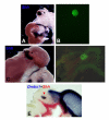
Prechordal-epichordal interactions anterior to the MHB result in ZLI induction in vivo. (A) Ectopic induction of Shh following transplantation of quail pro-Pth tissue into chick midbrain at stage HH10. (B) QCPN antibody staining allowed for localisation of the graft (asterisk in (A)). (C) Ectopic induction of Shh following transplantation of quail telencephalic tissue into chick midbrain at stage HH10. (D) QCPN antibody staining allowed for localisation of the graft (asterisk in (C)). (E) A graft of quail midbrain tissue into a chick pro-Th at stage HH10 maintains midbrain identity (red arrowhead; Dmbx1 expression in blue), but does not result in ectopic induction of Shh (red).
Figure 6
Figure 6
The ZLI is necessary and sufficient for the expression of thalamic differentiation genes in explant culture. (A) Pro-Pth and (B) pro-ZLI + pro-Pth explants were isolated from stage HH10 embryos, cultured for 72 hours and analysed for the expression of Shh (red) and Dlx2 (blue). (C) Pro-Th and (D) pro-ZLI + pro-Th explants were isolated from stage HH10 embryos, cultured for 72 hours and analysed for the expression of Shh (red) and Gbx2 (blue).
Figure 7
Figure 7
Dorsal diencephalic tissue suppresses ZLI formation. (A, B) Whole embryos and (C-F) explants were analysed by double in situ hybridisation for expression of Shh (red) and Wnt3a (blue). (A) Embryo at stage HH10, corresponding to the time when explants were dissected. Note expression of Wnt3a along the dorsal midline of the diencephalon and midbrain (black arrowheads). (B) Embryo at stage HH23, correponding to the developmental stage of explants after culture for 72 hours. Note expression of Wnt3a in a dorsal triangluar domain flanking the ZLI posteriorly (black arrowhead). (C) Explants excluding dorsal diencephalic tissue express only little Wnt3a at the time of explantation (pre-cult.) and form a ZLI after 72 hours in culture (D) (post-cult.). (E) Explants including dorsal diencephalic tissue express high levels of Wnt3a at the time of dissection and after culture but fail to express Shh after culture (F).
Figure 8
Figure 8
CYP1B1 is expressed in the dorsal diencephalon and is able to suppress ZLI formation. (A) Stage HH15 chick head, in situ hybridisation for expression of CYP1B1. Note expression in the dorsoanterior part of the eye, anterior to the MHB and in the epithalamus (red arrowhead). (B-D) Stage HH16 embryos were electroporated with CYP1B1-IRES-GFP, cultured to stage HH21/22 and analysed by in situ hybridisation for expression of Shh followed by immunochemical detection of green flourescent protein (GFP; lateral views of hemisected brains, anterior to the left, dorsal to the top). (B) Non-electroporated control side (co). (C, D) Electroporated side (ep); (D) an overlay with the anti-GFP fluorescent signal in green. Note reduced Shh expression in the ZLI even in areas where only few cells are transfected (red arrowhead), suggesting a non-autonomous effect.
Figure 9
Figure 9
Schematic model for ZLI establishment. Morphogenesis of the Lfng-negative wedge and Shh expression in the definitive ZLI are positively regulated by planar signals from the adjacent Lfng-positive areas (pro-Pth, Irx3-negative; pro-Th, Irx3-positive). CYP1B1 is expressed in the dorsal diencephalon and generates RA that inhibits Shh expression in the ZLI.

Similar articles

Cited by

References

    1. Stern CD. Induction and initial patterning of the nervous system - the chick embryo enters the scene. Curr Opin Genet Dev. 2002;12:447–451. doi: 10.1016/S0959-437X(02)00324-6. - DOI - PubMed
    1. Niehrs C. Regionally specific induction by the Spemann-Mangold organizer. Nat Rev Genet. 2004;5:425–434. doi: 10.1038/nrg1347. - DOI - PubMed
    1. Wilson SW, Houart C. Early steps in the development of the forebrain. Dev Cell. 2004;6:167–181. doi: 10.1016/S1534-5807(04)00027-9. - DOI - PMC - PubMed
    1. Echevarria D, Vieira C, Gimeno L, Martinez S. Neuroepithelial secondary organizers and cell fate specification in the developing brain. Brain Res Brain Res Rev. 2003;43:179–191. doi: 10.1016/j.brainresrev.2003.08.002. - DOI - PubMed
    1. Kiecker C, Lumsden A. Compartments and their boundaries in vertebrate brain development. Nat Rev Neurosci. 2005;6:553–564. doi: 10.1038/nrn1702. - DOI - PubMed

Publication types

MeSH terms

LinkOut - more resources