

Structural consequences of diffuse traumatic brain injury: a large deformation tensor-based morphometry study
- PMID: 17999940
- PMCID: PMC2323832
- DOI: 10.1016/j.neuroimage.2007.10.005
Structural consequences of diffuse traumatic brain injury: a large deformation tensor-based morphometry study
Abstract
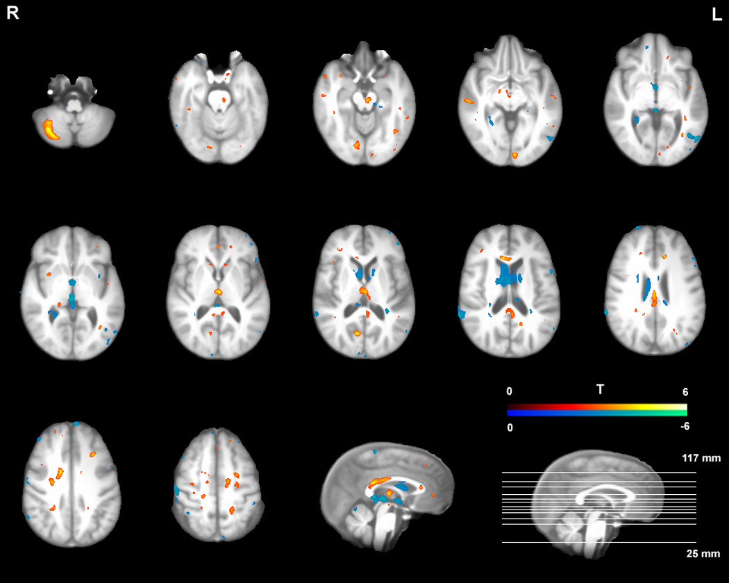
Traumatic brain injury (TBI) is one of the most common causes of long-term disability. Despite the importance of identifying neuropathology in individuals with chronic TBI, methodological challenges posed at the stage of inter-subject image registration have hampered previous voxel-based MRI studies from providing a clear pattern of structural atrophy after TBI. We used a novel symmetric diffeomorphic image normalization method to conduct a tensor-based morphometry (TBM) study of TBI. The key advantage of this method is that it simultaneously estimates an optimal template brain and topology preserving deformations between this template and individual subject brains. Detailed patterns of atrophies are then revealed by statistically contrasting control and subject deformations to the template space. Participants were 29 survivors of TBI and 20 control subjects who were matched in terms of age, gender, education, and ethnicity. Localized volume losses were found most prominently in white matter regions and the subcortical nuclei including the thalamus, the midbrain, the corpus callosum, the mid- and posterior cingulate cortices, and the caudate. Significant voxel-wise volume loss clusters were also detected in the cerebellum and the frontal/temporal neocortices. Volume enlargements were identified largely in ventricular regions. A similar pattern of results was observed in a subgroup analysis where we restricted our analysis to the 17 TBI participants who had no macroscopic focal lesions (total lesion volume >1.5 cm(3)). The current study confirms, extends, and partly challenges previous structural MRI studies in chronic TBI. By demonstrating that a large deformation image registration technique can be successfully combined with TBM to identify TBI-induced diffuse structural changes with greater precision, our approach is expected to increase the sensitivity of future studies examining brain-behavior relationships in the TBI population.
Figures


Similar articles
-
Resting cerebral blood flow alterations in chronic traumatic brain injury: an arterial spin labeling perfusion FMRI study.J Neurotrauma. 2010 Aug;27(8):1399-411. doi: 10.1089/neu.2009.1215. J Neurotrauma. 2010. PMID: 20528163 Free PMC article.
-
Multivariate analysis of structural and diffusion imaging in traumatic brain injury.Acad Radiol. 2008 Nov;15(11):1360-75. doi: 10.1016/j.acra.2008.07.007. Acad Radiol. 2008. PMID: 18995188 Free PMC article.
-
Heterogeneity of brain lesions in pediatric traumatic brain injury.Neuropsychology. 2013 Jul;27(4):438-51. doi: 10.1037/a0032837. Neuropsychology. 2013. PMID: 23876117
-
Imaging assessment of traumatic brain injury.Postgrad Med J. 2016 Jan;92(1083):41-50. doi: 10.1136/postgradmedj-2014-133211. Epub 2015 Nov 30. Postgrad Med J. 2016. PMID: 26621823 Review.
-
Neuropathology of Mild Traumatic Brain Injury: Correlation to Neurocognitive and Neurobehavioral Findings.In: Kobeissy FH, editor. Brain Neurotrauma: Molecular, Neuropsychological, and Rehabilitation Aspects. Boca Raton (FL): CRC Press/Taylor & Francis; 2015. Chapter 31. In: Kobeissy FH, editor. Brain Neurotrauma: Molecular, Neuropsychological, and Rehabilitation Aspects. Boca Raton (FL): CRC Press/Taylor & Francis; 2015. Chapter 31. PMID: 26269912 Free Books & Documents. Review.
Cited by
-
Structural brain atlases: design, rationale, and applications in normal and pathological cohorts.J Alzheimers Dis. 2012;31 Suppl 3(0 3):S169-88. doi: 10.3233/JAD-2012-120412. J Alzheimers Dis. 2012. PMID: 22647262 Free PMC article. Review.
-
Multimodal surface-based morphometry reveals diffuse cortical atrophy in traumatic brain injury.BMC Med Imaging. 2009 Dec 31;9:20. doi: 10.1186/1471-2342-9-20. BMC Med Imaging. 2009. PMID: 20043859 Free PMC article.
-
Effects of traumatic brain injury on a virtual reality social problem solving task and relations to cortical thickness in adolescence.Neuropsychologia. 2011 Feb;49(3):486-97. doi: 10.1016/j.neuropsychologia.2010.12.007. Epub 2010 Dec 13. Neuropsychologia. 2011. PMID: 21147137 Free PMC article.
-
Volume Change in Frontal Cholinergic Structures After Traumatic Brain Injury and Cognitive Outcome.Front Neurol. 2020 Aug 13;11:832. doi: 10.3389/fneur.2020.00832. eCollection 2020. Front Neurol. 2020. PMID: 32903569 Free PMC article.
-
Resting cerebral blood flow alterations in chronic traumatic brain injury: an arterial spin labeling perfusion FMRI study.J Neurotrauma. 2010 Aug;27(8):1399-411. doi: 10.1089/neu.2009.1215. J Neurotrauma. 2010. PMID: 20528163 Free PMC article.
References
-
- Adams JH, Doyle D, Ford I, Gennarelli TA, Graham DI, McLellan DR. Diffuse axonal injury in head injury: definition, diagnosis and grading. Histopathology. 1989;15(1):49–59. - PubMed
-
- Adams JH, Graham DI, Jennett B. The neuropathology of the vegetative state after an acute brain insult. Brain. 2000;123(Pt 7):1327–1338. - PubMed
-
- Ashburner J, Friston KJ. Voxel-based morphometry--the methods. Neuroimage. 2000;11(6 Pt 1):805–821. - PubMed
Publication types
MeSH terms
Grants and funding
LinkOut - more resources
Full Text Sources
Other Literature Sources
