

Coloration using higher order optical interference in the wing pattern of the Madagascan sunset moth
- PMID: 17999945
- PMCID: PMC2607392
- DOI: 10.1098/rsif.2007.1268
Coloration using higher order optical interference in the wing pattern of the Madagascan sunset moth
Abstract
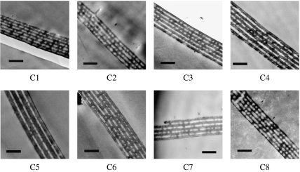
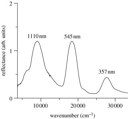
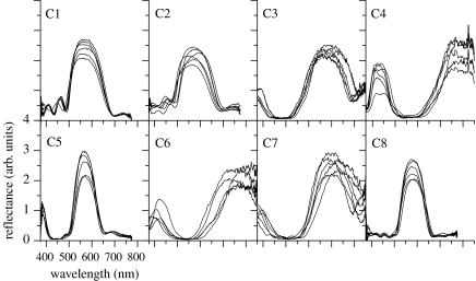
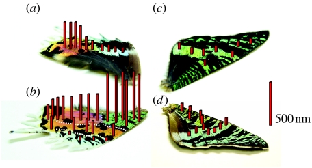
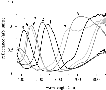
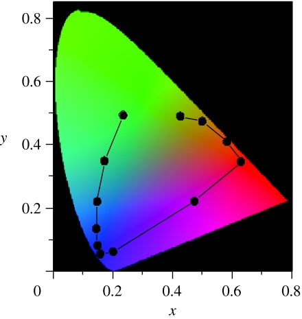
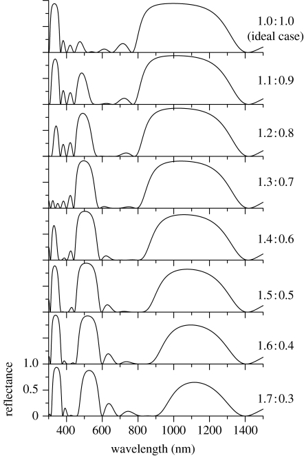
Colour patterns of animals' bodies are usually produced by the spatial distribution of pigments with different colours. However, some animals use the spatial variation of colour-producing microstructures. We have studied one distinctive example of such structurally produced colour patterns, the wing of the Madagascan sunset moth, to clarify the physical rules that underlie the colour variation. It is known that the iridescent wing scale of the sunset moth has the alternate air-cuticle multilayer structure that causes optical interference. The microscopic and optical investigations of various parts of the wing have confirmed that the thickness of the cuticle layers within the scale largely varies to produce the colour pattern. However, it varies in very different ways between the dorsal and ventral sides of the hind wing; the thickness gradually varies on the dorsal side from scale to scale, while the abrupt changes are found on the ventral side to form distinctive borders between differently coloured areas. It is also revealed that an unusual coloration mechanism is involved in the green part of the ventral hind wing: the colour is caused by higher order optical interference of the highly non-ideal multilayer structure. The physical mechanism of the colour pattern formation is briefly discussed with the several mathematical models proposed so far.
Figures
Similar articles
-
Polarization-sensitive color mixing in the wing of the Madagascan sunset moth.Opt Express. 2007 Mar 5;15(5):2691-701. doi: 10.1364/oe.15.002691. Opt Express. 2007. PMID: 19532506
-
Remarkable iridescence in the hindwings of the damselfly Neurobasis chinensis chinensis (Linnaeus) (Zygoptera: Calopterygidae).Proc Biol Sci. 2004 Mar 22;271(1539):595-601. doi: 10.1098/rspb.2003.2595. Proc Biol Sci. 2004. PMID: 15156917 Free PMC article.
-
Ultrastructural variation tune wing coloration of a moth Asota caricae Fabricius, 1775.Tissue Cell. 2017 Dec;49(6):648-656. doi: 10.1016/j.tice.2017.09.001. Epub 2017 Sep 13. Tissue Cell. 2017. PMID: 28935358
-
Evo-devo of wing colour patterns in beetles.Curr Opin Genet Dev. 2021 Aug;69:97-102. doi: 10.1016/j.gde.2021.02.007. Epub 2021 Mar 18. Curr Opin Genet Dev. 2021. PMID: 33744509 Review.
-
Iridescence: a functional perspective.J R Soc Interface. 2009 Apr 6;6 Suppl 2(Suppl 2):S115-32. doi: 10.1098/rsif.2008.0395.focus. J R Soc Interface. 2009. PMID: 19336344 Free PMC article. Review.
Cited by
-
Iridescent structural colour production in male blue-black grassquit feather barbules: the role of keratin and melanin.J R Soc Interface. 2009 Apr 6;6 Suppl 2(Suppl 2):S203-11. doi: 10.1098/rsif.2008.0460.focus. Epub 2009 Jan 13. J R Soc Interface. 2009. PMID: 19141431 Free PMC article.
-
Fine nanostructural variation in the wing pattern of a moth Chiasmia eleonora Cramer (1780).J Biosci. 2018 Sep;43(4):673-684. J Biosci. 2018. PMID: 30207313
-
Photonic Crystal Structure and Coloration of Wing Scales of Butterflies Exhibiting Selective Wavelength Iridescence.Materials (Basel). 2012 Apr 30;5(5):754-771. doi: 10.3390/ma5050754. Materials (Basel). 2012. PMID: 28817007 Free PMC article. Review.
-
Classical lepidopteran wing scale colouration in the giant butterfly-moth Paysandisia archon.PeerJ. 2018 Apr 11;6:e4590. doi: 10.7717/peerj.4590. eCollection 2018. PeerJ. 2018. PMID: 29666756 Free PMC article.
-
Mechanism of variable structural colour in the neon tetra: quantitative evaluation of the Venetian blind model.J R Soc Interface. 2011 Jan 6;8(54):56-66. doi: 10.1098/rsif.2010.0253. Epub 2010 Jun 16. J R Soc Interface. 2011. PMID: 20554565 Free PMC article.
References
-
- Bard J.B.L, French V. Butterfly wing patterns: how good a determining mechanism is the simple diffusion of a single morphogen? J. Embryol. Exp. Morphol. 1984;84:255–274. - PubMed
-
- Berthier S, Boulenguez J, Bálint Z. Multiscaled polarization effects in Suneve coronata (Lepidoptera) and other insects: application to anti-counterfeiting of banknotes. Appl. Phys. A. 2006;86:123–130. doi: 10.1007/s00339-006-3723-9. - DOI
-
- Durrer H. Schillerfarben beim Pfau (Pavo cristatus L.) Verh. Naturforsch. Ges. Basel. 1962;73:204–224.
-
- Hogue R. ‘Praying Mantis’ diffuse reflectance accessory for UV–Vis–NIR spectroscopy. Fresenius J. Anal. Chem. 1991;339:68–69. doi: 10.1007/BF00324507. - DOI
Publication types
MeSH terms
LinkOut - more resources
Full Text Sources
Other Literature Sources
