. 2008 Jan;36(1):94-109.
doi: 10.1093/nar/gkm1004. Epub 2007 Nov 13.

The process of displacing the single-stranded DNA-binding protein from single-stranded DNA by RecO and RecR proteins

Affiliations

The process of displacing the single-stranded DNA-binding protein from single-stranded DNA by RecO and RecR proteins

Jin Inoue et al. Nucleic Acids Res. 2008 Jan.

Abstract

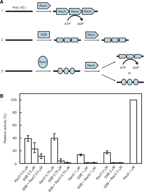
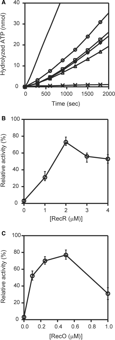
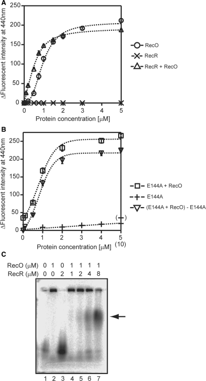
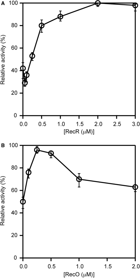
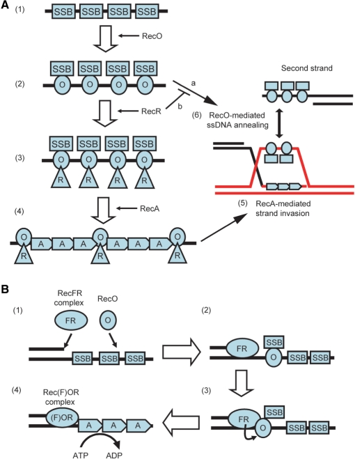
The regions of single-stranded (ss) DNA that result from DNA damage are immediately coated by the ssDNA-binding protein (SSB). RecF pathway proteins facilitate the displacement of SSB from ssDNA, allowing the RecA protein to form protein filaments on the ssDNA region, which facilitates the process of recombinational DNA repair. In this study, we examined the mechanism of SSB displacement from ssDNA using purified Thermus thermophilus RecF pathway proteins. To date, RecO and RecR are thought to act as the RecOR complex. However, our results indicate that RecO and RecR have distinct functions. We found that RecR binds both RecF and RecO, and that RecO binds RecR, SSB and ssDNA. The electron microscopic studies indicated that SSB is displaced from ssDNA by RecO. In addition, pull-down assays indicated that the displaced SSB still remains indirectly attached to ssDNA through its interaction with RecO in the RecO-ssDNA complex. In the presence of both SSB and RecO, the ssDNA-dependent ATPase activity of RecA was inhibited, but was restored by the addition of RecR. Interestingly, the interaction of RecR with RecO affected the ssDNA-binding properties of RecO. These results suggest a model of SSB displacement from the ssDNA by RecF pathway proteins.


Figures

Figure 1.
Figure 1.
Purification of ttRecF, ttRecO, ttRecR, ttRecA and ttSSB. The indicated samples (0.5 μg) were subjected to electrophoresis on 12.5% polyacrylamide gels containing 0.1% sodium dodecyl sulfate (SDS) under reducing conditions and visualized by staining with Coomassie Brilliant Blue R-250. The lanes contained the following samples: 1, Molecular mass markers (97, 66, 42, 30, 20 and 14 kDa); 2, ttRecF (37.8 kDa); 3, ttRecO (24.7 kDa); 4, ttRecR (21.2 kDa); 5, ttRecA (36.3 kDa); 6, ttSSB (29.8 kDa).
Figure 2.
Figure 2.
Analysis of protein–protein interactions among RecF pathway proteins using native-PAGE. (A) Analysis of the interaction between RecO and SSB. SSB (10 μM) was incubated with 1, 2, 5, 10, 20 and 50 μM RecO, and the complexes were resolved by electrophoresis (lanes 4–9). SSB was also incubated with 50 μM lysozyme as a control (lane 10). 10 μM SSB, 10 μM RecO and 10 μM lysozyme were loaded in lanes 1, 2 and 3, respectively. (B) Analysis of the interaction between RecO and RecR. RecR (10 μM) was incubated with 1, 2, 5, 10, 20 and 50 μM RecO (lanes 4–9). RecR was also incubated with 50 μM lysozyme as a control (lane 10). 10 μM RecR, 10 μM RecO and 10 μM lysozyme were loaded in lanes 1, 2 and 3, respectively. (C) Analysis of the interaction between RecR and RecF. RecR (10 μM) was incubated with 1, 2, 5 and 7 μM RecF (lanes 4–7). RecR was also incubated with 10 μM lysozyme as a control (lane 8). 10 μM RecR, 10 μM RecF and 10 μM lysozyme were loaded in lanes 1, 2 and 3, respectively.
Figure 3.
Figure 3.
A ssDNA-binding assay using fluorophotometry and etheno-modified ssDNA. (A) Fluorescence spectral changes. The indicated concentrations of protein were incubated with 10 μM εDNA at 25°C for 5 min. The emission spectra (excitation wavelength of 305 nm) were measured using a 5 mm × 5 mm cell at 25°C. The ordinate axis represents the difference in fluorescence emission intensity at 440 nm between εDNA in the presence and absence of each protein. The data represents the average of three independent experiments. (B) Competition-binding experiments using RecO and SSB. 4 μM SSB or RecO was incubated with 10 μM εDNA at 25°C for 3 min prior to the addition of the competing protein. The indicated concentration of the competing protein was added to the solution for an additional 5 min before measuring the change in fluorescence.
Figure 4.
Figure 4.
The observation of the protein-ssDNA complexes by electron microscopy. (A, G) The images of SSB in the presence of M13 ssDNA. (B, C) The images of RecO in the presence of M13 ssDNA. (D) The image of SSB-RecO in the presence of M13 ssDNA. M13 ssDNA was incubated with SSB for 10 min, and then RecO was added to the solution. (E) The image of RecO-SSB in the presence of M13 ssDNA. M13 ssDNA was incubated with RecO for 10 min, and then SSB was added to the solution. (F) The wide-ranging image of SSB-RecO in the presence of M13 ssDNA. The arrowheads indicate the protein-ssDNA complexes. Scales of each image are indicated by the white bars. Parentheses indicate the classified shapes in Table 1 (a, b and d).
Figure 5.
Figure 5.
The measurement of the ssDNA-dependent ATPase activity of RecA. (A) Schematic representation of the ATPase activity of RecA. 1. RecA hydrolyzes ATP in an ssDNA-dependent manner. 2. When SSB binds to the ssDNA prior to RecA, RecA ATPase activity is inhibited. 3. The effect of RecO binding to ssDNA prior to RecA on ATPase activity is not known. (B) The ssDNA-dependent ATPase activity of RecA in the presence of RecO and/or SSB. The ATPase activity of 1 μM RecA in the absence of RecO and SSB was defined as 100%. The ATPase activity of RecA in the presence of the indicated concentrations of SSB and/or RecO was measured. Each sample was incubated with a 340-mer poly (dC) prior to RecA, then 1 μM of RecA was added and the ATPase activity was measured. The data represent the average of three independent experiments.
Figure 6.
Figure 6.
The pull-down assay of the ssDNA-bound proteins. (A) The indicated concentrations of the proteins were incubated with ssDNA-immobilized streptavidine sepharose. After three washes, ssDNA-bound proteins were analyzed by SDS-PAGE. The asterisk indicates the band derived from the streptavidine sepharose. A band corresponding to BSA was observed in all lanes because it was present in the washing buffer. (B) Effect of pH on the ssDNA-binding properties of SSB and RecO. The buffers contained 50 mM MES-NaOH (pH 6.5, lanes 1–6), 50 mM Tris-HCl (pH 7.5, lanes 7–12) or 50 mM Tris-HCl (pH 8.5, lanes 13–18), together with 1 mM DTT, 0.15% Tween20 and 0.2 mg/ml BSA. The pull-down assays were performed as described in Figure 7. (C) The pull-down assay was performed in buffer containing 50 mM Tris-HCl (pH 8.5), 1 mM DTT, 0.15% Tween20 and 0.2 mg/ml BSA.
Figure 7.
Figure 7.
The removal of inhibition of RecA by RecR. (A) The ordinate axis is the amount of hydrolyzed ATP. The ssDNA-dependent ATPase activity of RecA was measured in the presence of 1 μM RecA (line), 1 μM SSB and 1 μM RecO (cross); 1 μM RecA, 1 μM SSB, 1 μM RecO and 1 μM RecR (triangle); 1 μM RecA, 1 μM SSB, 1 μM RecO and 2 μM RecR (circle); 1 μM RecA, 1 μM SSB, 1 μM RecO and 3 μM RecR (square); and 1 μM RecA, 1 μM SSB, 1 μM RecO and 4 μM RecR (diamond). Proteins were incubated with 340-mer poly (dC) in following order: SSB, RecO and RecR; RecA was then added to the mixture and the ATPase activity was measured. The data represent the averages of three independent experiments. (B) The proteins were incubated with a 340-mer poly (dC) in the following order: 1 μM SSB, 1 μM RecO and the indicated concentrations of RecR and then 1 μM RecA was added to the mixture, and ssDNA-dependent ATPase activity was measured. The activity relative to the activity of RecA alone (defined as 100%) was plotted against RecR concentration. (C) The proteins were incubated with a 340-mer poly (dC) in the following order: 1 μM SSB, the indicated concentrations of RecO, and 1 μM RecR, and then 1 μM RecA was added to the mixture and the ssDNA-dependent ATPase activity was measured. The ATPase activity was plotted against RecO concentration as described in (B).
Figure 8.
Figure 8.
The effect of RecR on the ssDNA-binding activity of RecO. (A) The indicated concentrations of RecO were incubated with 10 μM εDNA at 25°C for 5 min in the absence (circle) or presence (triange) of 10 μM RecR. The effect of RecR alone is represented by a cross. The measurement conditions were the same as described for Figure 4. (B) The indicated concentrations of RecO were incubated with 10 μM εDNA at 25°C for 5 min in the presence of 10 μM RecR_E144A (square). The effect of RecR_E144A alone (0–5 µM) is represented by a plus, and the effect of 10 μM RecR_E144A is shown by a bracketed plus. The reverse triangle represents the binding curve of RecO to ssDNA with the ΔF of the 10 μM RecR_E144A alone (∼35) subtracted. (C) An agarose gel retardation assay of the RecO-RecR complex. A 5′-33P labeled 66-mer ssDNA was incubated with the indicated concentrations of RecO and RecR for 5 min at 50°C, and then the samples were analyzed by electrophoresis on a 2.5% agarose gel.
Figure 9.
Figure 9.
The effect of changing the molar ratio of RecO and RecR on the activity of RecA in the absence of SSB. (A) The proteins were incubated with a 340-mer poly (dC) in the following order: 0.25 μM RecO, the indicated concentrations of RecR and then 1 μM RecA was then added to the mixture and the ssDNA-dependent ATPase activity was measured. The RecA activity relative to that of RecA alone (defined as 100%) was plotted against the concentration of RecR. The data represent the average of three independent experiments. (B) The proteins were incubated with a 340-mer poly (dC) in the following order: the indicated concentrations of RecO, 1 μM of RecR and then 1 μM RecA was added to the mixture and the ssDNA-dependent ATPase activity was measured. RecA activity was plotted as described in (A).
Figure 10.
Figure 10.
A model for the displacement of SSB by RecO, RecR and RecA. (A): (1) ssDNA regions are coated by SSB immediately. (2) RecO interacts with SSB, remodels the SSB-ssDNA complex and binds to ssDNA directly. The displaced SSB interacts with RecO in the RecO-ssDNA complex. If a complementary ssDNA strand exists, the ssDNAs are annealed by RecO (a). (3) RecR interacts with RecO in the SSB-RecO-ssDNA complex. Then RecR modulates the ssDNA-binding mode of RecO and inhibits the ssDNA annealing activity of RecO (b). At this time, SSB may be released from RecO. (4) RecO and RecR remain on ssDNA as the RecOR complex, where they assist the formation of the RecA filament. RecA makes nucleoprotein filament on ssDNA, resulting in SSB dissociation. The RecOR complex may stabilize the RecA filament probably by binding to the end of the RecA filament. (5) RecA-mediated strand invasion occurs. The strand which is displaced by RecA is captured by SSB and RecO like in (1) and (2). (6) The displaced strand is annealed with a second strand by SSB and RecO. (B): (1) When dsDNA-ssDNA junctions are generated as a consequence of DNA damage, the RecFR complex binds to the dsDNA-ssDNA junction, and RecO interacts with the SSB-coated ssDNA. (2) RecO displaces SSB on the SSB-coated ssDNA. The released SSB remains on the ssDNA indirectly via its interaction with RecO. (3) RecR, which is in a RecFR complex at the dsDNA-ssDNA junction site, interacts with RecO that attaches SSB to the ssDNA at this site. (4) The RecOR complex, which may also associate with RecF, facilitates the displacement of SSB from the ssDNA region and the loading of RecA onto the ssDNA. This may be enhanced by extension of RecA filaments.

Similar articles

Cited by

References

    1. Kuzminov A. Recombinational repair of DNA damage in Escherichia coli and bacteriophage lambda. Microbiol. Mol. Biol. Rev. 1999;63:751–813. - PMC - PubMed
    1. Krogh BO, Symington LS. Recombination proteins in yeast. Annu Rev. Genet. 2004;38:233–271. - PubMed
    1. Yang H, Li Q, Fan J, Holloman WK, Pavletich NP. The BRCA2 homologue Brh2 nucleates RAD51 filament formation at a dsDNA-ssDNA junction. Nature. 2005;433:653–657. - PubMed
    1. Song B, Sung P. Functional interactions among yeast Rad51 recombinase, Rad52 mediator, and replication protein A in DNA strand exchange. J. Biol. Chem. 2000;275:15895–15904. - PubMed
    1. Sugiyama T, Kowalczykowski SC. Rad52 protein associates with replication protein A (RPA)-single-stranded DNA to accelerate Rad51-mediated displacement of RPA and presynaptic complex formation. J. Biol. Chem. 2002;277:31663–31672. - PubMed

Publication types

MeSH terms