. 2007 Nov 20;104(47):18724-9.
doi: 10.1073/pnas.0709276104. Epub 2007 Nov 13.

Poxvirus DNA primase

Affiliations

Poxvirus DNA primase

Frank S De Silva et al. Proc Natl Acad Sci U S A. .

Abstract

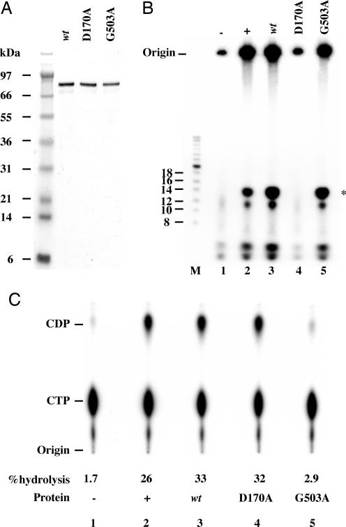
Poxviruses are large enveloped viruses that replicate in the cytoplasm of vertebrate or invertebrate cells. At least six virus-encoded proteins are required for synthesis and processing of the double-stranded DNA genome of vaccinia virus, the prototype member of the family. One of these proteins, D5, is an NTPase that contains an N-terminal archaeoeukaryotic primase domain and a C-terminal superfamily III helicase domain. Here we report that individual conserved aspartic acid residues in the predicted primase active site were required for in vivo complementation of infectious virus formation as well as genome and plasmid replication. Furthermore, purified recombinant D5 protein synthesized oligoribonucleotides in vitro. Incorporation of label from [alpha-(32)P]CTP or [alpha-(32)P]UTP into a RNase-sensitive and DNase-resistant product was demonstrated by using single-stranded circular bacteriophage DNA templates and depended on ATP or GTP and a divalent cation. Mutagenesis studies showed that the primase and NTPase activities of the recombinant D5 protein could be independently inactivated. Highly conserved orthologs of D5 are present in all poxviruses that have been sequenced, and more diverged orthologs are found in members of all other families of nucleocytoplasmic large DNA viruses. These viral primases may have roles in initiation of DNA replication or lagging-strand synthesis and represent potential therapeutic targets.


Conflict of interest statement

The authors declare no conflict of interest.

Figures

Fig. 1.
Fig. 1.
DNA replication depends on conserved amino acids in the primase domain of D5. (A) Virus complementation. BS-C-1 cells were infected in triplicate with Cts24 at a multiplicity of five plaque-forming units (PFU) per cell at 31°C. At 3 h after infection, cells were transfected with empty plasmid vector or plasmid that expressed WT D5, D5D70A, D5D72A, D5D170A, or D5G503A. Two hours later, the cells were shifted to 40°C or maintained at 31°C, as indicated. Cells were harvested at 24 h after infection and virus yields determined by plaque assay at 31°C. Error bars indicate range of one standard deviation. (B) Genome replication. Cells were infected in triplicate with Cts24, transfected and harvested as above. Total DNA was isolated and 2 μg transferred to a nylon membrane. The blot was hybridized to 32P-labeled VACV DNA and radioactivity was quantified with a PhosphorImager. (C) Viral origin-independent plasmid replication. Cells were uninfected or infected with Cts24 and transfected with plasmid p716 in addition to empty vector or D5 plasmids as above. At 5 h after infection, the temperature was raised to 40°C and at 24 h, the cells were harvested. Total DNA was isolated, digested with DpnI, and p716 replication was measured by real-time PCR.
Fig. 2.
Fig. 2.
Primase activity of purified recombinant D5. (A) Affinity purification of D5. HeLa cells were infected with a recombinant VACV containing an inducible copy of D5 with a C-terminal 10-histidine tag. After 24 h, cell extracts were prepared, and the tagged D5 was purified on Ni-NTA beads. Samples were analyzed by SDS/PAGE and staining with Coomassie blue. Lanes: 1, cell extract; 2, flowthrough; 3, 20 mM imidazole wash; 4, 40 mM imidazole wash; and 5–9 fractions eluting with 200 mM imidazole. Molecular masses of protein markers (M) are shown on the left. (B) Comparison of herpes simplex virus (HSV) and VACV (D5) primase activities. Recombinant histidine-tagged D5 (1.1 pmols), HSV primase complex (3.2 pmols), or the equivalent volume of Ni-NTA eluate from cells infected with parental VACV (mock) was incubated in a reaction mixture containing rNTPs including [α-32P]CTP and 0.57 pmols φX174 DNA at 37°C for 30 min. Products were digested with calf intestinal alkaline phosphatase, denatured, and resolved by electrophoresis. Positions of 32P-end-labeled oligonucleotide markers (M) are indicated on the left. Asterisk indicates position of the reaction product migrating with 14-nt phosphorylated marker.
Fig. 3.
Fig. 3.
Primase and NTPase activities of WT and mutated D5 proteins. (A) Analysis of recombinant D5 proteins expressed by recombinant alphaviruses. Proteins were affinity purified on a Ni-NTA column, resolved by SDS/PAGE, and stained with Coomassie blue. Lanes containing histidine tagged WT D5, D5D170A, and D5G503A are indicated. Protein molecular mass standards are indicated at the left. (B) Oligoribonucleotide synthesis. Reaction conditions were the same as in Fig. 2B. Positions of end-labeled oligonucleotide markers (M) are indicated on the left. Lanes: 1, (−) Ni-NTA eluate from uninfected BHK cell extracts; 2, (+) 1.1 pmols WT D5 expressed by recombinant VACV; 3, 1.3 pmols WT D5 expressed by alphavirus; 4, 1.3 pmols D5D170A expressed by alphavirus; 5, 1.3 pmols D5G503A expressed by alphavirus. Asterisk indicates position of product migrating with 14-nt marker. (C) NTPase activity. Autoradiograph of thin-layer chromatography plate with positions of CDP and CTP markers. Percent hydrolysis of CTP to CDP is indicated. Lanes are the same as in B.
Fig. 4.
Fig. 4.
Characterization of primase reaction and products. (A) Primase reaction time course. Primase reactions were carried out under standard conditions for the times indicated. (B) Effect of nuclease treatments on product. Primase reactions were carried out under standard conditions and the product was treated with calf intestinal phosphatase and (lane 1) nothing, (lane 2) nuclease P1, (lane 3) RNase A and T1, and (lane 4) DNase I. (C) Omission of reaction components. Primase reaction was carried out under standard conditions or with indicated component(s) missing using WT D5 expressed by alphavirus vector and φX174 DNA template. Asterisks indicate position of product migrating with the 14-nt marker. End-labeled oligonucleotide markers are on the left.

References

    1. Moss B. In: Fields Virology. Knipe DM, editor. Philadelphia: Lippincott Williams & Wilkins; 2007. pp. 2905–2946.
    1. Harrison SC, Alberts B, Ehrenfeld E, Enquist L, Fineberg H, McKnight SL, Moss B, O'Donnell M, Ploegh H, Schmid SL, et al. Proc Natl Acad Sci USA. 2004;101:11178–11192. - PMC - PubMed
    1. Moss B, De Silva F. In: DNA Replication and Human Disease. DePamphilis ML, editor. Cold Spring Harbor, NY: Cold Spring Harbor Laboratory Press; 2006. pp. 707–727.
    1. Challberg MD, Englund PT. J Biol Chem. 1979;254:7812–7819. - PubMed
    1. McDonald WF, Traktman P. J Biol Chem. 1994;269:31190–31197. - PubMed

Publication types

LinkOut - more resources