. 2007 Nov 20;104(47):18648-53.
doi: 10.1073/pnas.0709198104. Epub 2007 Nov 13.

Protection against simian/human immunodeficiency virus (SHIV) 89.6P in macaques after coimmunization with SHIV antigen and IL-15 plasmid

Affiliations

Protection against simian/human immunodeficiency virus (SHIV) 89.6P in macaques after coimmunization with SHIV antigen and IL-15 plasmid

Jean D Boyer et al. Proc Natl Acad Sci U S A. .

Abstract

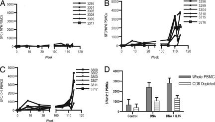
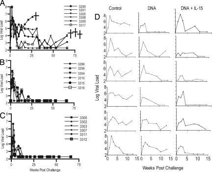
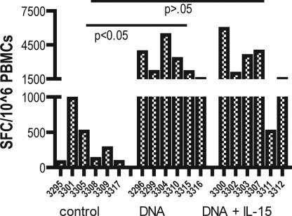
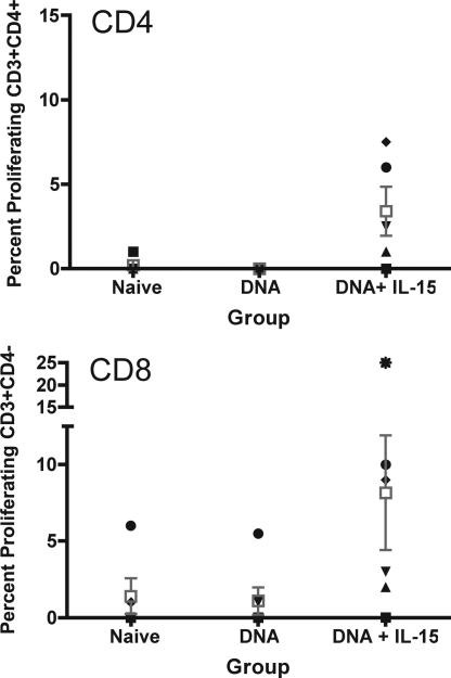
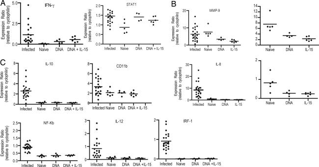
The cell-mediated immune profile induced by a recombinant DNA vaccine was assessed in the simian/HIV (SHIV) and macaque model. The vaccine strategy included coimmunization of a DNA-based vaccine alone or in combination with an optimized plasmid encoding macaque IL-15 (pmacIL-15). We observed strong induction of vaccine-specific IFN-gamma-producing CD8(+) and CD4(+) effector T cells in the vaccination groups. Animals were subsequently challenged with 89.6p. The vaccine groups were protected from ongoing infection, and the IL-15 covaccinated group showed a more rapidly controlled infection than the group treated with DNA vaccine alone. Lymphocytes isolated from the group covaccinated with pmacIL-15 had higher cellular proliferative responses than lymphocytes isolated from the macaques that received SHIV DNA alone. Vaccine antigen activation of lymphocytes was also studied for a series of immunological molecules. Although mRNA for IFN-gamma was up-regulated after antigen stimulation, the inflammatory molecules IL-8 and MMP-9 were down-regulated. These observed immune profiles are potentially reflective of the ability of the different groups to control SHIV replication. This study demonstrates that an optimized IL-15 immune adjuvant delivered with a DNA vaccine can impact the cellular immune profile in nonhuman primates and lead to enhanced suppression of viral replication.


Conflict of interest statement

The authors declare no conflict of interest.

Figures

Fig. 1.
Fig. 1.
IFN-γ-producing cells after the primary and secondary sets of immunization. Samples were taken 2 weeks after each injection and assessed for an SIVgag antigen-specific response by ELISpot. (A–C) The number of cells able to secrete IFN-g after SIVgag in vitro stimulation of PBMCs isolated from naïve macaques (A) pCSIVgag (B), pCSIVgag, and pmacIL15 immunized macaques (C) is presented as SFC per 1 million PBMCs. (D) After the final immunization, we evaluated the contribution of the CD8 T cells to the observed population of cells from macaques secreting IFN-γ.
Fig. 2.
Fig. 2.
Viral load after challenge of cynomologous macaques with 100 MID of SHIV89.6p. (A–C) Viral load is presented for control (A), SIV DNA (B), SIV DNA + pmacIL15-immunized (C) macaques. The assay has a threshold sensitivity of 200 RNA copies per milliliter of plasma with interassay variations averaging 0.5 log10. (D) The viral loads for the first 15 weeks for each individual macaque. Several animals were killed because of AIDS-like syndrome.
Fig. 3.
Fig. 3.
IFN-γ-producing cells after IV challenge with SHIV89.6p virus. Samples were taken 12 weeks after challenge. PBMCs were isolated by a standard Percoll separation technique and assessed for a gag antigen-specific response by ELISpot. The data represent the number of cells able to secrete IFN-γ after SIVgag in vitro stimulation of PBMCs isolated from vaccine-naïve macaques, pCSIVgag-vaccinated, and pCSIVgag- and pmacIL15-immunized macaques.
Fig. 4.
Fig. 4.
T cell proliferative responses to SIVgag. PBMCs were stained with CFSE and stimulated with SIVgag peptides for 5 days. Standard surface staining protocol was followed for CD3/CD4-positive cells.
Fig. 5.
Fig. 5.
Gene expression after SIV gag stimulation, quantitative RT-PCR. Five million PBMCs were taken from the macaques 4 weeks after the sixth and final immunization stimulated in vitro with SIVgag peptides, and mRNA was extracted. Data are presented for effector function (IFN-g and STAT1) (A), inflammatory response (MMP-9 and IL-8) (B), and to assess other immunological markers of T cell activation (C). Data presented are mRNA levels compared with control mRNA of cyclophilin.

References

    1. Cao H, Kanki P, Sankale JL, Dieng-Sarr A, Mazzara GP, Kalams SA, Korber B, Mboup S, Walker BD. J Virol. 1997;71:8615–8623. - PMC - PubMed
    1. Koup RA, Safrit JT, Cao Y, Andrews CA, McLeod G, Borkowsky W, Farthing C, Ho DD. J Virol. 1994;68:4650–4655. - PMC - PubMed
    1. Rinaldo C, Huang XL, Fan ZF, Ding M, Beltz L, Logar A, Panicali D, Mazzara G, Liebmann J, Cottrill M. J Virol. 1995;69:5838–5842. - PMC - PubMed
    1. Rowland-Jones S, Sutton J, Ariyoshi K, Dong T, Gotch F, McAdam S, Whitby D, Sabally S, Gallimore A, Corrah T, et al. Nat Med. 1995;1:59–64. - PubMed
    1. Rowland-Jones SL, Dong T, Dorrell L, Ogg G, Hansasuta P, Krausa P, Kimani J, Sabally S, Ariyoshi K, Oyugi J, et al. Immunol Lett. 1999;66:9–14. - PubMed

Publication types

MeSH terms