. 2007 Nov 20;104(47):18754-9.
doi: 10.1073/pnas.0704908104. Epub 2007 Nov 13.

Selective inhibition of NF-kappaB activation prevents dopaminergic neuronal loss in a mouse model of Parkinson's disease

Affiliations

Selective inhibition of NF-kappaB activation prevents dopaminergic neuronal loss in a mouse model of Parkinson's disease

Anamitra Ghosh et al. Proc Natl Acad Sci U S A. .

Abstract

Parkinson's disease (PD) is the second most common neurodegenerative disorder. Despite intense investigations, no effective therapy is available to stop its onset or halt its progression. The present study evaluates the ability of peptide corresponding to the NF-kappaB essential modifier-binding domain (NBD) of IkappaB kinase alpha (IKKalpha) or IKKbeta to prevent nigrostriatal degeneration in the 1-methyl-4-phenyl-1,2,3,6-tetrahydropyridine (MPTP) mouse model of PD and establish a role for NF-kappaB in human parkinsonism. First, we found that NF-kappaB was activated within the substantia nigra pars compacta of PD patients and MPTP-intoxicated mice. However, i.p. injection of wild-type NBD peptide reduced nigral activation of NF-kappaB, suppressed nigral microglial activation, protected both the nigrostriatal axis and neurotransmitters, and improved motor functions in MPTP-intoxicated mice. These findings were specific because mutated NBD peptide had no effect. We conclude that selective inhibition of NF-kappaB activation by NBD peptide may be of therapeutic benefit for PD patients.


Conflict of interest statement

The authors declare no conflict of interest.

Figures
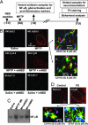
Fig. 1.
Fig. 1.
Activation of NF-κB in ventral midbrain of MPTP-intoxicated mice and PD patients. (A) Treatment schedule of MPTP-intoxicated mice with NBD peptide. (B) Twenty-four hours after the last injection of MPTP, ventral midbrain sections were immunostained for p65. (Magnification, ×10.) NF-κB p65-positive cells counted in four nigral sections (two images per slide) of each of four mice in an Olympus IX81 fluorescence microscope using the MicroSuite imaging software are mentioned as cells per square millimeter at the upper left corner of each image. Midbrain sections of MPTP-intoxicated mice were also double-labeled for p65 and glial cell markers (GFAP for astrocytes and CD11b for microglia). (Magnification, ×60.) DAPI was used to visualize nucleus. (C) EMSA was carried out with nuclear extracts prepared from ventral midbrain tissues. (D) Midbrain sections of PD patients and age-matched controls were immunostained for p65. (Magnification, ×10.) Number of p65-positive cells (cells per square millimeter) is mentioned at the upper left corner of each image. Midbrain sections of PD patients were also double-labeled for p65 and glial cell markers (GFAP and CD11b). (Magnification, ×60.) The arrows indicate colocalization of p65 with DAPI. Results represent analysis of four different sections from each of four different brains.
Fig. 2.
Fig. 2.
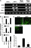
NBD peptide inhibits the expression of proinflammatory molecules in ventral midbrain of MPTP-intoxicated mice. Mice receiving wtNBD and mNBD peptide (0.75 mg/kg body weight per day) from 1 day before MPTP insult were killed 24 h after the last injection of MPTP. The mRNA expression of iNOS, IL-1β, and TNF-α was analyzed by RT-PCR (A) and quantitative real-time PCR (B). Data are means ± SEM of five to six mice per group. (C) Ventral midbrain sections were immunolabeled for iNOS. (D) Cells positive for iNOS were counted in four nigral sections (two images per slide) of each of four mice as described above. **, P < 0.001 vs. the MPTP group.
Fig. 3.
Fig. 3.
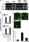
NBD peptide inhibits the activation of glial cells in ventral midbrain of MPTP-intoxicated mice. Mice receiving wtNBD and mNBD peptides (0.75 mg/kg body weight per day) from 1 day before MPTP insult were killed 24 h after the last injection of MPTP. The mRNA expression of GFAP and CD11b was analyzed by RT-PCR (A) and quantitative real-time PCR (B). Data are means ± SEM of five to six mice per group. (C) Ventral midbrain sections were immunolabeled for CD11b. (D) CD11b-positive cells were counted in four nigral sections (two images per slide) of each of four mice. **, P < 0.001 vs. the MPTP group.
Fig. 4.
Fig. 4.
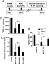
NBD peptide protects dopaminergic neurons in MPTP-intoxicated mice. Mice receiving wtNBD and mNBD peptides (0.75 mg/kg body weight per day) from 1 day before MPTP insult were killed 7 days after the last injection of MPTP followed by TH immunostaining of striatum (Left) and SNpc (Right) (A), counting of TH-positive neurons in SNpc (B), quantification of TH-positive fibers in striatum (C), and assay of neurotransmitters in striatum (D). Data are means ± SEM of five to six mice per group. **, P < 0.001 vs. the indicated group.
Fig. 5.
Fig. 5.
NBD peptide suppresses disease progression in the MPTP mouse model. Mice were treated with MPTP and NBD peptides (A) followed by counting of TH-positive neurons in SNpc (B), quantification of TH-positive fibers in striatum (C), and assay of neurotransmitters in striatum (D). Data are means ± SEM of five to six mice per group. **, P < 0.001 vs. the indicated group.

References

    1. Hardy J, Cai H, Cookson MR, Gwinn-Hardy K, Singleton A. Ann Neurol. 2006;60:389–398. - PubMed
    1. Fahn S, Przedborski S. In: Merritt's Neurology. Rowland LP, editor. New York: Lippincott Williams & Wilkins; 2000. pp. 679–693.
    1. Dauer W, Przedborski S. Neuron. 2003;39:889–909. - PubMed
    1. Olanow CW, Tatton WG. Annu Rev Neurosci. 1999;22:123–144. - PubMed
    1. Gao HM, Liu B, Zhang W, Hong JS. Trends Pharmacol Sci. 2003;24:395–401. - PubMed

Publication types

MeSH terms