Review
. 2007 Dec 1;6(23):2859-68.
doi: 10.4161/cc.6.23.5018. Epub 2007 Sep 4.

TLR8: an innate immune receptor in brain, neurons and axons

Affiliations
Review

TLR8: an innate immune receptor in brain, neurons and axons

Yinghua Ma et al. Cell Cycle. .

Abstract

Toll-like receptors (TLRs) play essential roles in generating innate immune responses, and are evolutionarily conserved across species. In mammals, TLRs specifically recognize the conserved microbial structural motifs referred to as pathogen-associated molecular patterns (PAMPs). Ligand recognition by TLRs activates signaling cascades that culminate in proinflammatory cytokine production and eventual elimination of invading pathogens. Although TLRs in mammals are expressed predominantly in the immune system, certain TLRs with poorly characterized function are also found in neurons. We recently profiled TLR8 expression during mouse brain development and established its localization in neurons and axons. We uncovered a novel role for TLR8 as a suppressor of neurite outgrowth as well as an inducer of neuronal apoptosis, and found that TLR8 functions in neurons through an NF-kappaB-independent mechanism. These findings add a new layer of complexity for TLR signaling, and expand the realm of mammalian TLR function to the CNS beyond the originally discovered immune context. Herein, we complement our earlier report with additional data, discuss their biological and mechanistic implications in central nervous system (CNS) developmental and pathological processes, and thus further our perspective on TLR signaling and potential physiological roles in mammals.


Figures

Figure 1
Figure 1
TLR8 expression and localization in cultured cortical neurons. (A) TLR8 (red) is expressed intracellularly in cultured neurons and is enriched in their growth cones (arrowheads). (B) TLR8 (green)- positive punctuate staining is distributed along axons (neurofilament 200 KD, red) and is concentrated in growth cones (arrowheads). Cortical neurons are cultured from embryonic day 17 (E17) mice, fixed at day-in-vitro (DIV) 3 with methanol at -20°C for 10 minutes, followed by staining with anti-TLR8 polyclonal antibody alone (A), or together with anti-neurofilament 200 KD monoclonal antibody (B).
Figure 2
Figure 2
Neurons do not express TLR7. (A-B) Immunostaining with anti-TLR7 polyclonal antibody on sagittal sections of E18 brain shows that TLR7 (red) is expressed only in the choroid plexus (arrowheads) and ependymal cells in the lateral ventricle (A) and fourth ventricle (B). (C) In the adult mouse spinal cord, TLR7 (red) is specifically expressed in ependymal cells (arrowheads) of the central canal. (D) Immunostaining of cortical neuron cultures from E18 mouse confirms that TLR7 is absent in neurons (MAP2, green; cell nuclei DAPI-stained, blue) but rather expressed in a few non-neuronal cells (red).
Figure 3
Figure 3
The amino acid sequences of the TIR domains of human TLR8, murine TLR8, and murine MyD88. The sequences were identified with the SMART (simple modular architecture research tool) program (smart.embl-heidelberg.de/); and the alignment analysis was performed with the MegAlign program (DNASTAR, Inc.) using the ClustalW method. Box 1, Box 2 along with the BB loop, and Box 3 are framed, and residues of the BB loop are shown in red. The residues within the three boxes of human and murine TLR8 are shown in bold. βA, B, C, D, E and αA, B, C, D, E at the top indicate five β-strands and α-helices, respectively, of the secondary structure of the TIR domain. The Genbank accession numbers assigned to the sequences are NP_057694 for human TLR8, NP_573475 for murine TLR8, and NP_034981 for murine MyD88.
Figure 4
Figure 4
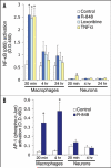
TLR8 stimulation in neurons activates neither the transcription factor NFκB (A) nor AP-1 (B). ELISA assays for NFκB (p65) and AP-1 (phospho-c-Jun) transactivation using nuclear extracts from the cortical neurons treated with 100 μM R-848, 500 μM loxoribine, 10 ng/ml TNFα for the indicated times. Note the robust activation of both NFκB and AP-1 detected in macrophages in response to R-848 stimulation. Data is presented as the mean ± SEM (n = 3). *p < 0.05, Student's t-Test.
Figure 5
Figure 5
R-848 effects on neurons do not require MAP kinase activity. (A) Representative micrographs of cortical neurons that were untreated (control, left column) or treated with 100 μM R-848 for 24 hours (right column), in the presence of vehicle control (DMSO, upper row) or a MAPKK inhibitor (50 μM PD98059, lower row). (B and C) Quantitative analyses of neurite length (B) and percent of the cleaved caspase3-positive cells (C) show that blocking MAPKK did not prevent the R-848-induced neurite outgrowth suppression and cell apoptosis. Data are presented as the mean ± SEM (n = 12 fields). **p < 0.01, Student's t-Test.
Figure 6
Figure 6
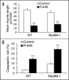
R-848 effects on neurons are independent of MyD88. Cortical neurons cultured from E17 Myd88−/− and wild-type littermate (WT) embryos were treated with PBS (control) or 100 μM R-848 for 24 hours. Quantitative analyses reveal that deficiency of MyD88 does not confer resistance to the R-848-induced inhibition of neurite outgrowth (A) and cell apoptosis (B). Data are presented as the mean ± SEM (n = 12 fields). **p < 0.01, Student's t-Test.
Figure 7
Figure 7
Cortical neurons lack IRAK1 expression. Neurons cultured from E17 mouse and Raw264.7 macrophages were treated with 100 μM R-848 for the indicated times. Equal amounts of protein lysates were subjected to Western blotting using anti-IRAK1 and anti-IRAK-M polyclonal antibodies. β-actin serves as the loading control.
Figure 8
Figure 8
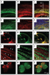
TLR8 is expressed in developing and adult axons. (A–F) Dual-immunolabeling of TLR8 (green) and the neuronal/axonal marker βIII-tubulin (red) on sagittal sections of E18 mouse cortex. Note that TLR8 is specifically expressed in the cortical intermediate zone (IMZ) where thalamocortical and corticofugal axons are enriched. Additional zones shown in (C) include marginal zone (MZ), cortical plate (CP), ventricular zone (VZ), and lateral ventricle (V). (D–F) Corresponding higher magnification views of the IMZ regions of (A-C). (G-I) Dual-immunolabeling of TLR8 (red) and the axonal marker growth-associated protein 43 (GAP43, green) on coronal sections of E18 mouse brain. Arrowheads indicate the IMZ and arrows point to the fimbra (an axonal bundle) of the hippocampus. (J-L) Dual-immunolabeling of TLR8 (red) and the neuronal dendrite marker microtubule-associated protein 2 (MAP2, green) on sagittal sections of three-month-old mouse brain caudae/putamen, showing striatal axonal bundles, with arrowheads pointing to individual axons. Asterisks indicate neuronal somas co-stained with TLR8 and MAP2. (M-O) Dual-immunolabeling of TLR8 (red) and βIII-tubulin (green) on three-month-old mouse spinal cord sections. Note that TLR8 is highly expressed in the axons in the dorsal funiculus (arrows) and the dorsal column (asterisk).

References

    1. Lemaitre B, Nicolas E, Michaut L, Reichart JM, Hoffmann JA. The dorsoventral regulatory gene cassette spatzle/Toll/cactus controls the potent antifungal response in Drosophila adults. Cell. 1996;86:973–83. - PubMed
    1. Medzhitov R, Preston-Hurlburt P, Janeway CA., Jr A human homologue of the Drosophila Toll protein signals activation of adaptive immunity. Nature. 1997;388:394–7. - PubMed
    1. Rock FL, Hardiman G, Timans JC, Kastelein RA, Bazan JF. A family of human receptors structurally related to Drosophila Toll. Proc Natl Acad Sci USA. 1998;95:588–93. - PMC - PubMed
    1. Akira S, Uematsu S, Takeuchi O. Pathogen recognition and innate immunity. Cell. 2006;124:783–801. - PubMed
    1. Lanzavecchia A, Sallusto F. Toll-like receptors and innate immunity in B-cell activation and antibody responses. Curr Opin Immunol. 2007;19:268–74. - PubMed

Publication types

Substances

LinkOut - more resources