. 2007 Nov 19;97(10):1344-53.
doi: 10.1038/sj.bjc.6604025. Epub 2007 Nov 13.

R306465 is a novel potent inhibitor of class I histone deacetylases with broad-spectrum antitumoral activity against solid and haematological malignancies

Affiliations

R306465 is a novel potent inhibitor of class I histone deacetylases with broad-spectrum antitumoral activity against solid and haematological malignancies

J Arts et al. Br J Cancer. .

Abstract

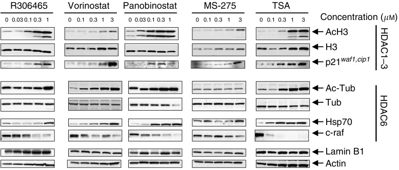
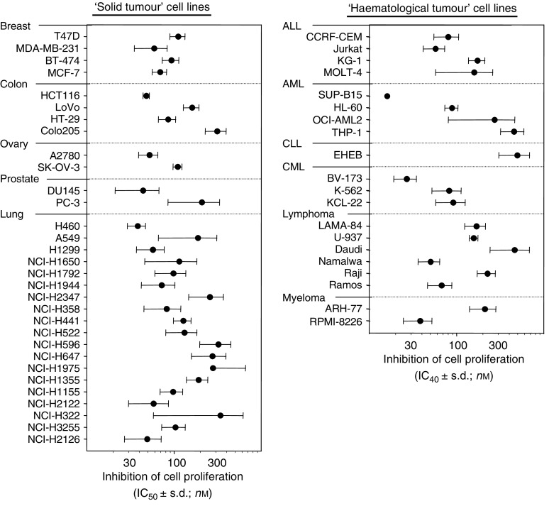
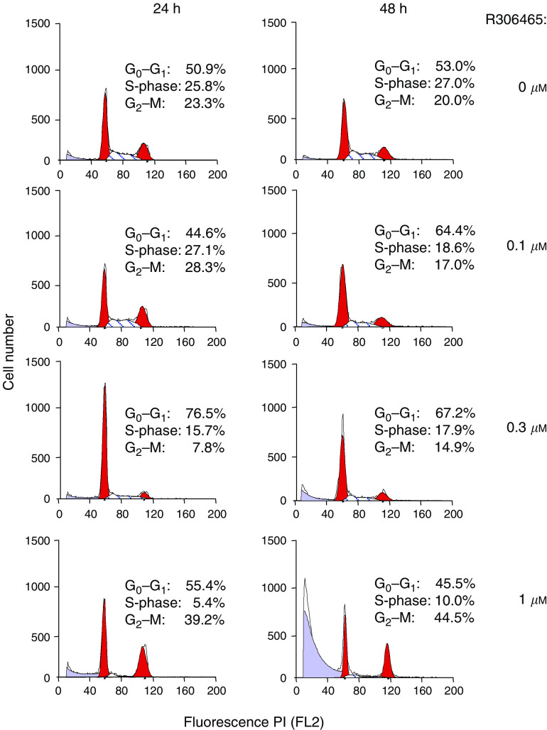
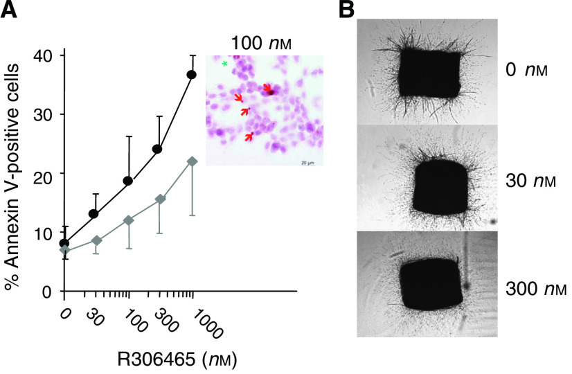
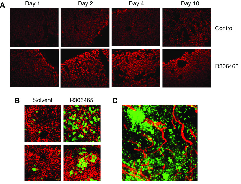
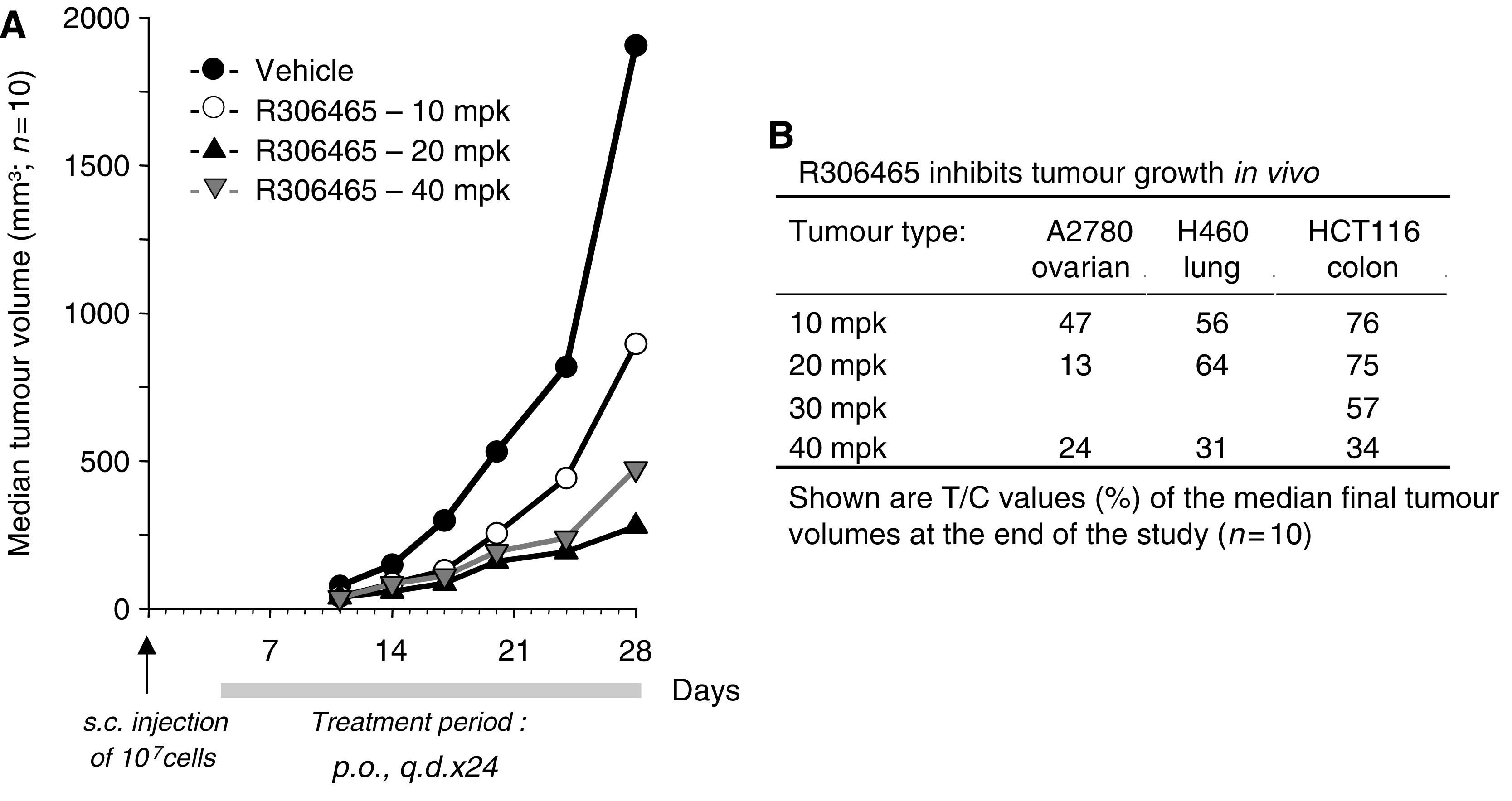
R306465 is a novel hydroxamate-based histone deacetylase (HDAC) inhibitor with broad-spectrum antitumour activity against solid and haematological malignancies in preclinical models. R306465 was found to be a potent inhibitor of HDAC1 and -8 (class I) in vitro. It rapidly induced histone 3 (H3) acetylation and strongly upregulated expression of p21waf1,cip1, a downstream component of HDAC1 signalling, in A2780 ovarian carcinoma cells. R306465 showed class I HDAC isotype selectivity as evidenced by poor inhibition of HDAC6 (class IIb) confirmed by the absence of downregulation of Hsp90 chaperone c-raf protein expression and tubulin acetylation. This distinguished it from other HDAC inhibitors currently in clinical development that were either more potent towards HDAC6 (e.g. vorinostat) or had a broader HDAC inhibition spectrum (e.g. panobinostat). R306465 potently inhibited cell proliferation of all main solid tumour indications, including ovarian, lung, colon, breast and prostate cancer cell lines, with IC50 values ranging from 30 to 300 nM. Haematological cell lines, including acute lymphoblastic leukaemia, acute myeloid leukaemia, chronic lymphoblastic leukaemia, chronic myeloid leukaemia, lymphoma and myeloma, were potently inhibited at a similar concentration range. R306465 induced apoptosis and inhibited angiogenesis in cell-based assays and had potent oral in vivo antitumoral activity in xenograft models. Once-daily oral administration of R306465 at well-tolerated doses inhibited the growth of A2780 ovarian, H460 lung and HCT116 colon carcinomas in immunodeficient mice. The high activity of R306465 in cell-based assays and in vivo after oral administration makes R306465 a promising novel antitumoral agent with potential applicability in a broad spectrum of human malignancies.


Figures

Figure 1
Figure 1
Chemical structure of R306465.
Figure 2
Figure 2
Substrate selectivity of R306465. Human A2780 ovarian carcinoma cells were incubated with the indicated concentrations of R306465, vorinostat, panobinostat, MS-275 or Trichostatin A (TSA) for 24 h. Total cell lysates were prepared and analysed by SDS–PAGE. Levels of acetylated H3 and tubulin, and of total levels of H3 and tubulin and of p21waf1, cip1 and Hsp70 and c-raf protein, were detected using rabbit polyclonal and mouse monoclonal antibodies, followed by ECL detection as indicated in the Methods section. To control for equal loading, blots were stripped and re-probed with antibodies against actin and the nuclear protein Lamin B1. A representative experiment out of three is shown.
Figure 3
Figure 3
Antiproliferative activity of R306465. Human tumour cell lines were seeded at low cell density and after 24 h cells were incubated with R306465 at 3 × 10⁻⁹, 10−8, 3 × 10⁻⁷, 10−6, 3 × 10⁻⁶ and 10−5M. The number of viable cells after a 4-day incubation period was assessed using a standard MTT colorimetric assay or Alamar Blue assay as indicated in the Methods section. Values represent mean±s.d. of three independent experiments. For haematological tumour cell lines, IC40 values (concentrations leading to 40% inhibition of cell proliferation) were determined for technical reasons: (ALL) acute lymphoblastic leukaemia; (AML) acute myeloid leukaemia; (CLL) chronic lymphoblastic leukaemia – EHEB (chronic B cell leukaemia); and (CML) chronic myeloid leukaemia.
Figure 4
Figure 4
R306465 affects cell-cycle distribution. Human A2780 ovarian carcinoma cells were incubated with the indicated concentrations of R306465 for 24 or 48 h. DNA content of nuclei was evaluated using propidium iodide staining followed by FACS analysis, and the number of cells in sub-G1, G1, S and G2M phase was calculated as a percentage of control. Results are expressed as % of total cells and a representative experiment out of three is shown.
Figure 5
Figure 5
R306465 induces apoptosis in human A2780 ovarian carcinoma cells and inhibits angiogenesis. (A) A2780 ovarian carcinoma cells were incubated with the indicated concentrations of R306465 for 48 h. Cells were stained with Annexin V (Pharmingen) and propidium iodide, and analysed by FACS. Apoptotic cells show Annexin V binding on the plasma membrane, but cells are intact and therefore do not stain for propidium iodide. Membrane-damaged cells were defined as both Annexin V- and propidium iodide-positive. Shown are mean values±variation for two independent experiments. Inset: cells incubated with R306465 (100 nM) were stained for DNA breaks using TUNEL dUTP labelling followed by peroxidase staining. Green asterisks represent mitotic cells, while red arrows point at specific DNA fragmentation. (B) Rat Aortic Ring Assays were performed as described in the Methods section. Aortic rings were incubated with either solvent or with the indicated concentrations of R306465 for 8 days and the average microvessel area for five independent rings was quantified.
Figure 6
Figure 6
R306465 induces histone acetylation and activates the p21waf1,cip1 promoter in vivo. Nude mice were injected s.c. with A2780 ovarian carcinoma cells and subsequently treated p.o. once daily with vehicle (20% HP-β-CD, control) or R306465 at 40 mpk (R306465). (A) Tumours were harvested 4 h after the last dose. Nuclei are stained with Hoechst, while (CY3) immunofluorescent labelling was performed to visualise acetylated H3. Representative sections from four independent mice are shown. (B) and (C) Tumours were collected after 24 h using transcardial perfusion fixation with 4% paraformaldehyde. (B) Bodipy 558/568-phalloidin staining was performed on whole mounts to visualise actin (background cell staining). (C) CD-31 (CY3) immunofluorescent labelling was performed on whole mounts to visualise the endothelium of blood vessels. Mounted samples were observed with the LSM510 laser scanning microscope.
Figure 7
Figure 7
R306465 inhibits tumour growth in vivo after oral administration. Nude mice were injected s.c. with A2780 ovarian, H460 lung or HCT116 colon carcinoma cells (107 cells per mouse) and subsequently treated p.o. with vehicle (control group, 20% HP-β-CD; •) or R306465 at either 10 (○), 20 (▴) or 40 mpk (▿). Mice were treated orally once daily (q.d.) between day 4 and 28 (A2780) or between day 4 and 32 (H460 and HCT116). (A) A2780 ovarian tumours were measured at least twice a week throughout the study, and results are represented as the median tumour size, expressed in mm3, of each individual group (n=10). (B) Mice were treated orally q.d. at the indicated doses. Results are represented as the treated/control values (%) of the median tumour size (n=10) at the end of the study.

References

    1. Arts J, De Schepper S, Van Emelen K (2003) Histone deacetylase inhibitors: from chromatin remodeling to experimental cancer therapeutics. Curr Med Chem 10(22): 2343–2350 - PubMed
    1. Bali P, Pranpat M, Bradner J, Balasis M, Fiskus W, Guo F, Rocha K, Kumaraswamy S, Boyapalle S, Atadja P, Seto E, Bhalla K (2005) Inhibition of histone deacetylase 6 acetylates and disrupts the chaperone function of heat shock protein 90. J Biol Chem 280: 26729–26734 - PubMed
    1. Belien A, De Schepper S, Floren W, Janssens B, Marien A, King P, Van Dun J, Andries L, Voeten J, Bijnens L, Janicot M, Arts J (2006) Real-time gene expression analysis in human xenografts for evaluation of histone deacetylase inhibitors. Mol Cancer Ther 5(9): 2317–2323 - PubMed
    1. Blagosklonny MV, Robey R, Sackett DL, Du L, Traganos F, Darzynkiewicz Z, Fojo T, Bates SE (2002) Histone deacetylase inhibitors all induce p21 but differentially cause tubulin acetylation, mitotic arrest, and cytotoxicity. Mol Cancer Ther 1: 937–941 - PubMed
    1. Butler LM, Agus DB, Scher HI, Higgins B, Rose A, Cordon-Cardo C, Thaler HT, Rifkind RA, Marks PA, Richon VM (2000) Suberoylanilide hydroxamic acid, an inhibitor of histone deacetylase, suppresses the growth of prostate cancer cells in vitro and in vivo. Cancer Res 60(18): 5165–5170 - PubMed

MeSH terms