. 2007 Nov 14;2(11):e1182.
doi: 10.1371/journal.pone.0001182.

RNA is an integral component of chromatin that contributes to its structural organization

Affiliations

RNA is an integral component of chromatin that contributes to its structural organization

Antonio Rodríguez-Campos et al. PLoS One. .

Abstract

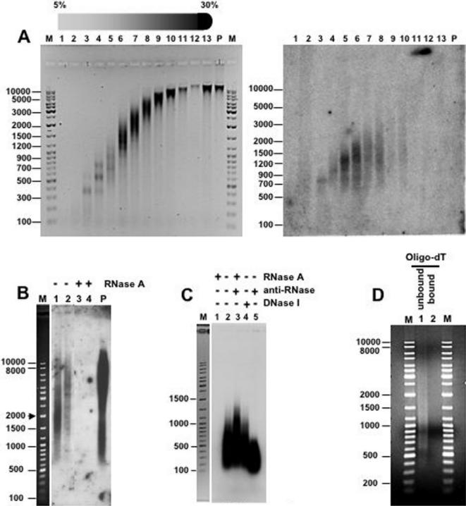
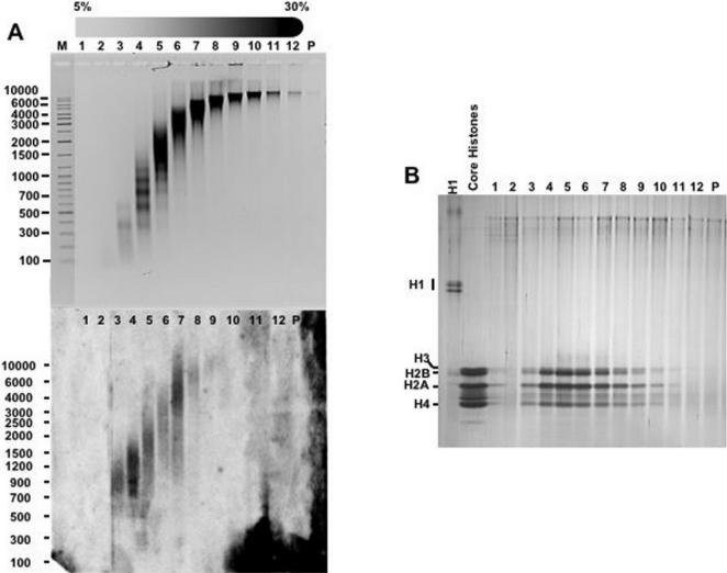
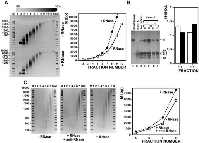
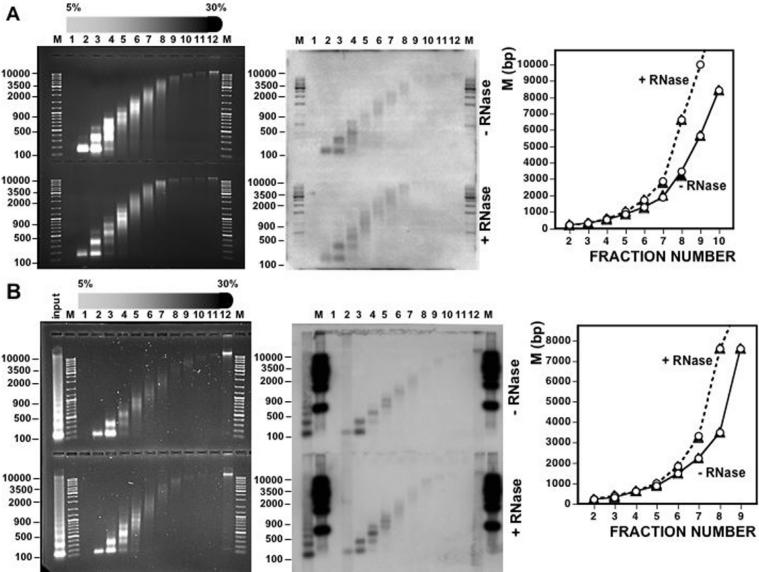
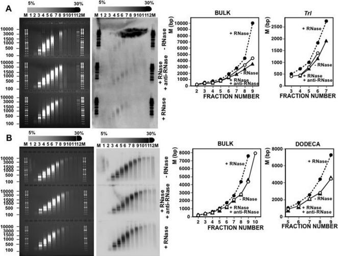
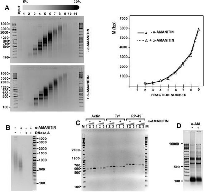
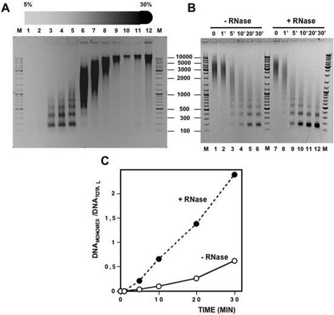
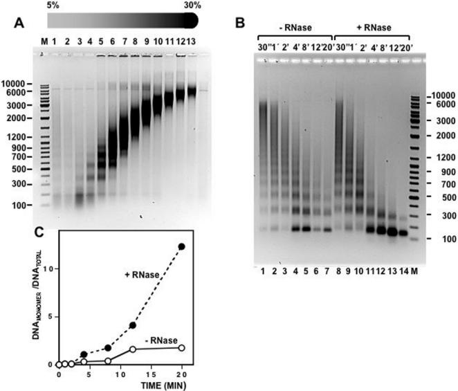
Chromatin structure is influenced by multiples factors, such as pH, temperature, nature and concentration of counterions, post-translational modifications of histones and binding of structural non-histone proteins. RNA is also known to contribute to the regulation of chromatin structure as chromatin-induced gene silencing was shown to depend on the RNAi machinery in S. pombe, plants and Drosophila. Moreover, both in Drosophila and mammals, dosage compensation requires the contribution of specific non-coding RNAs. However, whether RNA itself plays a direct structural role in chromatin is not known. Here, we report results that indicate a general structural role for RNA in eukaryotic chromatin. RNA is found associated to purified chromatin prepared from chicken liver, or cultured Drosophila S2 cells, and treatment with RNase A alters the structural properties of chromatin. Our results indicate that chromatin-associated RNAs, which account for 2%-5% of total chromatin-associated nucleic acids, are polyA(-) and show a size similar to that of the DNA contained in the corresponding chromatin fragments. Chromatin-associated RNA(s) are not likely to correspond to nascent transcripts as they are also found bound to chromatin when cells are treated with alpha-amanitin. After treatment with RNase A, chromatin fragments of molecular weight >3.000 bp of DNA showed reduced sedimentation through sucrose gradients and increased sensitivity to micrococcal nuclease digestion. This structural transition, which is observed both at euchromatic and heterochromatic regions, proceeds without loss of histone H1 or any significant change in core-histone composition and integrity.


Conflict of interest statement

Competing Interests: The authors have declared that no competing interests exist.

Figures

Figure 1
Figure 1. Purified chicken liver chromatin contains RNA.
A) Chicken liver chromatin was prepared by micrococcal nuclease digestion of purified nuclei and then subjected to sedimentation through a linear 5%–30% sucrose gradients. After centrifugation, 1ml fractions were collected, subjected to total nucleic acids extraction and analyzed by electrophoresis in 1% agarose-TBE gels (left panel). In parallel, chromatin fractions were subjected to RNA extraction using Ultraspec™ RNA Isolation System (Biotecx) and analyzed by Northern blotting in a glyoxal-1% agarose gel using high molecular weight genomic chicken DNA as a probe (right panel). Fraction numbers are indicated. Lanes M correspond to molecular weight markers. B) Chromatin fractions 7 and 10 of the gradient shown in A) were subjected to RNA extraction as indicated above, treated with RNase A (lanes 3 and 4) or not (lanes 1 and 2), and analyzed by Northern blotting as in A). Lane M, corresponds to molecular weight markers. Lane P, corresponds to the probe used. C) Chromatin from a mixture of fractions 7, 8 and 9 of the gradient shown in A) were subjected to RNA extraction and either untreated (lane 2), treated with RNase A in the absence (lane 1) or in the presence of anti-RNase (Ambion) (lane 3), or treated with DNase I (Roche) (lane 4). After phenol extraction and isopropanol precipitation, samples were 32P-labeled by reverse transcription with Omniscript® RT Kit (Qiagen) (2 h at 37°C) using a mixture of hexanucleotides of random sequence. Samples were then analyzed in a 1% agarose-TBE gel, blotted and the membrane directly exposed. Lane M, corresponds to molecular weight markers. D) Chromatin-associated RNA was purified and incubated with oligo-dT immobilized resin (Oligotex™ mRNA Purification System, QIAGEN). After elution, bound (lane 2) and unbound material (lane1) were analyzed in a glyoxal-1% agarose, 10 mM sodium phosphate (pH 6,8) gel. Lanes M correspond to molecular weight markers.
Figure 2
Figure 2. RNA remains associated to chromatin at high ionic strength.
A) Chicken liver chromatin was prepared by micrococcal nuclease digestion of purified nuclei and then subjected to sedimentation through a linear 5%–30% sucrose gradients containing 0,65 M NaCl. After centrifugation, fractions were collected and their DNA (top) and RNA (bottom) content determined as in Figure 1. Fraction numbers are indicated. Lane M, corresponds to molecular weight markers. B) The histone content of each fraction was analyzed by SDS-PAGE (lanes 1-P). As controls, H1 from calf thymus and hydroxylapatite-purified core histones from chicken liver are also presented. The gel was stained with silver.
Figure 3
Figure 3. Treatment with RNase A alters the sedimentation behavior of purified chicken liver chromatin.
A) Chicken liver chromatin, prepared by micrococcal nuclease digestion of purified nuclei, was subjected to sedimentation through a linear 5%–30% sucrose gradient after treatment with RNase A (bottom panel) or not (top panel). After centrifugation and fractionation, samples were subjected to total nucleic acids extraction and analyzed by electrophoresis in 1% agarose-TBE gels. Fraction numbers are indicated. Lanes M correspond to molecular weight markers. Quantitation of the results is shown on the right where the average molecular weight (M) of the chromatin fragments contained in each fraction, expressed as bp of DNA, is presented as a function of the fraction number: (°) untreated chromatin, (•) chromatin treated with RNase A. B) Histone content of chromatin fractions 11 and 12 of the gradients shown in A) was analyzed by SDS-PAGE: untreated chromatin (lanes 4 and 5), chromatin treated with RNase A (lanes 6 and 7) and, as controls, H1 from calf thymus (lane 1), RNase A (lane 2) and hydroxylapatite-purified core histones from chicken liver (lane 3). The gel was stained with silver. Quantitation of the results is shown on the right where the H1/H2A ratio of fractions 11 and 12 is presented before (white columns) and after (black columns) treatment with RNase A. C) The sedimentation behavior of chicken liver chromatin was determined before (left panel) and after treatment RNase A either in the presence of anti-RNase (Ambion) (central panel) or in the absence of any added inhibitor (right panel). Fraction numbers are indicated. Lanes M, correspond to molecular weight markers. Quantitation of the results is shown on the right: (°) untreated chromatin, (▴) chromatin treated with RNase A in the presence of anti-RNase, (•) chromatin treated with RNase A in the absence of any added inhibitor.
Figure 4
Figure 4. Treatment with RNase A alters the sedimentation behavior of chicken liver chromatin at specific genomic regions.
The effect of treatment with RNase A on the sedimentation behavior of chromatin was determined for bulk chicken liver chromatin (left panels) and for chromatin at specific genomic locations (right panels) by Northern analysis of the gels on the left using specific probes for the Pax3 locus A) and the chicken 41–42 bp centromeric satellite B). Fraction numbers are indicated. Lanes M, correspond to molecular weight markers. Quantitation of the results is shown on the right of each panel: (°) bulk chromatin, (▴) chromatin of the Pax3 locus A) or the chicken 41–42 bp centromeric satellite B).
Figure 5
Figure 5. Treatment with RNase A alters the sedimentation behavior of purified Drosophila S2 chromatin.
The effect of treatment with RNase A on the sedimentation behavior of chromatin prepared from Drosophila S2 cells was determined for bulk chromatin (left panels) and for chromatin at specific genomic locations (right panels) by Southern analysis of the gels on the left using specific probes for the Trl locus A) and the centromeric Drosophila dodeca-satellite B). Fraction numbers are indicated. Lanes M, correspond to molecular weight markers. Quantitation of the results is shown on the right of each panel for bulk chromatin and chromatin of the Trl locus A) or the centromeric dodeca-satellite B): untreated chromatin (°); chromatin treated with RNase A in the presence of anti-RNase (Ambion) (▴) or in the absence of any added inhibitor (•).
Figure 6
Figure 6. Treatment with α-amanitin does not alter the sedimentation behavior of purified Drosophila S2 chromatin.
Prior to nuclei isolation and chromatin purification, S2 cells were either treated for 36 h with 0,2 µg/ml α-amanitin or not. A) S2 chromatin from untreated (top) and treated cells (bottom) was purified by micrococcal nuclease digestion of purified nuclei and subjected to sedimentation through linear 5%–30% sucrose gradients as described above. Quantitation of the results is shown on the right where the average molecular weight (M) of the chromatin fragments contained in each fraction, expressed as bp of DNA, is presented as a function of the fraction number for chromatin prepared from untreated (▴) and treated cells (Δ). B) Chromatin fractions 4, 5 and 6 of the gradients shown in A) obtained from cells treated with α-amanitin (lanes 2 and 4) or not (lanes 1 and 3), were subjected to RNA extraction and analised in a glyoxal-1% agarose-sodium phosphate gel before (lanes 1 and 2) and after treatment with RNase A (lanes 3 and 4). Lane M corresponds to molecular weight markers. C and D) Analysis of the efficiency of treatment with α-amanitin. D) Total RNA was prepared from treated (lane 1) or untreated cells (lanes 2), and 5 µg of each were analysed in a 1% agarose-TBE native gel. Lane M corresponds to molecular weight markers. C) Determination of the levels of nascent transcripts encoding GAGA (Trl), actin 5C and RP-49. Increasing amounts of total RNA (0,1, 0,2 and 0,5 µg, lanes 1-3), prepared from untreated (lanes -) and treated cells (lanes +), were analysed by RT-PCR (Omniscript® RT Kit, QIAGEN) as indicated under Materials and Methods using appropriate primers to specifically amplify fragments of the Actin 5C (585 bp), Trl (662 bp) and RP-49 (702 bp) genes. Amplified fragments were analysed in a 1% agarose-TBE gel. Lanes M correspond to molecular weight markers.
Figure 7
Figure 7. Treatment with RNase A increases the sensitivity of chicken liver chromatin to cleavage by micrococcal nuclease.
A) Preparative 5%–30% linear sucrose gradient of chicken liver chromatin. Fraction numbers are indicated. Lanes M, correspond to molecular weight markers. B) Fraction 8 of the gradient shown in A) was treated with RNase A (lanes 7–12) or not (lanes 1–6) and, then, digested at 37°C with 0.4 units of micrococcal nuclease (Sigma) at increasing times as indicated. After micrococcal nuclease digestion, samples were deproteinized and analyzed by electrophoresis in 1% agarose-TBE gels. Lanes M correspond to molecular weight markers. C) Quantitation of the results shown in B). The ratio of mononucleosomal DNA versus total DNA is presented as a function of the digestion time for untreated chromatin (°) and chromatin treated with RNase A (•).
Figure 8
Figure 8. Treatment with RNase A increases the sensitivity of purified Drosophila S2 chromatin to cleavage by micrococcal nuclease.
A) Preparative 5%–30% linear sucrose gradient of chromatin from Drosophila S2 cells. Fraction numbers are indicated. Lane M corresponds to molecular weight markers. B) Fraction 13 of the gradient shown in A) was treated RNase A (lanes 8 to 14) or not (lanes 1–7), and, then, digested at 37°C with 0,4 units of micrococcal nuclease (Sigma) at increasing times as indicated. After micrococcal nuclease digestion, samples were deproteinized and analyzed by electrophoresis in 1% agarose-TBE gels. Lane M corresponds to molecular weight markers. C) Quantitation of the results shown in B). The ratio of mononucleosomal DNA versus total DNA is presented as a function of the digestion time for untreated chromatin (°) and chromatin treated with RNase A (•).

References

    1. Kornberg RD. Structure of chromatin. Annu Rev Biochem. 1977;46:931–954. - PubMed
    1. McGhee JD, Felsenfeld G. Nucleosome structure. Annu Rev Biochem. 1980;49:1115–1156. - PubMed
    1. Finch T, Lutter LC, Rhodes D, Brown AS, Rushton B, et al. Structure of nucleosome core particle. Nature (London) 1977;269:29–36. - PubMed
    1. Richmond TJ, Finch JT, Rushton B, Rhodes D, Klug A. Structure of nucleosome core particle at 7Å resolution. Nature (London) 1984;311:532–537. - PubMed
    1. Luger K, Mäder AW, Richmond RK, Sargent DF, Richmond TJ. Crystal structure of the nucleoosme core particle at 2,8Å resolution. Nature. 1997;389:251–260. - PubMed