Case Reports
. 2007 Dec 15;143A(24):2981-3008.
doi: 10.1002/ajmg.a.32040.

Neuroimaging findings in macrocephaly-capillary malformation: a longitudinal study of 17 patients

Affiliations
Case Reports

Neuroimaging findings in macrocephaly-capillary malformation: a longitudinal study of 17 patients

Robert L Conway et al. Am J Med Genet A. .

Abstract

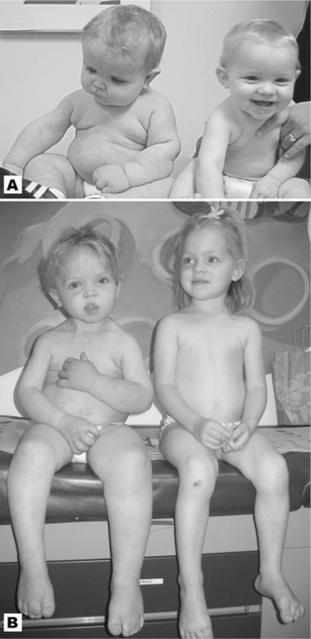
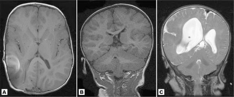
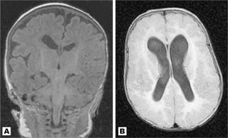
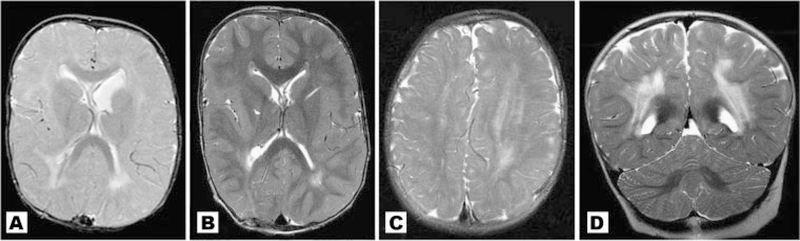
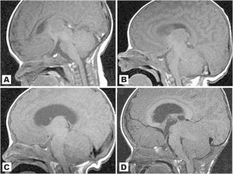
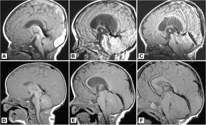
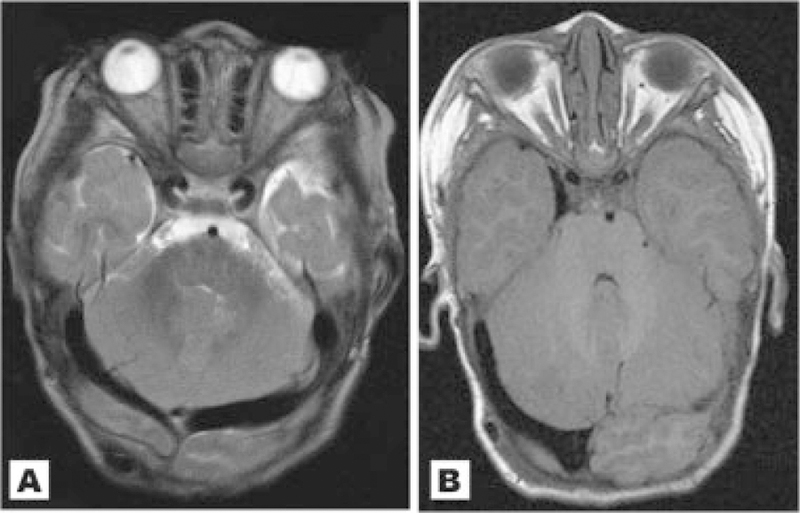
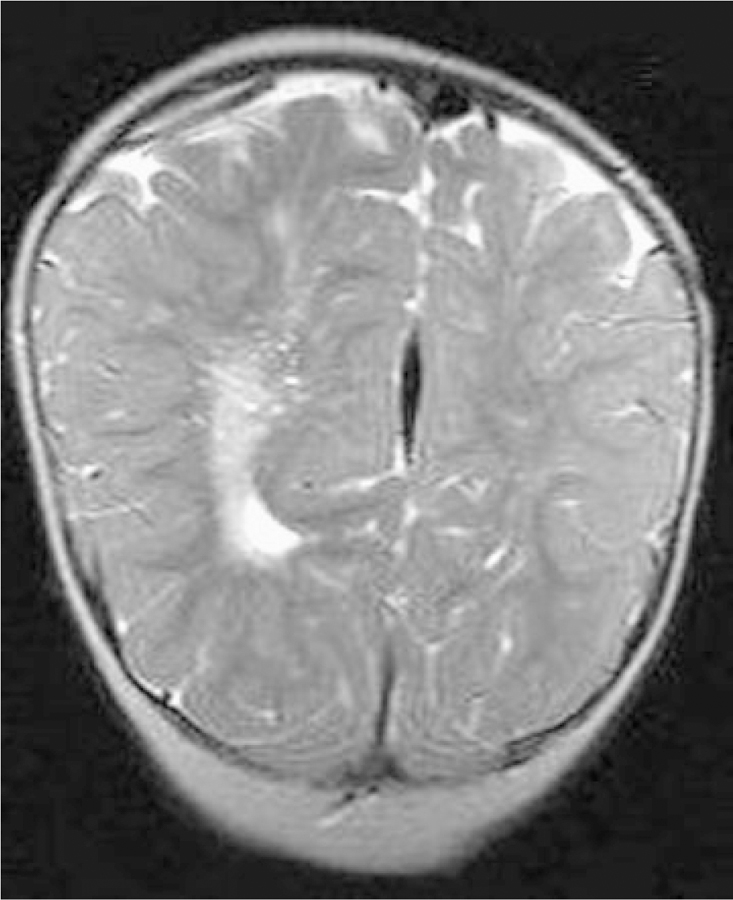
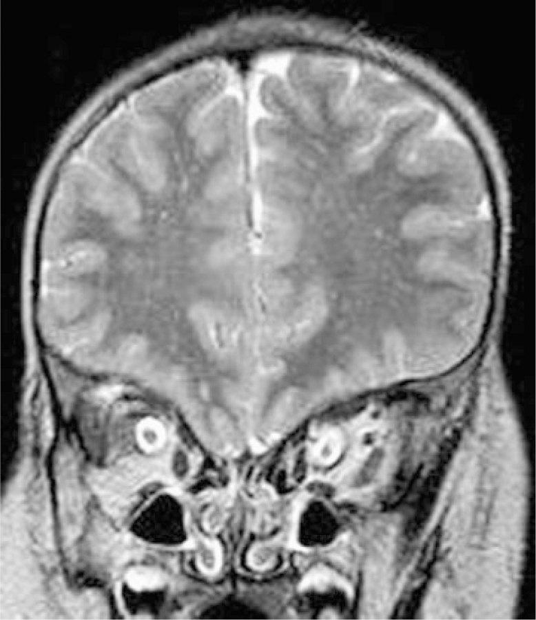
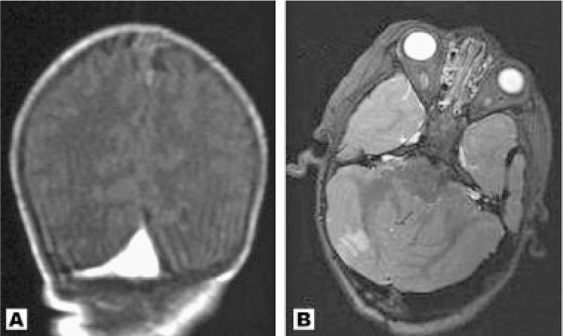
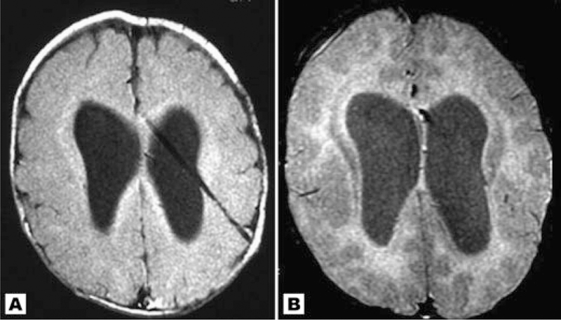
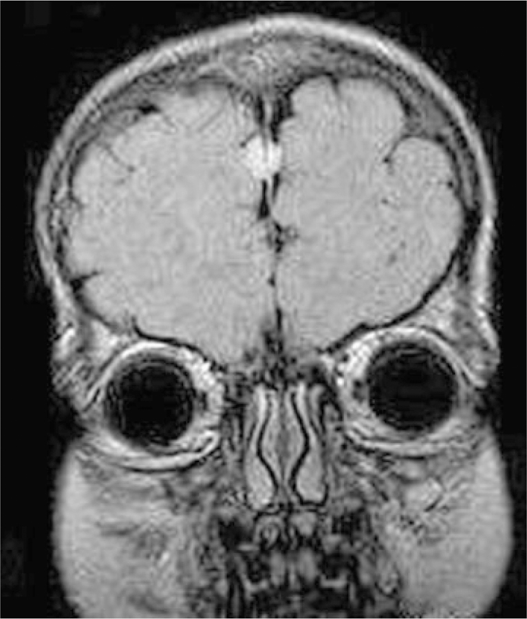
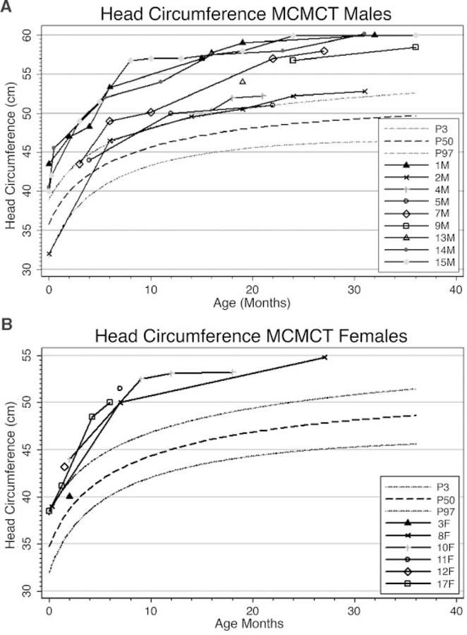
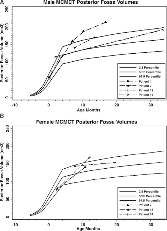
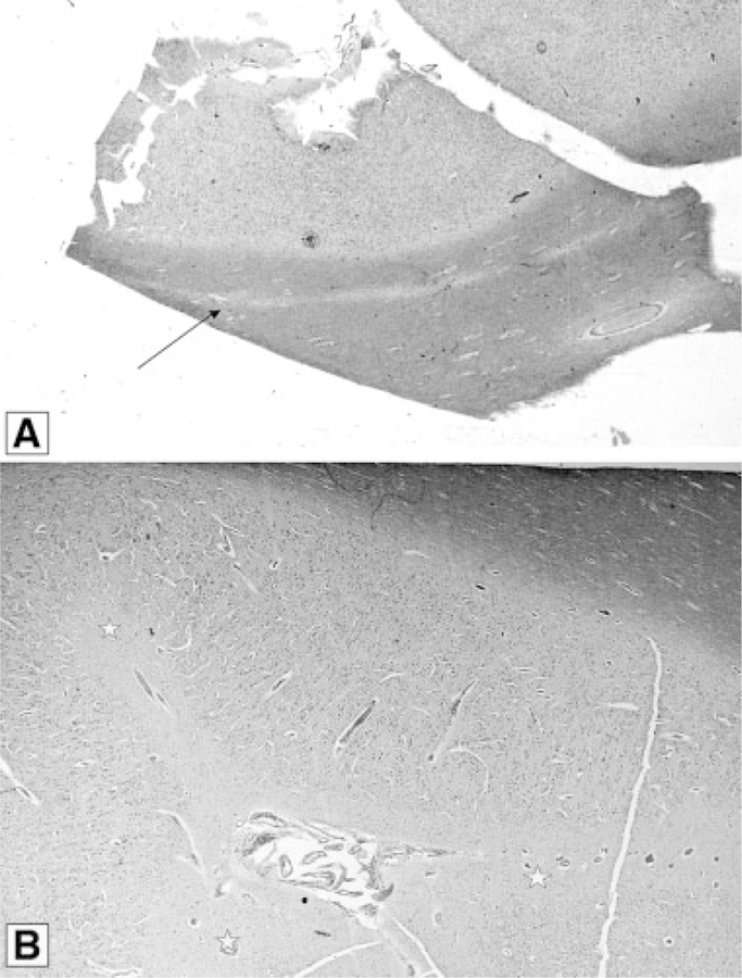
Here, we report the neuroimaging findings and neurological changes in 17 unpublished patients with Macrocephaly-Capillary Malformation (M-CM). This syndrome has been traditionally known as Macrocephaly-Cutis Marmorata Telangiectatica Congenita (M-CMTC), but we explain why M-CM is a more accurate term for this overgrowth syndrome. We analyzed the 17 patients with available brain MRI or CT scans and compared their findings with features identified by a comprehensive review of published cases. White matter irregularities with increased signal on T2-weighted images were commonly observed findings. A distinctive feature in more than half the patients was cerebellar tonsillar herniation associated with rapid brain growth and progressive crowding of the posterior fossa during infancy. In four such cases, we confirmed that the tonsillar herniation was an acquired event. Concurrently, with the development of these findings, ventriculomegaly (frequently obstructive) and dilated dural venous sinuses were observed in conjunction with prominent Virchow-Robin spaces in many of those in whom cerebellar tonsil herniation had developed. We postulate that this constellation of unusual features suggests a dynamic process of mechanical compromise in the posterior fossa, perhaps initiated by a rapidly growing cerebellum, which leads to congestion of the venous drainage with subsequently compromised cerebrospinal fluid reabsorption, all of which increases the posterior fossa pressure and leads to acquired tonsillar herniation. We make a distinction between congenital Chiari I malformation and acquired cerebellar tonsil herniation in this syndrome. We also observed numerous examples of abnormal cortical morphogenesis, including focal cortical dysplasia, polymicrogyria which primarily involved the perisylvian and insular regions, and cerebral and/or cerebellar asymmetric overgrowth. Other findings included a high frequency of cavum septum pellucidum or vergae, thickened corpus callosum, prominent optic nerve sheaths and a single case of venous sinus thrombosis. One patient was found to have a frontal perifalcine mass resembling a meningioma at age 5 years. This is the second apparent occurrence of this specific tumor in M-CM.


Figures

F<sc>ig</sc>. 1.
Fig. 1.
Patient 5 (left) and his unaffected twin sister at ages 12 months (A) and 40 months (B). The boy has classic features of M–CM including left sided hemihyperplasia, typical facial features, toe syndactyly, and connective tissue involvement with loose, redundant skin. [Color figure can be viewed in the online issue, which is available at www.interscience.wiley.com.]
F<sc>ig</sc>. 2.
Fig. 2.
A: Study of patient 1 at 27 months showing mild cerebral asymmetry with focal enlargement of the occipital-temporal region, which was less noticeable when younger. The bright spot on the right hemisphere represents artifact from a shunt. B: Coronal MRI of Patient 5 showing moderate cerebral and cerebellar asymmetry. This patient also has total left-sided hyperplasia from the midline. C: T2 weighted coronal image of patient 17 showing marked right sided brain asymmetry similar to true hemimegalencephaly. The enlarged hemisphere shows an abnormal gyral pattern with a deep sylvian fissure, perisylvian polymicrogyria and focal cortical dysplasia resembling pachygyria.
F<sc>ig</sc>. 3.
Fig. 3.
A: Coronal T1 MRI of Patient 12 at 2 months with right sided incomplete opercularization and perisylvian polymicrogyria with cortical irregularity. In addition, there is ventricular asymmetry. B: Axial image of Patient 14 demonstrating extensive cortical dysplasia with thick and irregular cortex in the bilateral frontal and parietal regions. This patient suffers from severe mental retardation and a partially refractive seizure disorder. Cavum septum pellucidum with mild cavum vergae are also seen.
F<sc>ig</sc>. 4.
Fig. 4.
A,B: Patient 1 at different ages. A: 9 months with demonstration of increased T2 weighted signal in deep and periventricular white matter; (B) 22 months with improved but persistent white matter abnormalities in same distribution; (C) Patient 5. The asymmetrically enlarged left hemisphere demonstrates more prominent white matter signal changes than the right; (D) Patient 3 with dramatic bilateral white matter signal abnormalities. A diagnosis of leukodystrophy was initially considered, but the patient was neurologically stable and remained mildly and statically delayed.
F<sc>ig</sc>. 5.
Fig. 5.
A–D: Sagittal images of patient 14 at different ages: (A) At 8 days old there is a normal cerebellum, a noncrowded posterior fossa, and no CTH; (B) At approximately 1 month there is now early growth of the cerebellum. The brainstem and midbrain appears to be mildly compressed anteriorly; (C) The cerebellum appears bulkier with crowded posterior fossa and further anterior compression of the brainstem. There is now mild to moderate ventriculomegaly and mild CTH; (D) At age 15 months, after posterior fossa decompression, the cerebellum has continued to grow and there is further crowding of the posterior fossa and compression of the brainstem, which now appears kinked at the foramen magnum. There is clear worsening of the CTH.
F<sc>ig</sc>. 6.
Fig. 6.
A–C: Sagittal images of Patient 11 at different ages showing acquired cerebellar tonsil herniation; (A) at 4 weeks. The cerebellum appears normal without posterior fossa crowding. A sinus thrombosis is present; (B) at 3 months, there appears to be interval enlargement of the cerebellum with a new finding of ventriculomegaly. The brainstem appears anteriorly deviated but motion artifact limits the study; (C) at 5.5 months, the cerebellum appears further enlarged. There is now inferior displacement of the cerebellar tonsils and herniation through the foramen magnum. The cerebellum is pushing anteriorly on the brainstem and midbrain, and the cerebellum appears to ramp up toward the tentorium and now rises as high as the superior margin of the midbrain. D–F: Sagittal scans of Patient 1 taken at different ages: (D) at 2 days there is a normal-sized cerebellum and no CTH. The brainstem is not compressed anteriorly; (E) by 6 months there is interval rapid growth of the cerebellum with new ventriculomegaly and early CTH. The straight sinus is enlarged; (F) after ventricular shunting the ventriculomegaly has resolved but the posterior fossa remains crowded by a large cerebellum. The CTH is still present.
F<sc>ig</sc>. 7.
Fig. 7.
A: Patient 14 at 22 months. Both transverse sinuses are dilated. B: Axial image through the posterior fossa of Patient 1 at 22 months showing a unilaterally enlarged right transverse venous sinus.
F<sc>ig</sc>. 8.
Fig. 8.
T2 weighted coronal image through the occipital region of Patient 12. Prominent Virchow–Robin spaces are more apparent in the ipsilateral enlarged cerebral hemisphere.
F<sc>ig</sc>. 9.
Fig. 9.
Coronal image of Patient 7 at approximately 3 years. Bilateral optic nerve sheaths are abnormally thick.
F<sc>ig</sc>. 10.
Fig. 10.
(A) Coronal image of Patient 11 taken at 4 weeks. There is a venous thrombosis at the junction of the transverse and sagittal sinuses; (B) Axial image of Patient 13 at 33 months showing an unexpected and asymptomatic right posterior inferior cerebellar artery infarction. There is also dilation of the transverse sinus.
F<sc>ig</sc>. 11.
Fig. 11.
Axial images of patient 11 at different ages. A: Age 1 month with normal gray-white matter differentiation for age; (B) age 5.5 months with appearance of possible band heterotopia.
F<sc>ig</sc>. 12.
Fig. 12.
Coronal scan of Patient 14 at age 5 years demonstrating a frontal perifalcine mass consistent with meningioma.
F<sc>ig</sc>. 13.
Fig. 13.
Head circumference curves for male (A) and female (B) cohort patients from age 0 to 36 months plotted against the normal standards. The graphs show the typical tendency for head circumference to cross centiles during infancy and early toddler years in this syndrome.
F<sc>ig</sc>. 14.
Fig. 14.
Graphic plots of digitally rendered posterior fossa volumes in 4 male (A) and three female (B) patients with M–CMTC compared to published normative data. The posterior fossa volumes tend to cross centiles over time in most patients.
F<sc>ig</sc>. 15.
Fig. 15.
Patient 11 autopsy findings. A: The arrow indicates a thin lamina of heterotopic grey matter within the white matter and well separated from overlying cortex (luxol fast bluecresyl violet stain). B: Abortive sulci (stars) with median vessels suggesting gyral fusion in polymicrogyric cerebral cortex. These fingers of molecular layer point perpendicularly away from the surface.

References

    1. Abeloff MD, Armitage JO, Niederhuber JE, Kastan MB, Gillies McKenna W. 2004. Clinical oncology. 3rd edition. New York: Churchill Livingstone.
    1. Achiron A, Faibel M. 2002. Sandlike appearance of Virchow-Robin spaces in early multiple sclerosis: A novel neuroradiologic marker. Am J Neuroradiol 23:376–380. - PMC - PubMed
    1. Ackar N, Adapinar B, Dinleyici C, Durak B, Ozkan IR. 2004. A case of macrocephaly-cutis marmorata telangiectatica congenita and review of neuroradiologic features. Ann Genet 47:261–265. - PubMed
    1. AnalyzeDirect. Analyze Versions 6.0 and 7.0.
    1. Artigas J, Poo P, Rovira A, Cardo E. 1999. Macrocephaly and dilated Virchow-Robin spaces in childhood. Pediatr Radiol 29:188–190. - PubMed

Publication types