. 2008 Jan;19(1):315-21.
doi: 10.1021/bc700196q. Epub 2007 Nov 15.

Ligand-modified aminobisphosphonate for linking proteins to hydroxyapatite and bone surface

Affiliations

Ligand-modified aminobisphosphonate for linking proteins to hydroxyapatite and bone surface

Robin S Ehrick et al. Bioconjug Chem. 2008 Jan.

Abstract

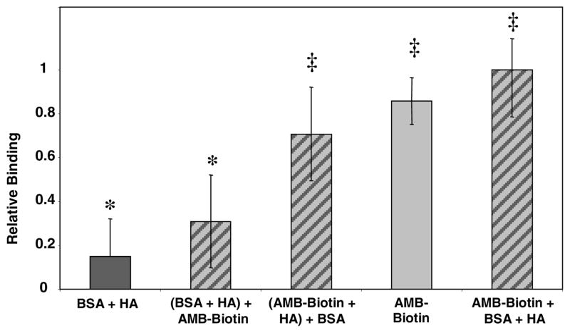
An increase in bone resorption is one of the main symptoms of osteoporosis, a disease that affects more and more individuals every day. Bisphosphonates are known to inhibit bone resorption and thus are being used as a treatment for osteoporosis. Aminobisphosphonates present a functionality that can be easily used for conjugation to other molecules, such as peptides, proteins, and ligands for protein recognition. In this study, an aminobisphosphonate conjugated with biotin was used as a model linker for protein attachment to bone. With this system, the interaction of biotinylated aminobisphosphonate with hydroxyapatite, a major mineral component of bone, was investigated. Quantification of the binding of aminobisphosphonate to hydroxyapatite was performed using a fluorescently labeled antibody for biotin. Additionally, the interaction of the biotinylated aminobisphosphonate with multiple treatments of cortical bone from the midshaft of a cow femur was studied. It was demonstrated that modified aminobisphosphonate reagents can bind hydroxyapatite and bone at high levels, while the biotin functionality is free to be recognized by the fluorescently labeled antibiotin antibody, suggesting that modified aminobisphosphonates could be used to link other peptides or proteins to the bone surface.


Figures

Figure 1
Figure 1
Binding of FITC-labeled monoclonal anti-biotin antibody to hydroxyapatite treated with AMB-Biotin and various controls. (A) Fluorescence signal of the supernatants (λex 490 nm) after removal of insoluble HA treated with AMB (•••) and AMB-Biotin (— • —), and untreated HA (— — —) compared to the spectrum of the FITC-labeled anti-biotin antibody solution used in the experiments (solid line). (B) Relative binding for untreated HA (n = 5), AMB treated HA (n = 6), and AMB-Biotin treated HA (n = 6). The * denotes statistically similar results. AMB-Biotin is statistically different from the AMB-treated and untreated HA samples at p > 0.01.
Figure 2
Figure 2
Binding measurements in the presence of free biotin quantified using a fluorescently labeled anti-biotin antibody. Relative binding for untreated HA, free biotin treated HA, AMB-Biotin and free biotin incubated together with HA, and HA treated with AMB-Biotin. For all samples, n equals three. The symbols * and † denote statistically similar results. There is no statistical difference between the Untreated HA, Free Biotin, and AMB-Biotin + Free Biotin + HA samples, nor is there a difference between the AMB-Biotin + Free Biotin + HA and AMB-Biotin samples. However, there is a statistical difference (p > 0.01 for each sample) between the Untreated HA, Free Biotin + HA, and the AMB-Biotin +HA samples.
Figure 3
Figure 3
Binding measurements of AMB-Biotin in the presence of BSA based on the loss in fluorescence of the supernatants after incubation with a FITC-labeled anti-biotin antibody. Relative binding for BSA, BSA preincubated with HA, AMB-Biotin preincubated with HA, AMB-Biotin alone on HA, and AMB-Biotin and BSA incubated together with HA. All samples were prepared in triplicate. The symbols * and ‡ denote statistically similar samples. The statistically significant difference between the samples denoted with the symbol * and those with the symbol ‡ was at least p > 0.05.
Figure 4
Figure 4
Binding measurements of AMB-Biotin and free biotin on multiple treatments of bone. The three treatments studied were untreated bone (A), demineralized bone (B), and ashed bone (C). Binding was quantified based on the binding of a fluorescently labeled anti-biotin antibody. For all samples, n equals three. The symbol * denotes statistically similar results. There is no statistical difference between the free biotin and AMB-Biotin samples on untreated and ashed bone. There is only a statistically significant (p > 0.025) difference between the AMB-Biotin and Free Biotin samples on Demineralized Bone.
Figure 5
Figure 5
Binding measurements of AMB-Biotin in the presence of casein. Binding was measured using FITC-labeled anti-biotin antibody. Relative binding on ashed bone of casein, casein and AMB-Biotin, AMB-Biotin preincubated with bone, and AMB-Biotin. Samples were prepared in triplicate. The symbol * denotes statistically similar samples. There is no statistical difference between the Casein + AMB-Biotin + Ash, (AMB-Biotin + Ash) + Casein, and AMB-Biotin + Ash samples. All are statistically different from the Casein + Ash sample by at least p > 0.05.
Scheme 1
Scheme 1
Synthesis of biotinyl-aminomethylenediphosphonic acid triethylammonium salt (AMB-Biotin).

Similar articles

Cited by

References

    1. Kiberstis P, Smith O, Norman C. Bone health in the balance. Science. 2000;289:1497.
    1. Ducy P, Schinke T, Karsenty G. The osteoblast: A sophisticated fibroblast under central surveillance. Science. 2000;289:1501–1504. - PubMed
    1. Teitelbaum SL. Bone Resorption by Osteoclasts. Science. 2000;289:1504–1508. - PubMed
    1. Rodan GA, Martin TJ. Therapeutic approaches to bone diseases. Science. 2000;289:1508–1514. - PubMed
    1. Finkelstein JS. Calcium plus vitamin D for postmenopausal women -bone appetit? N Engl J Med. 2006;354:750–751. - PubMed

Publication types