Review
. 2007 Nov 14:2:45.
doi: 10.1186/1750-1172-2-45.

Arrhythmogenic right ventricular cardiomyopathy/dysplasia

Affiliations
Review

Arrhythmogenic right ventricular cardiomyopathy/dysplasia

Gaetano Thiene et al. Orphanet J Rare Dis. .

Abstract

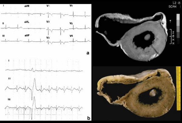
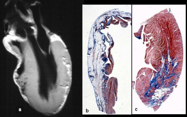
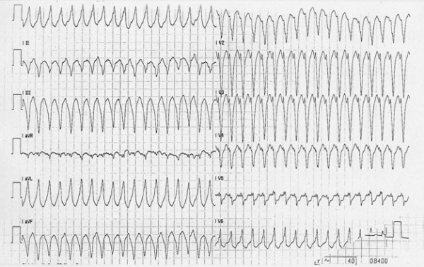
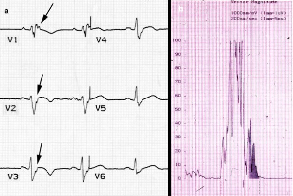
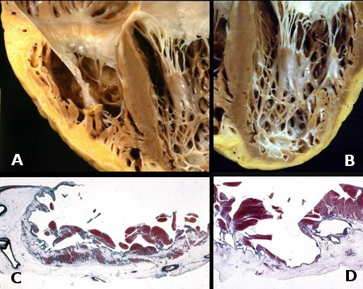
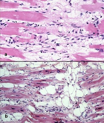
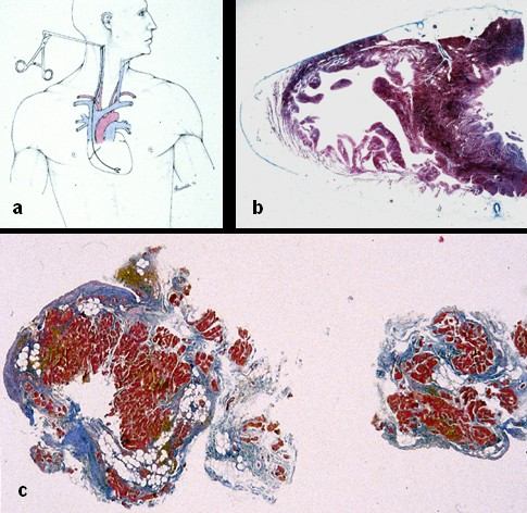
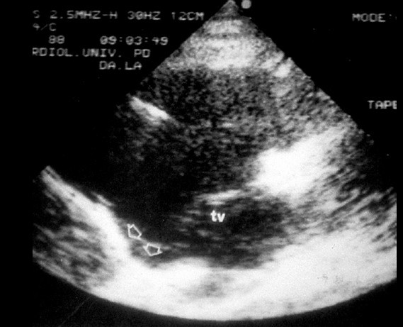
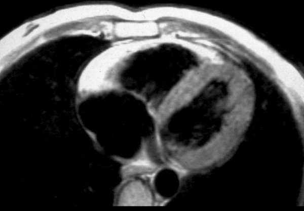
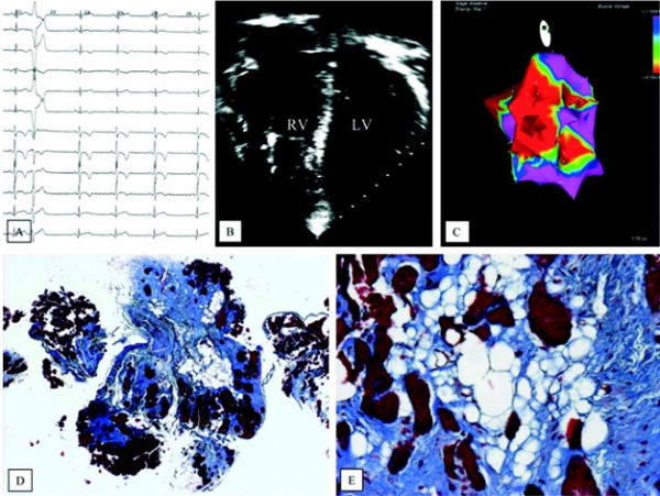
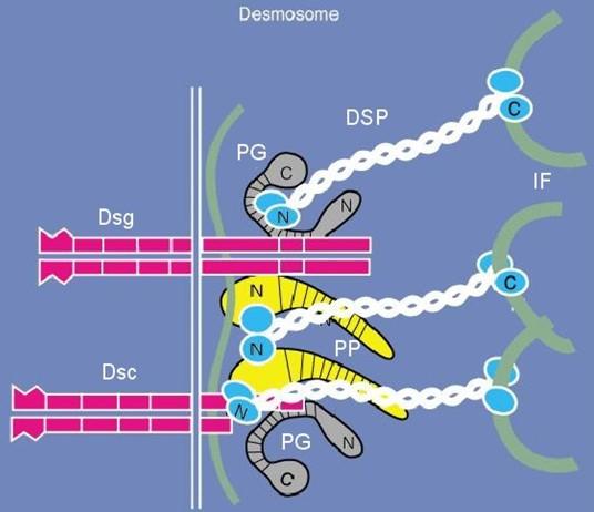
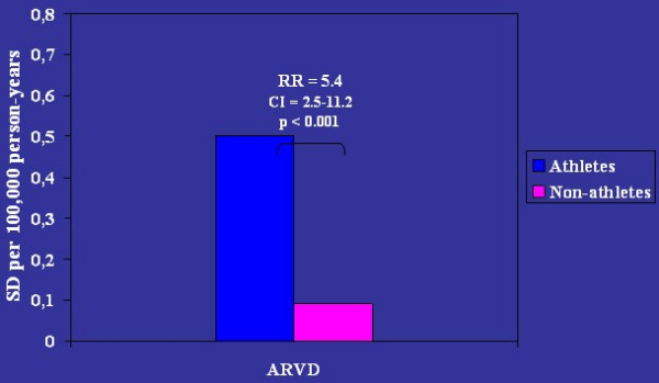
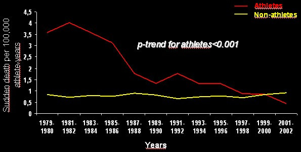
Arrhythmogenic right ventricular cardiomyopathy/dysplasia (ARVC/D) is a heart muscle disease clinically characterized by life-threatening ventricular arrhythmias. Its prevalence has been estimated to vary from 1:2,500 to 1:5,000. ARVC/D is a major cause of sudden death in the young and athletes. The pathology consists of a genetically determined dystrophy of the right ventricular myocardium with fibro-fatty replacement to such an extent that it leads to right ventricular aneurysms. The clinical picture may include: a subclinical phase without symptoms and with ventricular fibrillation being the first presentation; an electrical disorder with palpitations and syncope, due to tachyarrhythmias of right ventricular origin; right ventricular or biventricular pump failure, so severe as to require transplantation. The causative genes encode proteins of mechanical cell junctions (plakoglobin, plakophilin, desmoglein, desmocollin, desmoplakin) and account for intercalated disk remodeling. Familiar occurrence with an autosomal dominant pattern of inheritance and variable penetrance has been proven. Recessive variants associated with palmoplantar keratoderma and woolly hair have been also reported. Clinical diagnosis may be achieved by demonstrating functional and structural alterations of the right ventricle, depolarization and repolarization abnormalities, arrhythmias with the left bundle branch block morphology and fibro-fatty replacement through endomyocardial biopsy. Two dimensional echo, angiography and magnetic resonance are the imaging tools for visualizing structural-functional abnormalities. Electroanatomic mapping is able to detect areas of low voltage corresponding to myocardial atrophy with fibro-fatty replacement. The main differential diagnoses are idiopathic right ventricular outflow tract tachycardia, myocarditis, dialted cardiomyopathy and sarcoidosis. Only palliative therapy is available and consists of antiarrhythmic drugs, catheter ablation and implantable cardioverter defibrillator. Young age, family history of juvenile sudden death, QRS dispersion > or = 40 ms, T-wave inversion, left ventricular involvement, ventricular tachycardia, syncope and previous cardiac arrest are the major risk factors for adverse prognosis. Preparticipation screening for sport eligibility has been proven to be effective in detecting asymptomatic patients and sport disqualification has been life-saving, substantially declining sudden death in young athletes.


Figures

Figure 1
Figure 1
Graphic showing the various causes of juvenile sudden cardiac death in Northeast Italy. ARVC/D ranks second (13%) after atherosclerotic coronary artery disease. ARVC = arrhythmogenic right ventricular cardiomyopathy; ATS CAD = atherosclerotic coronary artery disease; DMC = dilated cardiomyopathy; HCM = hypertrophic cardiomyopathy; NonATS CAD = non-atherosclerotic coronary artery disease; Postop CHD = postoperative congenital heart disease.
Figure 2
Figure 2
A 17 year old asymptomatic male athlete who died suddenly during a soccer game. 12 lead ECG showing inverted T waves up to V4 (a) and isolated premature ventricular beats (b). In vitro MRI (c) and corresponding cross section of the heart (d) show RV dilatation with anterior and posterior aneurysms.
Figure 3
Figure 3
Same case of fig. 2. Note the biventricular involvement at long axis in vitro MRI (a), with transmural fibro-fatty replacement in the RV free wall (b) and focal subepicardial in the LV free wall (c).
Figure 4
Figure 4
12 lead ECG recording of VT with left bundle branch block (LBBB) morphology.
Figure 5
Figure 5
ECG recording: (a) post-excitation epsilon wave (arrows) in right precordial leads; (b) positive late potentials at signal-averaged electrocardiography (SAECG).
Figure 6
Figure 6
Heart specimen coming from heart transplantation. Note the biventricular involvement both at gross examination (a, b) and histology (c, d).
Figure 7
Figure 7
Typical histologic features of ARVC/D. Ongoing myocyte death (a) with early fibrosis and adipocytes infiltration (b).
Figure 8
Figure 8
In vivo tissue characterization by endomyocardial biopsy: a) transvenous jugular approach of the bioptome; b) cross section of the heart showing that the septum is spared to underlie the need to perform the biopsy at the level of the RV free wall; c) fibro-fatty replacement of two bioptic samples.
Figure 9
Figure 9
RV angiocardiography features of ARVC/D: RV dilatation with deep horizontal fissures in trabecular hypertrophy ("pile d'assiettes" profile) as well as subtricuspid aneurysm.
Figure 10
Figure 10
Two dimensional echocardiography findings in ARVC/D: note the presence of a typical inferior subtricuspid bulging (TV= tricuspid valve, parasternal long axis view of the RV).
Figure 11
Figure 11
MRI in a patient affected by ARVC/D (long axis view of the right ventricle): note the transmural diffuse bright signal in the RV free wall on spin echo T1 (a) due to massive myocardial atrophy with fatty replacement (b).
Figure 12
Figure 12
Invasive electro-anatomic mapping by CARTO. a) 12 lead ECG with inverted T waves up to V4 and LBBB premature ventricular beat; b) four chamber 2D echo showing RV dilatation and apical aneurysm; c) low voltages RV areas (red) by Carto mapping; d, e) extensive fibro-fatty replacement of the RV myocardium at endomyocardial biopsy (modified from Corrado et al., 2005) [39].
Figure 13
Figure 13
Scheme of the molecular structure of the desmosome, site of defective proteins in ARVC/D. PG = plakoglobin, DSP = desmoplakin, PP = plakophilin, DSG = desmoglein, DSC = desmocollin
Figure 14
Figure 14
Transmission electron microscopy of the intercellular junction between two adjacent myocytes in ARVC/D. Note the presence of abnormal desmosomes, either long (arrows) or short-repeated structures (insert) (modified from Basso et al., 2006) [31].
Figure 15
Figure 15
Efficacy rates of different antiarrhythmic drugs for treatment of ventricular tachycardia in ARVC/D (modified from Wichter et al., 2005) [76].
Figure 16
Figure 16
Long term follow-up after catheter ablation in ARVC/D (modified from Wichter et al., 2005) [77].
Figure 17
Figure 17
Long term follow-up after ICD implantation in ARVC/D patients for secondary prevention (modified from Corrado et al., 2003) [38].
Figure 18
Figure 18
Proposed algorithm for management of ARVC/D (modified from Wichter et al., 2005) [76].
Figure 19
Figure 19
Diagram illustrating the different levels for prevention of sudden death in ARVC/D.
Figure 20
Figure 20
Relative risk of sport-related sudden death in ARVC/D (modified from Corrado et al., 2003) [79].
Figure 21
Figure 21
Trends of sudden cardiac death incidence in athletes vs non-athletes, Veneto Region of Italy, 1979–2002: note the sharp decrease (modified from Corrado et al., 2006) [80].

References

    1. Marcus F, Fontaine G, Guirdaudon G, Frank R, Laurenceau JL, Malergue C, Grosgogeat Y. Right ventricular dysplasia: a report of 24 adult cases. Circulation. 1982;65:384–398. - PubMed
    1. Nava A, Thiene G, Canciani B, Scognamiglio R, Daliento L, Buja G, Martini B, Stritoni P, Fasoli G. Familial occurrence of right ventricular dysplasia: a study involving nine families. J Am Coll Cardiol. 1988;12:1222–1228. - PubMed
    1. Thiene G, Nava A, Corrado D, Rossi L, Pennelli N. Right ventricular cardiomyopathy and sudden death in young people. N Engl J Med. 1988;318:129–133. - PubMed
    1. Basso C, Thiene G, Corrado D, Angelini A, Nava A, Valente M. Arrhythmogenic right ventricular cardiomyopathy: dysplasia, dystrophy or myocarditis ? Circulation. 1996;94:983–991. - PubMed
    1. McKenna WJ, Thiene G, Nava A, Fontaliran F, Blomstrom-Lundqvist C, Fontaine G, Camerini F. Diagnosis of arrhythmogenic right ventricular dysplasia/cardiomyopathy. Task Force of the Working Group Myocardial and Pericardial Disease of the European Society of Cardiology and of the Scientific Council on Cardiomyopathies of the International Society and Federation of Cardiology. Br Heart J. 1994;71:215–218. - PMC - PubMed

Publication types

MeSH terms

LinkOut - more resources