. 2007 Dec;134(24):4357-67.
doi: 10.1242/dev.010678. Epub 2007 Nov 14.

SCAR/WAVE and Arp2/3 are crucial for cytoskeletal remodeling at the site of myoblast fusion

Affiliations

SCAR/WAVE and Arp2/3 are crucial for cytoskeletal remodeling at the site of myoblast fusion

Brian E Richardson et al. Development. 2007 Dec.

Abstract

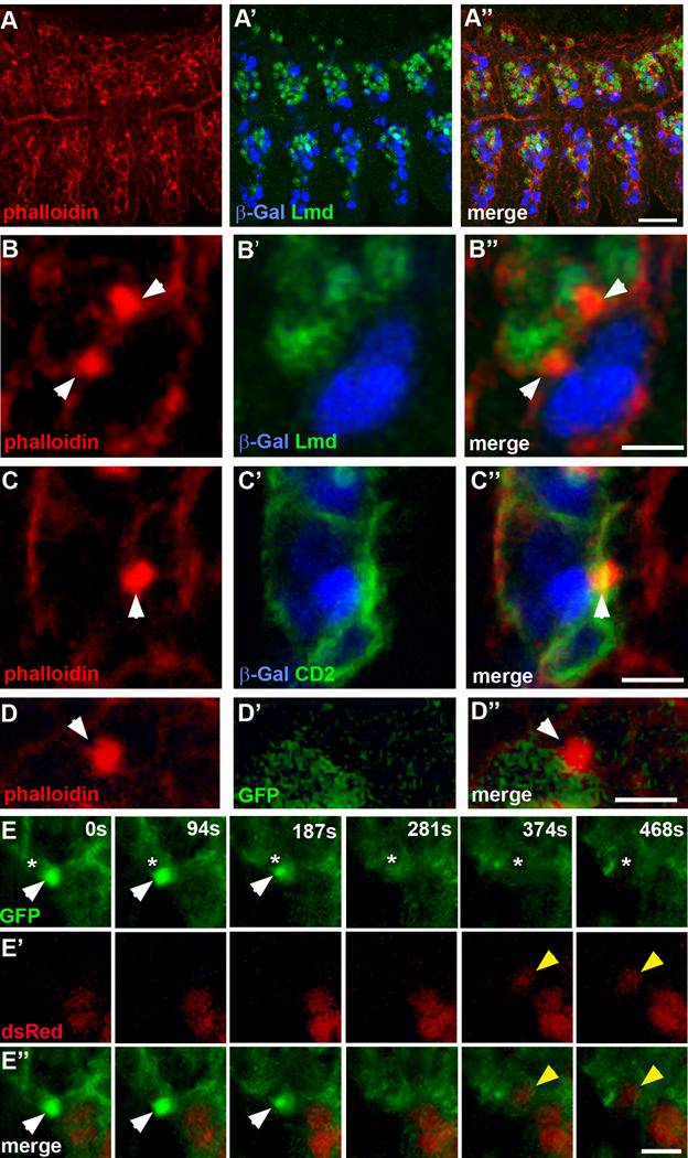
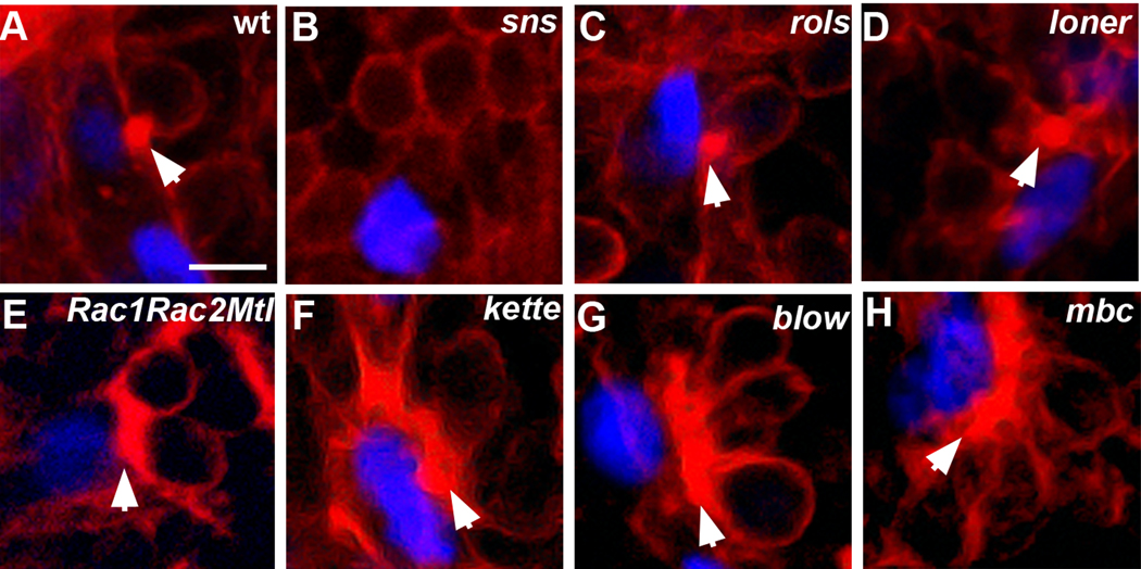
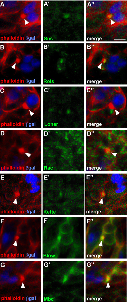
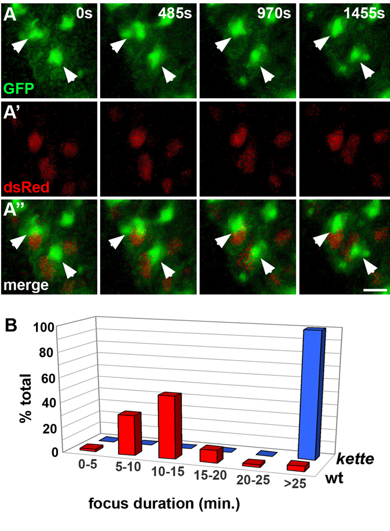
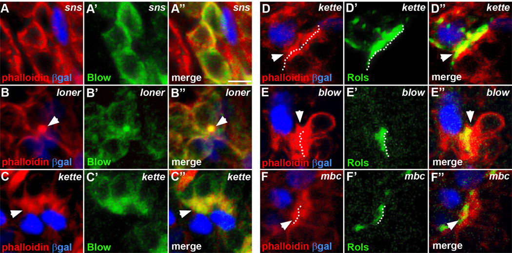
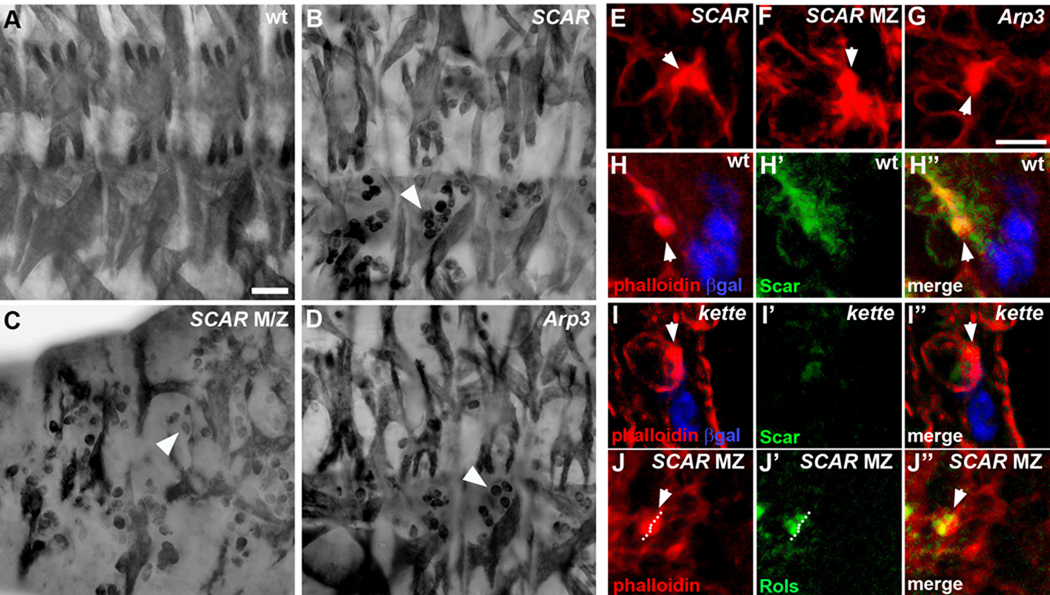
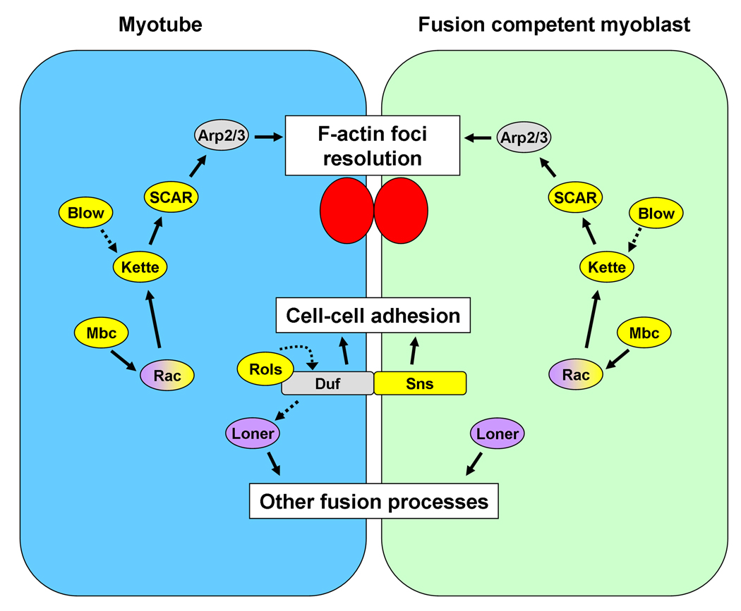
Myoblast fusion is crucial for formation and repair of skeletal muscle. Here we show that active remodeling of the actin cytoskeleton is essential for fusion in Drosophila. Using live imaging, we have identified a dynamic F-actin accumulation (actin focus) at the site of fusion. Dissolution of the actin focus directly precedes a fusion event. Whereas several known fusion components regulate these actin foci, others target additional behaviors required for fusion. Mutations in kette/Nap1, an actin polymerization regulator, lead to enlarged foci that do not dissolve, consistent with the observed block in fusion. Kette is required to positively regulate SCAR/WAVE, which in turn activates the Arp2/3 complex. Mutants in SCAR and Arp2/3 have a fusion block and foci phenotype, suggesting that Kette-SCAR-Arp2/3 participate in an actin polymerization event required for focus dissolution. Our data identify a new paradigm for understanding the mechanisms underlying fusion in myoblasts and other tissues.


Figures

Figure 1
Figure 1. Dynamic remodeling of the actin cytoskeleton during Drosophila myoblast fusion
Lateral views of stage 14 embryos. Scale bars = 20µm in A, 5µm in B–E. Phalloidin was used to label F-actin (red) in A–D. (A) rP298-lacZ embryo stained with phalloidin and antibodies against β-galactosidase to label FCs/myotubes (blue) and Lameduck to label FCMs (green). These images show the arrangement of myotubes and FCMs in one plane of focus and the occurrence of F-actin foci at this stage. F-actin is seen predominantly at the cell cortices. (B) Closeup of A. F-actin foci form at the adhesion sites between FCs/myotubes and FCMs (arrowheads). See Supplemental Movie 1 for 3D reconstruction of actin focus. (C) rP298-lacZ; twi-CD2 embryo stained with phalloidin and antibodies against β-galactosidase to label FCs/myotubes (blue) and CD2 to label mesoderm cell membranes (green). Actin focus is present in both FC and FCM, as evident by bisection of the focus with membrane staining (arrowhead). (D) apME-GFP embryo stained with phalloidin and antibody against GFP to label the cytoplasm of apterous-expressing FCs/myotubes (green). GFP does not leak from the apterous-expressing myotube into the adherent FCM when the F-actin focus is present. (E) Live twip-GFP-actin, apME-NLS-dsRed embryo. Each column of panels represents a time point from a time-lapse sequence. Each image is an optical projection displaying 9 µm of the Z-axis. The optical projection allows visualization of several cell layers simultaneously and tracking of all relevant cell movements. In this sequence, an actin focus (white arrowheads) forms at the adhesion site between an FCM and an apterous-labeled myotube. This focus dissolves, followed by fusion and addition of a labeled nucleus (yellow arrowhead) to the myotube. The nucleus of the fusing cell is indicated (asterisk). Additional actin accumulation in 468s panel may represent a new actin focus forming.
Figure 2
Figure 2. Roles of fusion proteins in actin remodeling
Lateral views of stage 14 rp298-lacZ embryos stained with phalloidin to label F-actin (red) and antibody against β-galactosidase to label FCs/myotubes (blue), except E, which is stained with antibody against Slouch to label a subset of FCs/myotubes (blue). Actin foci are indicated by arrowheads. Scale bar = 5µm. One optical slice is shown for each mutant. See Supplemental Figure S2 and Materials and Methods for details of focus size determination. (A) wild type (B–C) Class 1: no/fewer foci (B) snsXB3 embryo. (C) rolsT627 embryo. A focus of wild-type size is occasionally seen (arrowhead). (D) Class 2: wild-type actin focus : lonerT1032 embryo. (E–H) Class 3: enlarged actin focus. (E) Rac1J11, Rac2Δ, mtlΔ embryo. (F) ketteJ4–48 embryo. (G) blow1 embryo. (H) mbcC1 embryo.
Figure 3
Figure 3. Localization of fusion machinery with actin focus
Lateral views of stage 14 rp298-lacZ embryos stained with phalloidin to label F-actin (red) and antibodies against β-galactosidase to label FCs/myotubes (blue), and Sns (green, A), Rols (green, B), Loner (green, C), Rac1 (green, D), Kette (green, E), Blow (green, F), or Mbc (green, G). Channels are shown separately and then merged. Sns, Rols, Kette, Blow, and Mbc protein colocalize with F-actin foci (arrowheads, A, B, E, F, G), while Loner does not (C). Rac shows partial overlap with the F-actin foci (D). Scale bar = 5µm.
Figure 4
Figure 4. Kette regulates actin foci dissolution during Drosophila myoblast fusion
(A) Lateral views of live stage 14 twip-GFP-actin, apME-NLS-dsRed; ketteJ4–48 embryo. Each column of panels represents a time point from a time-lapse sequence. Each image is an optical projection displaying 9 µm of the Z-axis, allowing visualization of several cell layers simultaneously and tracking of all relevant cell movements. Large actin foci form (arrowheads) but do not dissolve as in wild type (compare to Fig. 1E). No incorporation of new red nuclei is seen, consistent with the kette fusion block. Scale bar = 5µm. (B) Actin foci persist significantly longer in ketteJ4–48 null mutants compared to wild type. Foci duration (mean ± standard deviation): wild type=10.9±6.9 minutes, ketteJ4–48=36.0±17.6 minutes. This difference is significant by a two-tail unpaired student t-test (P<0.0001, n=25 foci).
Figure 5
Figure 5. Localization of fusion machinery in different mutant classes
Lateral views of stage 14 rp298-lacZ embryos stained with phalloidin to label F-actin (red) and antibody against β-galactosidase to label FCs/myotubes (blue). Channels are shown separately and in a merge. Scale bar = 5µm. (A–C) Embryos stained with an antibody against Blow (green). (A) Class 1: snsXB3 mutant. Blow no longer has a polarized localization (compare to Fig. 3F) and instead is distributed cortically in FCMs. (B) Class 2: lonerT1032 mutant. Blow localization overlaps with the actin focus (arrowhead, compare to Fig. 3F). (C) Class 3: ketteJ4–48 mutant. Blow is polarized and distributed throughout large actin accumulations (arrowhead). (D–F) Embryos stained with antibody against Rols to label FCs/myotubes (green). (D) ketteJ4–48 mutant. (E) blow1 mutant. (F) mbcC1 mutant. Dotted lines indicate FC/myotube membranes and were drawn based on Rols localization. In each case, enlarged actin foci localize across both the myotube and FCM.
Figure 6
Figure 6. SCAR and Arp2/3 are required for myoblast fusion and regulate actin foci dissolution during fusion
(A–D) Lateral views of stage 16 embryos stained with antibody against myosin heavy chain to visualize body wall muscles. Scale bar = 20µm. (A) Wild-type embryo (B) SCARΔ37 embryo. Approximately 10–20 free myoblasts are seen in each hemisegment indicating a myoblast fusion defect (arrowhead). (C) Embryo from SCARk13211 germline clones with reduced levels of maternal and zygotic SCAR protein. Increased numbers of free myoblasts are seen, along with thinner muscles, indicating a more severe myoblast fusion defect (arrowhead). (D) Arp3EP3640 embryo. These embryos display a myoblast fusion defect, with approximately 10–20 free myoblasts seen in each hemisegment (arrowhead). (E–J) Lateral views of stage 14 embryos stained with phalloidin to label F-actin (red). F-actin labels the foci as well as cortical actin. Scale bar = 5µm. (E) SCARΔ37 embryo. Actin foci appear larger than in wild type (arrowhead). The focus shown is an example of the larger foci seen in these mutants, although the average focus size is similar to wildtype (Table 1). (F) SCARk13211 germline clone embryo with reduced levels of maternal and zygotic SCAR protein. Large accumulations of F-actin have formed at the site of adhesion between FC/Myotubes and FCMs (arrowhead). (G) Arp3EP3640 embryo. Actin foci appear larger than in wild type (arrowhead). The focus shown is an example of the larger foci seen in these mutants, although the average focus size is similar to wild type (Table 1). (H–I) Embryos stained with antibodies against β-galactosidase to label FCs/myotubes (blue) and SCAR (green). (H) rP298-lacZ embryo. SCAR protein partially colocalizes with F-actin foci (arrowhead) in both FCMs and FCs/Myotubes. (I) rP298-lacZ; ketteJ4–48 embryo. SCAR protein is virtually undetectable in this mutant background. Residual protein is mislocalized (compare to H). (J) SCARk13211 germline clone embryo stained against Rols (green). Dotted line indicates FC/myotube membrane and was drawn based on Rols localization. Rols partially overlaps with actin focus (arrowhead), indicating that enlarged actin focus localizes across both FC/myotube and FCM.
Figure 7
Figure 7. Model of Drosophila myoblast fusion
Updated model of myoblast fusion based on the known fusion genes following this work. Those proteins that colocalize with the F-actin foci are colored yellow, those that do not are purple. Rac’s localization partially overlaps with actin foci. Solid arrows between proteins indicate well-established biochemical interactions, while dashed arrows indicate genetic and/or suggested, but unsubstantiated biochemical interactions. See Discussion for details.

References

    1. Abmayr SM, Balagopalan L, Galletta BJ, Hong SJ. Cell and molecular biology of myoblast fusion. Int Rev Cytol. 2003;225:33–89. - PubMed
    1. Alvarez-Dolado M, Pardal R, Garcia-Verdugo JM, Fike JR, Lee HO, Pfeffer K, Lois C, Morrison SJ, Alvarez-Buylla A. Fusion of bone-marrow-derived cells with Purkinje neurons, cardiomyocytes and hepatocytes. Nature. 2003;425:968–973. - PubMed
    1. Artero RD, Castanon I, Baylies MK. The immunoglobulin-like protein Hibris functions as a dose-dependent regulator of myoblast fusion and is differentially controlled by Ras and Notch signaling. Development. 2001;128:4251–4264. - PubMed
    1. Barolo S, Carver LA, Posakony JW. GFP and beta-galactosidase transformation vectors for promoter/enhancer analysis in Drosophila. Biotechniques. 2000;29:726. 728, 730, 732. - PubMed
    1. Barolo S, Castro B, Posakony JW. New Drosophila transgenic reporters: insulated P-element vectors expressing fast-maturing RFP. Biotechniques. 2004;36:436–440. 442. - PubMed

Publication types

MeSH terms