. 2008 Apr;57(4):879-88.
doi: 10.2337/db07-1204. Epub 2007 Nov 14.

Proinflammatory effects of advanced lipoxidation end products in monocytes

Affiliations

Proinflammatory effects of advanced lipoxidation end products in monocytes

Narkunarajaa Shanmugam et al. Diabetes. 2008 Apr.

Abstract

Objective: The reactions of carbohydrate- or lipid-derived intermediates with proteins lead to the formation of Maillard reaction products, which subsequently leads to the formation of advanced glycation/lipoxidation end products (AGE/ALEs). Levels of AGE/ALEs are increased in diseases like diabetes. Unlike AGEs, very little is known about ALE effects in vitro. We hypothesized that ALEs can have proinflammatory effects in monocytes.

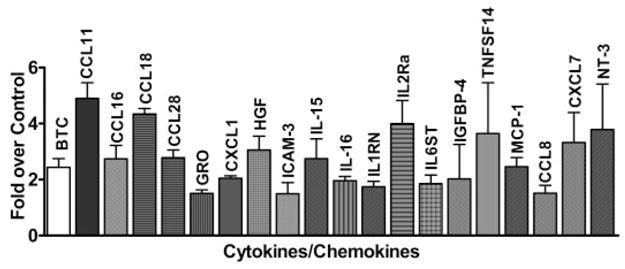
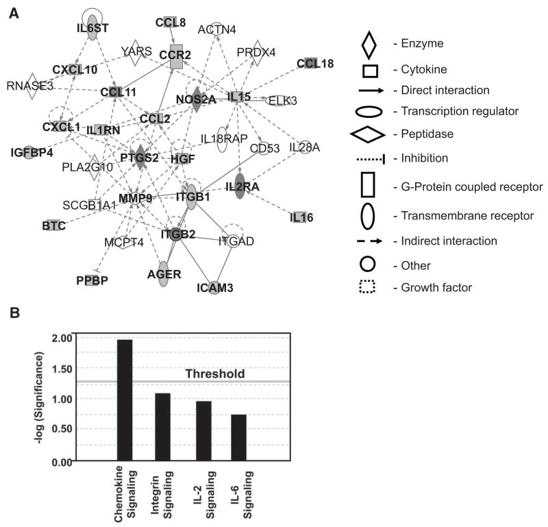
Research design and methods: In a profiling approach, conditioned media from THP-1 cells either cultured in normal glucose (5.5 mmol/l) or treated with MDA-Lys or MDA alone were hybridized to arrays containing antibodies to 120 known human cytokines/chemokines. Pathway analyses with bioinformatics software were used to identify signalling networks.

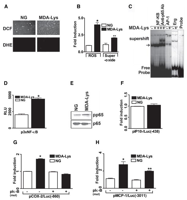
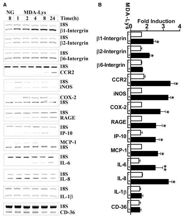
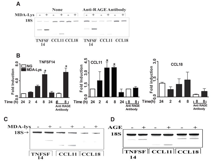
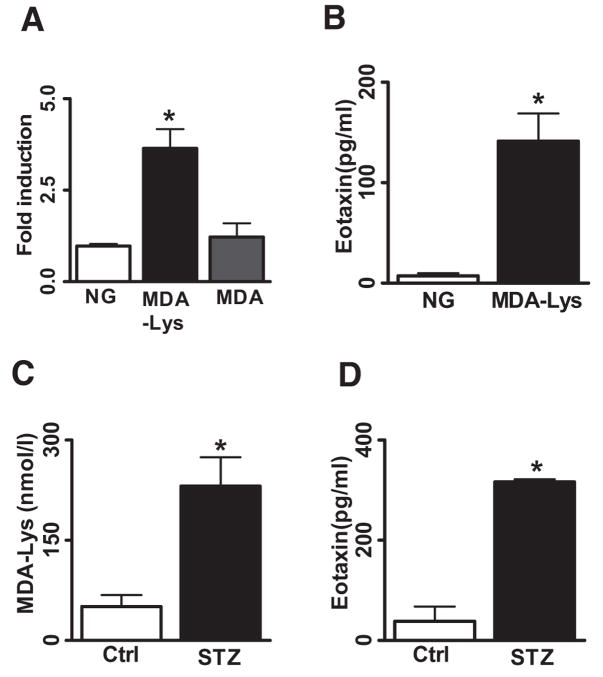
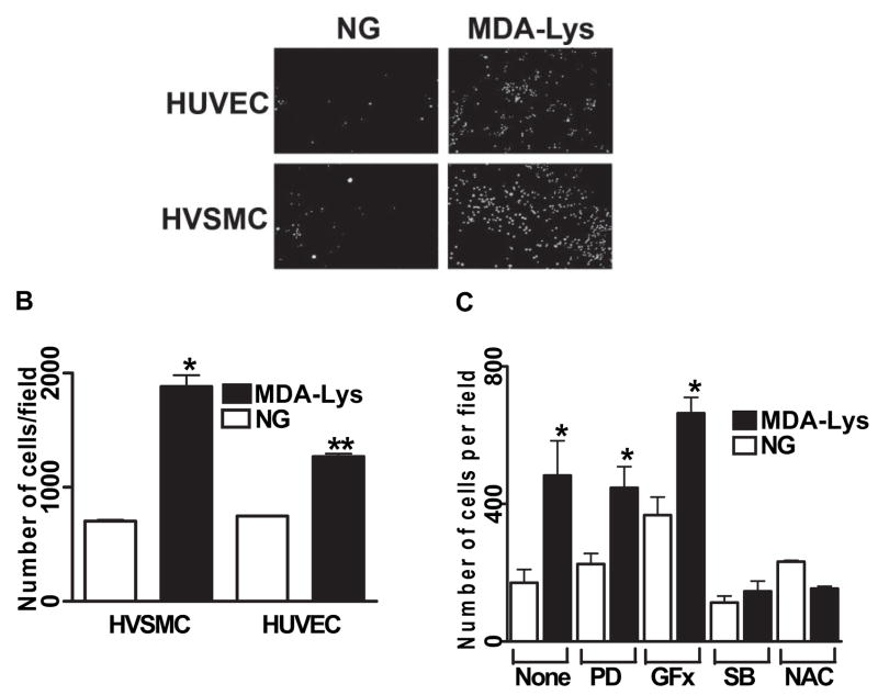
Results: Synthetic ALE (malondialdehyde-lysine [MDA-Lys]) (50 micromol/l) could induce oxidant stress and also activate the transcriptional factor nuclear factor-kappaB (NF-kappaB) in THP-1 monocytes. MDA-Lys also significantly increased the expression of key candidate proinflammatory genes, interferon-gamma-inducible protein-10, beta1- and beta2-integrins, cyclooxygenase-2 (COX-2), monocyte chemoattractant protein-1 (MCP-1), interleukin-6 and -8, and inducible nitric-oxide synthase, which are also associated with monocyte dysfunction. Several key target proinflammatory proteins were significantly induced by MDA-Lys relative to normal glucose or MDA alone, including MCP-1; tumor necrosis factor ligand superfamily member-14; chemokine CC motif ligand-11 (CCL11); growth-related oncogene-alpha, -beta, and -gamma; and chemokine CXC motif ligand-13. Bioinformatics analyses identified a network of chemokine signaling among MDA-Lys-regulated genes. MDA-Lys also increased monocyte binding to vascular smooth muscle and endothelial cells. Furthermore, plasma from diabetic rats showed significantly higher levels of MDA-Lys and CCL11.

Conclusions: These new results suggest that ALEs can promote monocyte activation and vascular complications via induction of inflammatory pathways and networks.


Figures

FIG. 1
FIG. 1
MDA-Lys induces oxidant stress and NF-κB activation in THP-1 cells. A: Fluorescence photomicrographs from control THP-1 cells (left panel) and THP-1 cells treated with 50 μmol/l MDA-Lys (right panel) for 30 min exposed to either 1 μmol/l H2DCFDA (DCF, for ROS, top panel) or 1 μmol/l DHE (forsuperoxide, bottom panel) for 15 min. B: Bar graph showing significant fourfold increase (*P < 0.05 vs. normal glucose) in ROS and twofold increase in superoxide formation (**P < 0.01 vs. normal glucose) by MDA-Lys (means ± SE, n = 3). C: EMSAs of nuclear extracts from normal glucose- or MDA-Lys–treated cells incubated with radiolabeled oligonucleotides containing NF-κB, AP-1, or Egr-1 consensus binding sites. Supershifts were performed with nuclear extracts of MDA-Lys treated cell extracts that were pretreated with p65 antibody. D: THP-1 cells were transiently transfected with 500 ng p3xNF-κB reporter plasmid carrying luciferase gene under control of NF-κB consensus elements. After 12 h, cells were treated with or without 50 μmol/l MDA-Lys and cultured for an additional 7 h. Results are from three individual experiments, each performed in triplicate. E: MDA-Lys induces phosphorylation of NF-κB p65. Lysates from THP-1 cells stimulated with MDA-Lys for 1 h were immunoblotted with antibodies to phospho-p65 or total p65 as internal control. F--H: Gene promoter transactivation by MDA-Lys. THP-1 cells were transfected with plasmids containing luciferase gene under the control of respective minimal human promoters of IP-10 (−438/+97) (F), COX-2 (−860/+127) alone or with pCMV-mIκB (G), or MCP-1 (−3,011 to +37) alone or with pCMV-mIκB (H). After overnight recovery, cells were treated with MDA-Lys for 8 h, and luciferase activities determined. Values shown are normalized to 50 μg protein. Means ± SE of three independent experiments (*P < 0.01 vs. normal glucose, **P < 0.05 vs. normal glucose).
FIG. 2
FIG. 2
Analysis of MDA-Lys-induced candidate genes. Relative RT-PCRs were performed with total RNA isolated from THP-1 cells treated with or without MDA-Lys for 1–24 h, using gene-specific primers (Supplemental Table S1). 18S RNA primers were included in each PCR reaction as internal control. A: Ethidium bromide-stained agarose gels of RT-PCR products. NG indicates control cell grown for 24 h. B: Bar graph showing significant induction of β1- and β2-integrins, CCR2, iNOS, COX-2, RAGE, IP-10, IL-6, IL-8, and MCP-1 mRNAs at 4, 8, or 24 h. Values shown are means ± SE of three independent experiments. *P < 0.001, **P < 0.05 vs. normal glucose.
FIG. 3
FIG. 3
Cytokine antibody array analysis of MDA-Lys–treated THP-1 cells. Conditioned medium from THP-1 monocytes cultured under either normal glucose (5.5 mmol/l) or MDA-Lys (50 μmol/l) conditions for 24 h were hybridized to human cytokine antibody arrays. Relative percentage of spot intensities in the membranes were measured and normalized to normal glucose. Values are average of data from three independent experiments. Cytokines showing significant increases (*P ≤ 0.05) are shown in the bar graph.
FIG. 4
FIG. 4
A: Role of RAGE receptor in MDA-Lys induced CCL11, CCL18, and TNFSF14 in mRNAs. THP-1 cells were pretreated with or without 70 μg/ml anti-RAGE antibody for 1 h followed by 50 μmol/l MDA-Lys for 4 h. mRNA levels analyzed by RT-PCR with 18S internal control. B: Bar graph showing time-course induction of CCL11, CCL18, and TNFSF14 mRNAs by real time quantitative RT-PCR using gene-specific primers and normalized with glyceraldehyde-3-phosphate dehydrogenase (GAPDH) as internal control. C: Effect of MDA-Lys on CCL11, CCL18, and TNFSF14 mRNA expression in peripheral blood primary human monocytes as analyzed by RT-PCR with 18S internal control. D: Agarose gel of RT-PCR products of TNFSF-14, CCL11, and CCL18 mRNA amplification from THP-1 cells treated with or without methylglyoxal modified bovine serum albumin (AGE) for 8 h.
FIG. 5
FIG. 5
Ingenuity pathway analysis identifies key interactive networks among proteins and genes regulated by MDA-Lys in THP-1 cells. A: The network is displayed schematically as nodes (genes/gene products) and edges (solid or dotted lines, depicting biological relationships between the nodes). Filled nodes are identified targets, and open ones are predicted pathway partners. Edges for protein regulations, such as phosphorylation and other modifications, are removed for simplicity. This network was generated by the software based on highest scores obtained from a total of 22 differentially expressed focus proteins noted in our experiments (Supplemental Table 2). In the current study, a score of 14 or higher was used to select significant biological networks regulated by MDA-Lys. A highly significant score of 56 was obtained (Table 1). B: The network score is next displayed as the negative log of the P value, indicating the likelihood of the focus molecules in a network being found together due to random chance. Therefore, −log values of two have at least 99% confidence of not being generated by chance alone as seen for chemokine signaling.
FIG. 6
FIG. 6
MDA-Lys induces specific and significant induction of CCL11. Total RNA from multiple sets of MDA-Lys, MDA, and normal glucose grown THP-1 cells were used to perform real-time quantitative PCRs using CCL11 specific primers and GAPDH primers. A: Eotaxin levels were calculated by normalizing to internal control GAPDH and results expressed as fold over normal glucose. Values shown are means ± SE of three independent experiments. *P < 0.012. B: Conditioned medium supernatants of THP-1 cells treated with normal glucose or MDA-Lys were assayed for secreted CCL11 (eotaxin) levels by specific ELISA. Results shown are means ± SE from three independent experiments run in triplicate. *P < 0.004. C: Quantification of protein cross-linked MDA in plasma from control and STZ-administered rats (n = 10). *P < 0.003. D: Rat plasma samples assayed for secreted CCL11 levels by specific ELISA. Results shown are mean ± SE from four independent rats run in triplicate. *P < 0.003.
FIG. 7
FIG. 7
A: MDA-Lys treatment increases monocytes adhesion to HVSMC and HUVECs. THP-1 cells were cultured with or without MDA-Lys for 12 h and then labeled with fluorescent dye Calcein-AM for 15 min at 37°C. Labeled THP-1 cells were allowed to adhere to either HUVEC or HVSMC monolayers in 24-well culture dishes. After careful washing, specifically bound monocytes were counted as described previously (19). Results are expressed as number of monocytes bound per high-power field. B: Bar graph shows means ± SE from three to five experiments (*P < 0.005, **P < 0.01 vs. respective controls). C: THP-1 cells were treated for 12 h with the indicated inhibitors or corresponding vehicle and then treated with or without MDA-Lys and then binding to HVSMC examined as above. Results shown are means ± SE (n = 4; *P < 0.01 vs. respective control). PD, 2′-amino-3′methoxyflavone (PD-98059); SB, 4-[4-(4-fluorophenyl)-5-(4-pyridinyl)-1H-imidazol-2-yl]phenol (SB202190).

References

    1. Thorpe SR, Baynes JW. Maillard reaction products in tissue proteins: new products and new perspectives. Amino Acids. 2003;25:275–281. - PubMed
    1. Portero-Otin M, Bellmunt MJ, Requena JR, et al. Protein modification by advanced Maillard adducts can be modulated by dietary polyunsaturated fatty acids. Biochem Soc Trans. 2003;31:1403–1405. - PubMed
    1. Requena JR, Fu MX, Ahmed MU, et al. Quantification of malondialdehyde and 4-hydroxynonenal adducts to lysine residues in native and oxidized human low-density lipoprotein. Biochem J. 1997;322:317–325. - PMC - PubMed
    1. Huang GS, Wang ZP, Wang SC, et al. Intracellular generation of MDA-LYS epitope in foam cells. Life Sci. 1999;65:285–296. - PubMed
    1. Parthasarathy S, Steinberg D, Witztum JL. The role of oxidized low-density lipoproteins in the pathogenesis of atherosclerosis. Annu Rev Med. 1992;43:219–225. - PubMed

Publication types

MeSH terms