

Case Reports
doi: 10.1128/JCM.01137-07.
Epub 2007 Nov 14.
Lasiodiplodia theobromae pneumonia in a liver transplant recipient
Affiliations
- PMID: 18003802
- PMCID: PMC2224279
- DOI: 10.1128/JCM.01137-07
Item in Clipboard
Case Reports
Lasiodiplodia theobromae pneumonia in a liver transplant recipient
J Clin Microbiol.
2008 Jan.
Abstract
We report a case of Lasiodiplodia theobromae pneumonia in a patient who died 14 days after cadaveric-liver transplantation. His condition was complicated by Enterococcus faecium peritonitis. Direct microscopy analysis of the bronchoalveolar lavage specimens showed septate hyphae. A dematiaceous mold was recovered and identified as L. theobromae by microscopic morphology and EF1alpha gene sequencing.
Figures
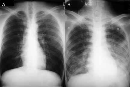
Chest radiographs of the patient's lungs taken 2 months before admission (A) and upon admission (B). The chest radiograph taken upon admission showed air space shadows over both lungs.
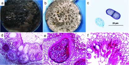
Phenotypic characterization of the mold. (a) Seven-day culture of the mold on SDA, showing dark gray cottony colonies. (b) Two-week culture of the mold on oatmeal agar incubated in sunlight, showing hairy, dark brown pycnidia. (c) Young, nonseptate, hyaline conidia and old, septate, brown conidia with longitudinal striations released from the pycnidia. (d) Periodic acid-Schiff-stained histological sections of the mold, showing aggregates of pycnidia. (e and f) Pycnidium with an obvious neck and conidiogenous cells and paraphyses (sterile filaments among conidia) lining the internal wall.
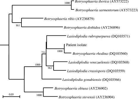
Phylogenetic tree showing the relationship of the mold from our patient to closely related species, inferred from EF1α gene sequence data (308 nucleotide positions) by the neighbor-joining method and rooted using Aspergillus clavatus (XM_001269545). The scale bar indicates the estimated number of substitutions per 10 bases. Numbers at nodes indicate levels of bootstrap support calculated from 1,000 trees. All names and accession numbers are given as cited in the GenBank database.
References
-
- Borderie, V. M., T. M. Bourcier, J. L. Poirot, M. Baudrimont, P. Prudhomme de Saint-Maur, and L. Laroche. 1997. Endophthalmitis after Lasiodiplodia theobromae corneal abscess. Graefe's Arch. Clin. Exp. Ophthalmol. 235259-261. - PubMed
-
- Burgess, T. I., P. A. Barber, S. Mohali, G. Pegg, W. de Beer, and M. J. Wingfield. 2006. Three new Lasiodiplodia spp. from the tropics, recognized based on DNA sequence comparisons and morphology. Mycologia 98423-435. - PubMed
-
- de Hoog, G. S., J. Guarro, J. Gene, and M. J. Figueras (ed.). 2000. Atlas of clinical fungi, 2nd ed., p. 325. Centraalbureau voor Schimmelcultures, Utrecht, The Netherlands.
-
- Ishibashi, Y., and Y. Matsumoto. 1984. Intravenous miconazole in the treatment of keratomycosis. Am. J. Ophthalmol. 97646-647. - PubMed
-
- Laverde, S., L. H. Moncada, A. Restrepo, and C. L. Vera. 1973. Mycotic keratitis: 5 cases caused by unusual fungi. Sabouraudia 11119-123. - PubMed
Publication types
MeSH terms
Substances
Associated data
- Actions
- Actions
LinkOut - more resources
Full Text Sources
Medical
