. 2007 Nov 20;104(47):18748-53.
doi: 10.1073/pnas.0709379104. Epub 2007 Nov 14.

The Parkinson's disease gene DJ-1 is also a key regulator of stroke-induced damage

Affiliations

The Parkinson's disease gene DJ-1 is also a key regulator of stroke-induced damage

Hossein Aleyasin et al. Proc Natl Acad Sci U S A. .

Abstract

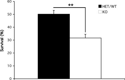
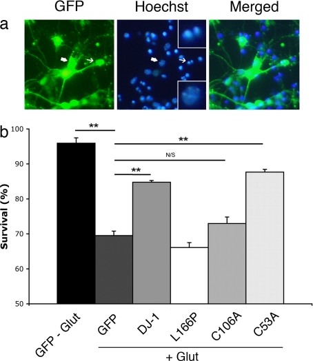
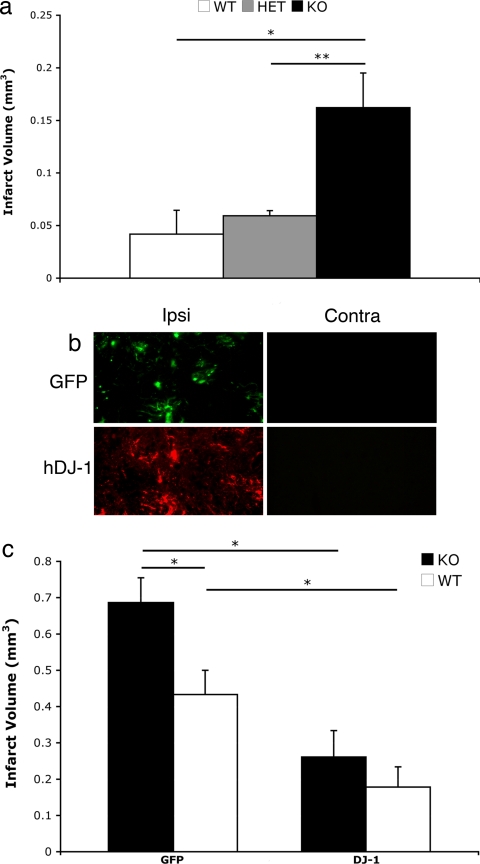
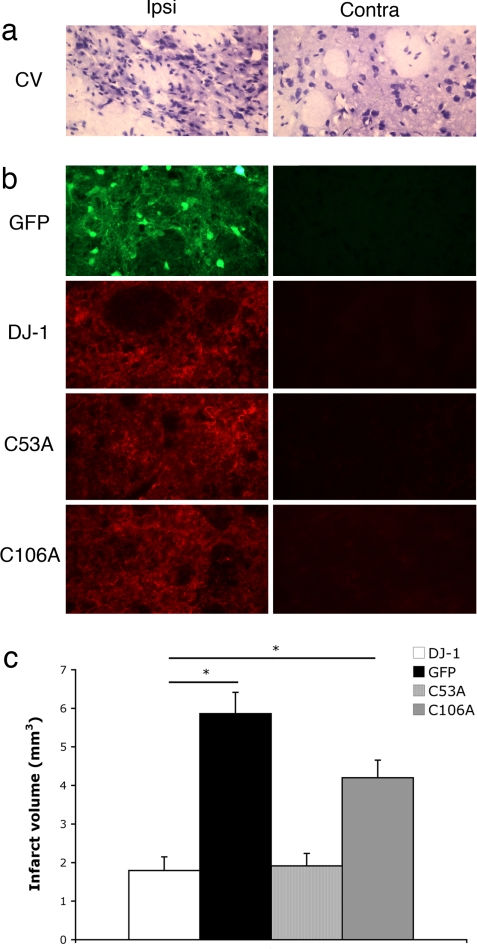
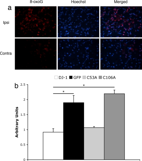
Recent evidence has indicated that common mechanisms play roles among multiple neurological diseases. However, the specifics of these pathways are not completely understood. Stroke is caused by the interruption of blood flow to the brain, and cumulative evidence supports the critical role of oxidative stress in the ensuing neuronal death process. DJ-1 (PARK7) has been identified as the gene linked to early-onset familial Parkinson's disease. Currently, our work also shows that DJ-1 is central to death in both in Vitro and in Vivo models of stroke. Loss of DJ-1 increases the sensitivity to excitotoxicity and ischemia, whereas expression of DJ-1 can reverse this sensitivity and indeed provide further protection. Importantly, DJ-1 expression decreases markers of oxidative stress after stroke insult in Vivo, suggesting that DJ-1 protects through alleviation of oxidative stress. Consistent with this finding, we demonstrate the essential role of the oxidation-sensitive cysteine-106 residue in the neuroprotective activity of DJ-1 after stroke. Our work provides an important example of how a gene seemingly specific for one disease, in this case Parkinson's disease, also appears to be central in other neuropathological conditions such as stroke. It also highlights the important commonalities among differing neuropathologies.


Conflict of interest statement

The authors declare no conflict of interest.

Figures

Fig. 1.
Fig. 1.
Increased sensitivity to glutamate-induced excitotoxicity in the absence of DJ-1. CGNs were harvested from DJ-1+/− or +/+ (n = 7) and DJ-1−/− (n = 4) 7- to 9-day-old mice pups. Seven days after plating, they were treated with 50 μM l-glutamate for 45 min, washed, and incubated in 37°C for 2 h. Then cells were lysed, and nuclei were fixed and counted on a hemocytometer slide to assess their viability. Each data point is the mean ± SEM of three independent cultures, **, P < 0.01. KO, knockout.
Fig. 2.
Fig. 2.
Cysteine-106 is essential for neuroprotective function of DJ-1 after glutamate-induced excitotoxicity in vitro. CGNs, harvested from CD-1 WT pups, were targeted by rAV vectors carrying the indicated constructs along with GFP, 5 days after plating, and treated with l-glutamate at day 7 in vitro (as described in Materials and Methods). (a) Viability of virally infected neurons was assessed based on nuclear morphology under UV microscopy (representative cells are shown). Condensed or fragmented nuclei (indicated by thin arrows) were scored as dead versus intact nuclei considered as alive (indicated by thick arrows). (Upper Inset) High magnification of fragmented nuclei. (Lower Inset) High magnification of intact nuclei. (b) Quantification of neuronal survival after treatment with l-glutamate compared with nontreated GFP-expressing neurons. Each data point is the mean ± SEM of three independent cultures (one-way ANOVA followed by Tukey's least significant difference test; **, P < 0.01; N/S, not significant).
Fig. 3.
Fig. 3.
DJ-1 protects brain tissue from endothelin-induced focal ischemia in vivo. Infarct volume in cortex and striatum of mouse brains after acute ischemic stroke is shown. (a) Nonvirally injected mice of DJ-1 colony were assessed for infarct volume after endothelin-induced focal ischemia and were analyzed according to their genotype (WT, n = 3; HET, n = 3; KO, n = 4). There is a significant difference between the animals of the DJ-1-null background vs. either the WT or HET group but no significant difference between the latter two. (b) Expression of either GFP or human DJ-1 was assessed in the ipsi- and contralateral sides of the brains of DJ-1 KO mice (compared with the injected side). (c) Mice of DJ-1-null or WT genotype from the DJ-1 colony were injected with adenoviral vectors expressing either GFP (KO, n = 5; WT, n = 7) or DJ-1 (KO, n = 6; WT, n = 8). Animals in each group received the same previously mentioned stroke model procedure. Seven days later, brain infarct volume was measured as described. There were significant differences between DJ-1-null + GFP-injected and DJ-1-null + DJ-1-injected as well as an additional protection seen in the WT overexpressing DJ-1 as opposed to overexpressing GFP. No significant changes were observed between the WT overexpressing GFP and the KO overexpressing DJ-1. Each data point represents mean ± SEM for a representative population of three to eight mice (one-way ANOVA followed by Tukey's least significant difference test; *, P < 0.05; **, P < 0.01).
Fig. 4.
Fig. 4.
Cysteine-106 is essential for neuroprotective function of DJ-1 after focal ischemia in vivo. Infarct volume in the striatum after injection of endothelin-1 in WT rats overexpressing AAVs was assessed. (a) Representative sections of damaged (ipsilateral to the endothelin-1 injection) and healthy (contralateral) cells were stained with cresyl violet, and damaged areas were characterized through shrunken/condensed nuclear staining (dead cell) vs. healthy neurons with smooth, round nuclei and visible nucleoli (alive). (b) Sections from the injected hemisphere overlapping the infarcted area were stained for respective virally expressing protein. (c) Infarct volume was measured by cresyl violet staining between different virally injected groups (n = 4 for GFP and DJ-1; n = 3 for C53A and C106A). Each data point represents mean ± SEM for a representative population of rats. There were significant differences between DJ-1 and GFP overexpressing rats as well as C53A and GFP but not C106A and GFP overexpressing rats (one-way ANOVA followed by Tukey's least significant difference test; *, significant). No significant changes were observed between DJ-1 and C53A overexpressing rats.
Fig. 5.
Fig. 5.
Cysteine-106 is crucial for the regulation of ROS end product, 8-oxoGafter focal ischemia in vivo. Rats subjected to AAV injection overexpressing DJ-1, GFP, C53A, and C106A, as indicated, were subjected to ischemic insult by endothelin-1 and were killed after 48 h. Brains were collected and processed to be stained for 8-oxoG as well as Hoechst 33258. Representative pictures were taken of sections surrounding injection area, and colocalizing 8-oxoG/Hoescht neurons (n >50) were evaluated by densitometry compared with the contralateral side. (a) Representative sections show expression of ROS marker 8-oxoG on the ipsi- and contralateral sides of the endothelin injection. (b) Quantification of densitometry results. Arbitrary units represent the ratio of signal density for the injected vs. noninjected (internal control) sides. Each data point represents mean ± SEM for a representative population of three or four rats per group. There is a significant difference between DJ-1 and GFP overexpressing groups as well as DJ-1 and C106A overexpressing rats (one-way ANOVA; followed by Tukey's least significant difference test; *, significant). No significant changes were observed between DJ-1 and C53A groups.

References

    1. Centers for Disease Control Prevention (CDC) MMWR Morb Mortal Wkly Rep. 2007;56:469–474. - PubMed
    1. Hunter DJW, Grant HJ, Purdue MPH, Spasoff RA, Dorland JL, Bains N. Chronic Dis Can. 2004;25:138–146. - PubMed
    1. Alexandrova M, Bochev P, Markova V, Bechev B, Popova M, Danovska M, Simeonova V. J Clin Neurosci. 2004;11:501–506. - PubMed
    1. Schaller B, Graf R. J Cereb Blood Flow Metab. 2004;24:351–371. - PubMed
    1. Adam-Vizi V. Antioxid Redox Signal. 2005;7:1140–1149. - PubMed

Publication types

MeSH terms