. 2007 Nov 20;104(47):18813-8.
doi: 10.1073/pnas.0705082104. Epub 2007 Nov 14.

Separate functions for nuclear and cytoplasmic cryptochrome 1 during photomorphogenesis of Arabidopsis seedlings

Affiliations

Separate functions for nuclear and cytoplasmic cryptochrome 1 during photomorphogenesis of Arabidopsis seedlings

Guosheng Wu et al. Proc Natl Acad Sci U S A. .

Abstract

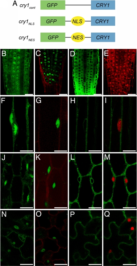
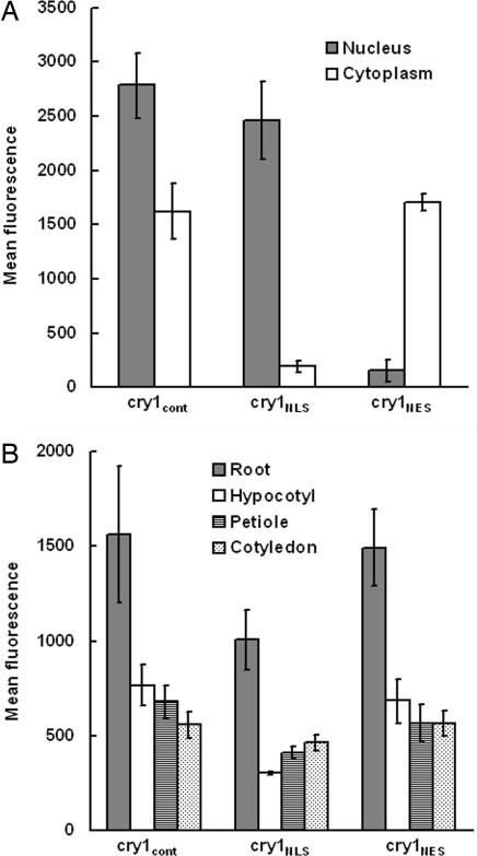
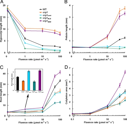
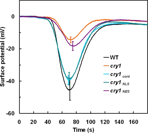
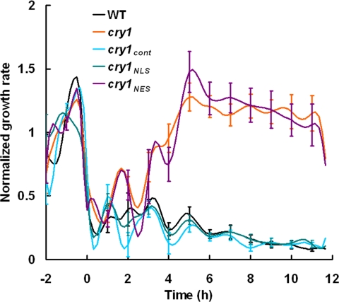
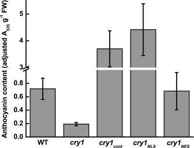
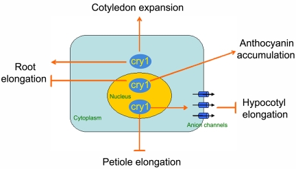
Cryptochrome blue-light receptors mediate many aspects of plant photomorphogenesis, such as suppression of hypocotyl elongation and promotion of cotyledon expansion and root growth. The cryptochrome 1 (cry1) protein of Arabidopsis is present in the nucleus and cytoplasm of cells, but how the functions of one pool differ from the other is not known. Nuclear localization and nuclear export signals were genetically engineered into GFP-tagged cry1 molecules to manipulate cry1 subcellular localization in a cry1-null mutant background. The effectiveness of the engineering was confirmed by confocal microscopy. The ability of nuclear or cytoplasmic cry1 to rescue a variety of cry1 phenotypes was determined. Hypocotyl growth suppression by blue light was assessed by standard end-point analyses and over time with high resolution by a custom computer-vision technique. Both assays indicated that nuclear, rather than cytoplasmic, cry1 was the effective molecule in these growth inhibitions, as was the case for the mechanistically linked membrane depolarization, which occurs within several seconds of cry1 activation. Petiole elongation also was inhibited by nuclear, but not cytoplasmic, cry1. Conversely, primary root growth and cotyledon expansion in blue light were promoted by cytoplasmic cry1 and inhibited by nuclear cry1. Anthocyanin production in response to blue light was strongly stimulated by nuclear cry1 and, to a lesser extent, by cytoplasmic cry1. An important step toward elucidation of cry1 signaling pathways is the recognition that different subcellular pools of the photoreceptor have different functions.


Conflict of interest statement

The authors declare no conflict of interest.

Figures

Fig. 1.
Fig. 1.
Schematics of CRY1 transgene constructs and subcellular localization of the proteins. (A) Schematics of the GFP-tagged chimeric genes with which cry1 was transformed to test the function of nuclear and cytoplasmically localized cry1. (B–D) Root tips of 4-day-old light-grown seedlings expressing cry1cont (B), cry1NLS (C), or cry1NES (D). (E) Propidium iodide staining highlights the nuclei in red. (F–H) Dark-grown hypocotyls expressing cry1cont (F), cry1NLS (G), or cry1NES (H). (I) Cell in H stained with Vybrant DyeCycle Orange to show the nucleus in red. (J–L) Light-grown hypocotyls expressing cry1cont (J), cry1NLS (K), or cry1NES (L). (M) Cell in L stained with Vybrant DyeCycle Orange to show the nucleus in red. (N–P) Light-grown cotyledons expressing cry1cont (N), cry1NLS (O), or cry1NES (P). (Q) Cell in P stained with Vybrant DyeCycle Orange to show the nucleus in red. Propidium iodide staining in C, G, K, and O shows the cell structure in red. (Scale bars: B–Q, 20 μm.)
Fig. 2.
Fig. 2.
GFP-cry1 quantification in the transgenic seedlings. (A) Nuclear and cytoplasmic GFP-cry1 levels in root apices measured as fluorescence intensity by confocal microscopy. (B) Overall GFP-cry1 levels in the indicated organs in the three transgenic lines. The roots were imaged with a C-Apochromat ×40 objective lens so their fluorescence levels in B are not comparable to the other organs, which were imaged with a Plan-Aprochromat ×20 objective lens. Each measurement in A or B is the mean of measurements obtained from four to seven seedlings. Error bars represent standard errors.
Fig. 3.
Fig. 3.
Phenotypic analysis of CRY1 transgenic plants. Plants were grown in darkness or under continuous blue light with various intensities for 7 days. Then hypocotyl lengths (A), cotyledon petiole lengths (B), root lengths (C), and cotyledon areas (D) were measured. (C Inset) Important differences in root lengths at 1 μmol·m−2·s−1 blue light. The mean values from >15 plants are shown. Error bars represent standard errors.
Fig. 4.
Fig. 4.
Blue light-induced membrane depolarizations in wild type, cry1, and transgenic plants. Membrane depolarization induced by a 20-s pulse of 200 μmol·m−2·s−1 blue light in hypocotyls started at 20 s and ended at 40 s. The mean values from more than eight responses were plotted. Error bars at the peak of each line were shown.
Fig. 5.
Fig. 5.
Morphometric analysis of seedling hypocotyl growth in blue light. The growth kinetics of 2-day-old etiolated wild type, cry1, cry1cont, cry1NLS, and cry1NES respond to 50 μmol·m−2·s−1 blue-light treatment. All growth rates were normalized to the average dark growth rate during the 2 h preceding blue-light treatment. Data represent the average of more than eight plants per genotype. Standard error was given every hour.
Fig. 6.
Fig. 6.
Anthocyanin levels in 7-day-old seedlings grown under 100 μmol·m−2·s−1 blue light.
Fig. 7.
Fig. 7.
Model integrating cry1 localization with cry1-mediated signal transduction induced by blue light.

References

    1. Lin C, Shalitin D. Annu Rev Plant Biol. 2003;54:469–496. - PubMed
    1. Ahmad M, Cashmore AR. Nature. 1993;366:162–166. - PubMed
    1. Jackson JA, Jenkins GI. Planta. 1995;197:233–239. - PubMed
    1. Neff MM, Chory J. Plant Physiol. 1998;118:27–35. - PMC - PubMed
    1. Bagnall DJ, King RW, Hangarter RP. Planta. 1996;200:278–280. - PubMed

Publication types