Comparative Study
. 2008 Mar;104(5):1233-48.
doi: 10.1111/j.1471-4159.2007.05054.x. Epub 2007 Nov 13.

Specificity proteins Sp1 and Sp3 interact with the rat GTP cyclohydrolase I proximal promoter to regulate transcription

Affiliations
Comparative Study

Specificity proteins Sp1 and Sp3 interact with the rat GTP cyclohydrolase I proximal promoter to regulate transcription

Nitya Sarath Chandran et al. J Neurochem. 2008 Mar.

Abstract

The role of the proximal promoter GC-box in regulating basal and cAMP-dependent GTP Cyclohydrolase I gene transcription was investigated using a variety of cell lines and techniques. These studies show that the GC-box is composed of a triad of cis-elements that in vitro bind specificity proteins Sp1 and Sp3. Sp1 and Sp3 were found associated with the native proximal promoter in PC12 cells but were not recruited to the promoter during cAMP-dependent transcription. Studies using Drosophila SL2 cells showed that Sp3 occupies two sites within the GC-box and enhances transcription when acting alone and synergistically when combined with nuclear factor-Y (NF-Y) and CCAAT/Enhancer-Binding Protein (C/EBP)beta, cognate binding proteins for the adjacent cAMP response element (CRE) and CCAAT-box cAMP response elements. In contrast, Sp1 bound only one site within the GC-box and did not enhance transcription unless combined with NF-Y and C/EBPbeta. Studies in SL2 cells also showed that Sp1 and Sp3 do not co-occupy the GC-box, and accordingly Sp1 competes for Sp3 binding to repress Sp3-dependent transcription. In PC12 cells, complete mutation of the GC-box reduced basal but not cAMP-dependent transcription, resulting in an overall increase in the cAMP response and demonstrating that formation of this enhanceosome does not require Sp1 or Sp3. Experiments in which the GC-box was replaced with a Gal4 element and the promoter challenged with Gal4 fusion proteins support this conclusion and a role for Sp3 in maintaining high levels of basal transcription in PC12 cells. Equivalent amounts of Sp1 and Sp3 were found associated with the native proximal promoter in PC12 and Rat2 cells, which differ 10-fold in basal transcription. Similar levels of methylation of CpG dinucleotides located within the GC-box were also observed in these two cells lines. These results suggest that Sp1 and Sp3 bound to the GC-box might help to preserve an open chromatin configuration at the proximal promoter in cells which constitutively express low levels of GTP Cyclohydrolase I.


Figures

Fig. 1
Fig. 1
The triad model of the rat GCH1 proximal promoter GC-box. (a) The rat GCH1 proximal promoter sequence spanning from −142 to −77 is shown with the GC-box, the CRE and the CCAAT-box labeled. TESS (Transcription Element Search System) analysis of the entire proximal promoter detected three high probability Sp1 binding sites clustered within the GC-box footprint spanning from −132 to −111. Site I (−134 GGGCGGGGCG −125), Site II (−130 GGGGCGGAGGG −121) and Site III ((−124 GAGGGGAGGGG (−114) are bracketed and labeled. These three sites are the basis for the triad model of the GC-box. (b) In the triad model of the GC-box each of the three sites within the GC-box is capable of binding Sp-proteins. Sites I and III can be occupied simultaneously by Sp-proteins because these two sites do not overlap. Because Sites I and III overlap with Site II occupation of Sites I or III would prevent binding to Site II. Similarly, because Site II overlaps Sites I and III occupation of Site II would block binding to Sites I and III.
Fig. 2
Fig. 2
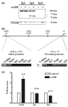
Sp1 and Sp3 protein expression and association with the native promoter in PC12 cells. (a) Three western blots of 5 μg of nuclear protein from control PC12 cells (−) and PC12 cells treated with 5 mmol/L 8-Br-cAMP for 4 h (cAMP, +). Proteins were separated on 4–12% gradient acrylamide gels and probed with antibodies directed against Sp1, Sp3 or Sp4. A single Sp1 protein of approximately 100 kDa was observed, as were three isoforms of Sp3 protein of approximately 100, 77, and 73 kDa. No Sp4 protein was detected. As a loading control the three blots were stripped and probed again with an antibody directed against the nuclear protein TFIID. (b) Schematic representation of 5812 bp of the rat GCH1 5′ flanking sequence showing the proximal (−167 to −27) and distal (−5439 to −5375) promoter regions amplified by PCR during ChIP analysis of the native promoter. Cross-linked and sheared DNA from control PC12 cells was precipitated using antibodies directed against Pol II, Sp1, Sp3 or control IgG, amplified by PCR and analyzed by agarose gel electrophoresis. Amplicons of the appropriate size were found in the proximal (141 bp) but not distal (65 bp) promoter reactions, indicating that under basal conditions Pol II, Sp1 and Sp3 are each associated with the GCH1 proximal promoter. (c) Control and 8Br-cAMP-treated (5 mmol/L for 1 h, cAMP) PC12 cells were analyzed by ChIP using antibodies directed against Pol II, Sp1 and Sp3 and precipitated proximal promoter DNA quantified by real-time PCR. Data were normalized relative to control immunoprecipitations and analyzed by ANOVA with post hoc Bonferroni tests. Only Pol II was significantly different between control and cAMP.
Fig. 3
Fig. 3
Binding of Sp1 and Sp3 to the GC-box in PC12 cells. (a) EMSA using 1 μg of nuclear protein from control PC12 cells (Ctrl) and PC12 cells treated with 5 mmol/L 8-Br-cAMP for 4 h (cAMP) identified six protein-DNA complexes (labeled 1–6) that could be competed away by excess unlabeled probe (+) and appeared unchanged by cAMP treatment. (b) EMSA using 1 μg of PC12 nuclear protein showed competition for binding of complexes 1–5 by a double-stranded oligonucleotide containing a wild-type Sp1 binding site (WT; ATTCGATCGGGGCGGGGCGAGC) but not a mutated Sp1 site (MT; ATTCGATCGGTTCGGGGCGAGC). (c) EMSA using 1 μg of nuclear protein from control (Ctrl) and 8-Br-cAMP treated (cAMP) PC12 cells combined with 1 μg of control IgG, anti-Sp1 or anti-Sp3 detected Sp1 in complex 4 and Sp3 in complexes 3 and 5 and probably in complexes 1 and 2. Complex 6 was not shifted by either antibody. (d) Analysis of EMSA band densities of the control lanes shown in Fig. 3(a)–(c) indicates that complex 1 contains 5%, complex 2 contains 10%, complex 3 contains 25%, complex 4 contains 40%, complex 5 contains 10% and complex 6 contains 10% of the total binding. (e) Densitometry traces of the three Ctrl lanes shown in Fig. 3(c). The solid line represents the IgG control and complexes 1–6 are identified with arrows. The dotted line represents the sample treated with anti-Sp1. Note the almost complete loss of complex 4. The dashed line represents the sample treated with anti-Sp3. Note the super-shift of complexes 3 and 5 and the probable shifts of complexes 1 and 2.
Fig. 4
Fig. 4
Sp1 and Sp3 binding to the GC-box and activation of transcription in SL2 cells. (a) Three simultaneously developed western blots of 5 and 10 μg of nuclear protein isolated from Drosophila SL2 cells transiently transfected with the pPac-0 empty vector, pPac-Sp1 or pPac-Sp3, harvested 48 h later and probed with antibodies to Sp1 and Sp3. The blot from cells transfected with the pPac-0 empty vector was probed with a combination of antibodies to Sp1 and Sp3. Note that no proteins were detected in the pPac-0 sample and that Sp1 and Sp3 proteins are produced at comparable levels with no small isoforms of Sp3 present. (b) EMSA using 1 μg of nuclear protein prepared from SL2 cells transiently transfected to express Sp1 or Sp3 shows that there are no proteins in SL2 cells which bind the GC-box (see pPac-0), that Sp1 forms a single complex (arrow) while Sp3 forms two complexes (arrows), and that Sp1 and Sp3 binding is competed away by excess unlabeled probe (+). (c) EMSA using 1 μg of nuclear protein prepared from SL2 cells transiently transfected to express Sp1 or Sp3 shows that anti-Sp1 super-shifts the Sp1 complex while anti-Sp3 super-shifts both the large and small Sp3 complexes. (d) EMSA using 1 μg of nuclear protein prepared from SL2 cells transiently transfected to express Sp1 or Sp3 shows that increasing amounts of unlabeled probe displaces Sp1 and Sp3 binding and that the large Sp3 complex is displaced at lower probe concentrations than is the small complex. (e) Luciferase assays of SL2 cells transiently co-transfected with 600 ng of p0.27-GCH1-GL3 or pGL3-basic along with 20 ng of pPac-Sp1, pPac-Sp3 or 20 ng pPac-Sp1 + 20 ng pPac-Sp3 and pPac-0 carrier DNA. Data were normalized relative to pPac-0 and analyzed by ANOVA with post hoc Bonferroni tests (*p ≤ 0.05 vs. pGL3-basic; #p ≤ 0.05 vs. pPac-Sp3). (f) EMSA using 1 μg of nuclear protein from SL2 cells expressing Sp1 or Sp3 or the combination of 0.5 μg of Sp1 and 0.5 μg of Sp3 nuclear protein shows that Sp1 competes to prevent formation of the large Sp3 complex.
Fig. 5
Fig. 5
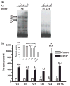
Mutation of the GC-box supports a triad model in which Sp1 and Sp3 interact with C/EBPβ and NF-Y to activate transcription. (a) Mutagenesis was performed based upon the triad model of the GC-box. In each case a GGG within a site was replaced with a TTT and is labeled above. Mutation M1 within Site I forces Sp-protein binding to Sites II or III while mutation M2 within Sites I and II forces binding to Site III. Similarly, mutation M3 within Sites II and III forces binding to Site I while mutation M4 within Site III forces binding to Sites I or II. Mutation M1234 in Sites I, II, and III eliminates Sp-protein binding completely. (b) EMSA using 1 μg of nuclear protein from SL2 cells transiently transfected to express Sp1 demonstrates that mutations M1, M2, M3 and M4 do not affect binding to the GC-box whereas mutation M1234 completely eliminates Sp1 binding. (c) EMSA using 1 μg of nuclear protein from SL2 cells transiently transfected to express Sp3 shows that mutations M1, M2, M3, and M4 prevent formation of the large Sp3 complex without affecting formation of the smaller complex whereas mutation M1234 completely eliminates Sp3 binding. (d) Luciferase assays of SL2 cells transiently co-transfected with 780 ng of wild-type (WT) p0.27GCH1-GL3 or the reporter constructs containing GC-box mutations shown in Fig. 5a, M1, M2, M3, M4 or M1234 along with 20 ng of pPac-0 or 20 ng of pPac-Sp3. Data were analyzed by ANOVA and post hoc Bonferroni tests (*p < 0.05 vs. WT pPac-Sp3). (e) Luciferase assays of SL2 cells transiently transfected with the 600 ng of wild-type p0.27-GCH1-GL3 reporter along with various combinations of 50 ng pPac-NF-YA, B and C, 50 ng pPac-C/EBPβ, 20 ng pPac-Sp3, 20 ng of pPac-Sp1 and varying amounts of pPac-0. Data were analyzed by ANOVA and post-hoc Bonferroni tests (*p < 0.05 vs. pPac-0; #p < 0.05 vs. pPac-C/EBPβ; @p < 0.05 vs. pPac-Sp3; !p < 0.05 vs. pPac-Sp3 + pPac-C/EBPβ; + p < 0.05 vs. pPac-Sp3 + pPac-NFY or pPac-C/EBPβ; &p < 0.05 vs. pPac-Sp3 + pPac-NF-Y + pPac-C/EBPβ).
Fig. 6
Fig. 6
Mutation of the GC-box decreases Sp3 binding yet can enhance cAMP-dependent transcription in PC12 cells. (a) EMSA using 1 μg of PC12 nuclear protein and 1 μg of control IgG, anti-Sp1 or anti-Sp3 shows that mutation M1 of the GC-box does not affect formation of the Sp1- and Sp3-containing complexes 3, 4 and 5 or complex 6 but does eliminate complexes 1 and 2, indicating that these complexes contain Sp3. Mutation M1234 completely eliminates PC12 nuclear protein binding to the GC-box. (b) Luciferase assays of PC12 cells transiently transfected with the wild-type (WT) 400 ng of p0.27GCH1-GL3 or reporter constructs containing the GC-box mutations M1, M2, M3, M4, or M1234, 40 ng of pRL-null and 360 ng of carrier plasmid DNA and then challenged with 5 mmol/L 8Br-cAMP (cAMP) for 4 h. The insert shows control activity plotted on a smaller scale. Data were analyzed by ANOVA with post hoc Bonferroni tests (*p < 0.05 vs. WT control or cAMP).
Fig. 7
Fig. 7
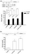
Replacement of the GC-box with a single Gal4 binding element and challenge with Gal4 fusion proteins supports a role for Sp3 in basal transcription. (a) A schematic diagram of the Gal4-0.27GCH1-GL3 luciferase reporter construct in which the 22 bp GC-box has been replaced with a single 19 bp Gal4 cis-element. Fusion proteins of the Gal4 DNA binding domain and the activation domains of Sp1, Sp3, and C/EBPβ are also shown. Luciferase assays of PC12 cells transiently transfected with 420 ng of Gal4-0.27GCH1-GL3, 40 ng of pRL-null, 150 ng Gal4-0, Gal4-Sp1, Gal4-Sp3 or Gal4-C/EBPβ or carrier plasmid DNA and then incubated with 5 mmol/L 8Br-cAMP (cAMP) for 4 h. The insert shows control activity plotted on a smaller scale. Data were analyzed by ANOVA with post hoc Bonferroni tests (*p < 0.05 vs. Gal4-0 control). (b) A schematic diagram of the 5XGal4-heterologous minimal luciferase reporter pFR-luc. Luciferase assays of PC12 cells transiently transfected with 420 ng 5XGal4-heterologous minimal luciferase reporter, 40 ng pRL-null, 150 ng Gal4-0, Gal4-Sp1 or Gal4-Sp3 and carrier plasmid DNA. Data were analyzed by ANOVA with post hoc Bonferroni tests (*p < 0.05 vs. Gal4-0).
Fig. 8
Fig. 8
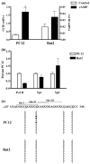
Cells that differ 10-fold in GCH1 gene expression have equal amounts of Sp1 and Sp3 associated with the native promoter and similar patterns of CpG methylation within the GC-box. (a) GCH1 mRNA levels in control cells (control) and cells treated with 5 mmol/L 8Br-cAMP for 4 h (cAMP). PC12 cells (left) and Rat2 cells (right) show 10-fold differences in basal and 47-fold differences in cAMP-stimulated transcription (note the scale change). Data were analyzed by ANOVA with post hoc Bonferroni tests (*p < 0.05 vs. PC12 control). (b) ChIP with quantitative real-time PCR analysis shows that equivalent amounts of Sp1 and Sp3 are associated with the GCH1 proximal promoter in PC12 and Rat2 cells but that significantly less Pol II is associated with the Rat2 promoter. Data are presented as percentages of values obtained for PC12 cells. Data were analyzed by ANOVA with post hoc Bonferroni tests (*p < 0.05 vs. PC12 Pol II). (c) The GCH1 GC-box showing Sites I, II, and III above and CpG dinucleotides underlined below. The four CpG dinucleotides within or adjacent to the GC-box are marked by four vertical lines. Bisulfite-treated GCH1 promoter DNA was amplified by methylation-specific PCR using primers which amplify a 288 bp product encompassing the GC-box, CRE and CCAAT-box. The ten vertical circles represent the ten PCR clones sequenced for each cell line. Filled circles represent CpG dinucleotides in which the cytosine was found to be methylated.

Similar articles

Cited by

References

    1. Abou-Donia MM, Wilson SP, Zimmerman TP, Nichol CA, Viveros OH. Regulation of guanosine triphosphate cyclohydrolase and tetrahydrobiopterin levels and the role of the cofactor in tyrosine hydroxylation in primary cultures of adrenomedullary chromaffin cells. J Neurochem. 1986;46:1190–1199. - PubMed
    1. Ahlgren R, Suske G, Waterman MR, Lund J. Role of Sp1 in cAMP-dependent transcriptional regulation of the bovine CYP11A gene. J Biol Chem. 1999;274:19422–19428. - PubMed
    1. Anastasiadis PZ, Bezin L, Gordon LJ, Imerman B, Blitz J, Kuhn DM, Levine RA. Vasoactive intestinal peptide induces both tyrosine hydroxylase activity and tetrahydrobiopterin biosynthesis in PC12 cells. Neuroscience. 1998;86:179–189. - PubMed
    1. Bauer M, Suppman S, Meyer M, Hesslinger C, Gasser T, Widner HR, Ueffing M. Glial cell line-derived neurotrophic factor up-regulates GTP-cyclohydrolase I activity and tetrahydrobiopterin levels in primary dopaminergic neurons. J Neurochem. 2002;82:1300–1310. - PubMed
    1. Billon N, Carlisi D, Datto MB, van Grunsven L, Watt A, Wang X-F, Rudkin BB. Cooperation of Sp1 and p300 in the induction of the CDK inhibitor p21WAF1/CIP1 during NGF-mediated neuronal differentiation. Oncogene. 1999;18:2872–2882. - PubMed

Publication types

MeSH terms