Review
. 2008 Feb;192(2):273-85.
doi: 10.1111/j.1748-1716.2007.01805.x. Epub 2007 Nov 15.

The PC12 cell as model for neurosecretion

Affiliations
Review

The PC12 cell as model for neurosecretion

R H S Westerink et al. Acta Physiol (Oxf). 2008 Feb.

Abstract

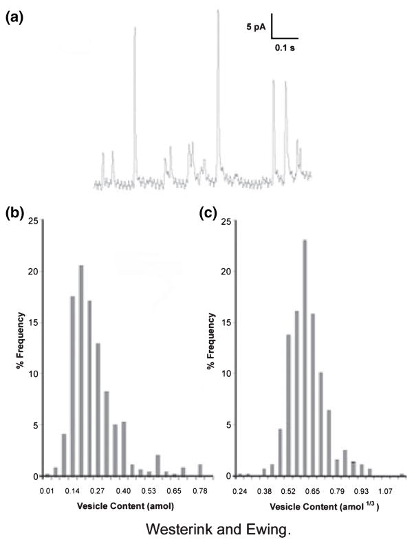
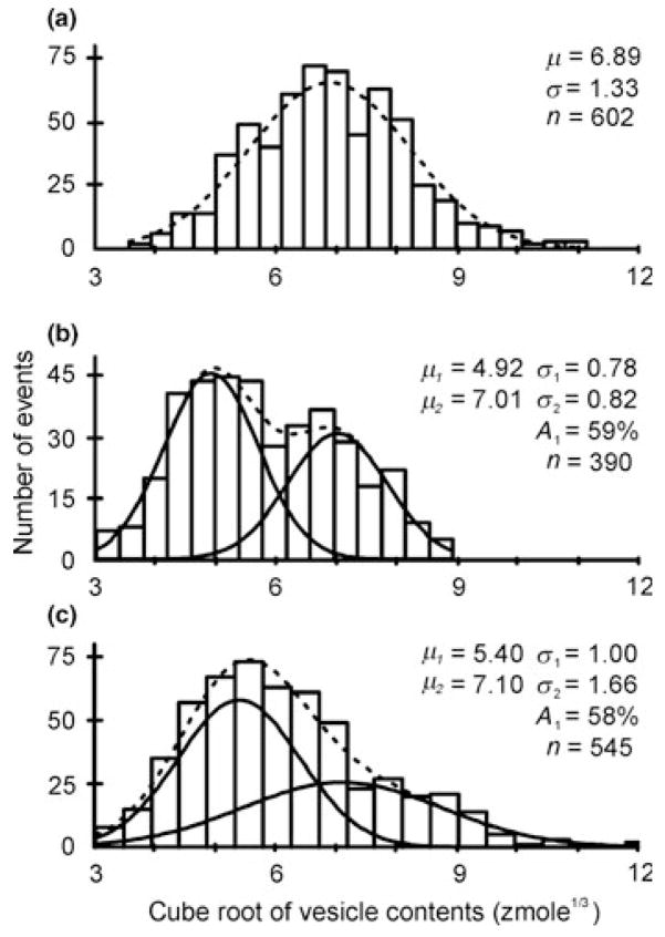
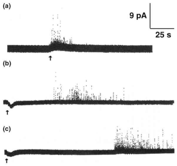
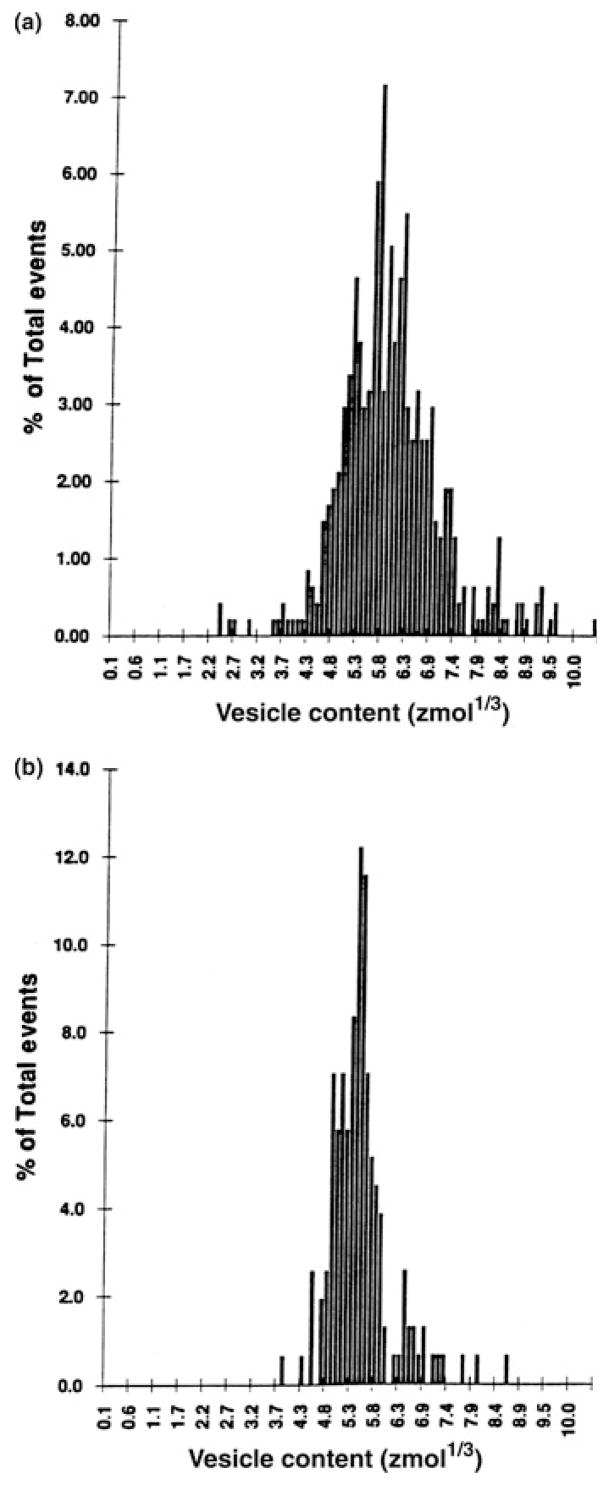
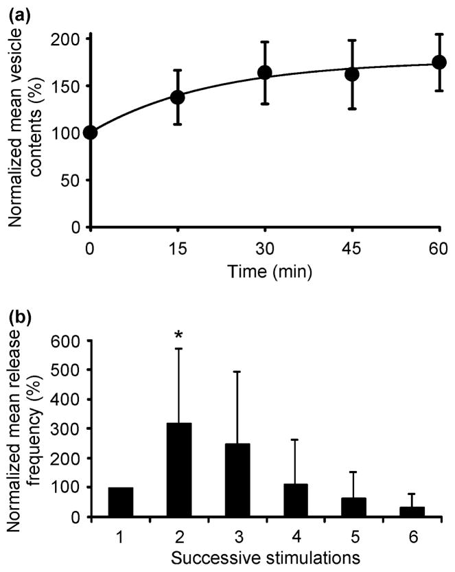
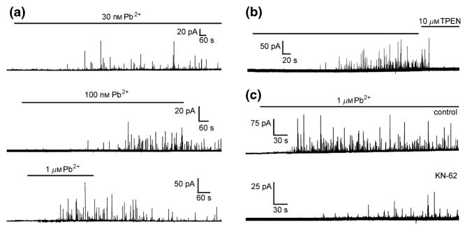
This review attempts to touch on the history and application of amperometry at PC12 cells for fundamental investigation into the exocytosis process. PC12 cells have been widely used as a model for neural differentiation and as such they have been used to examine the effects of differentiation on exocytotic release and specifically release at varicosities. In addition, dexamethasone-differentiated cells have been shown to have an increased number of releasable vesicles with increased quantal size, thereby allowing for an even broader range of applications including neuropharmacological and neurotoxicological studies. PC12 cells exhibiting large numbers of events have two distinct pools of vesicles, one about twice the quantal size of the other and each about half the total releasable vesicles. As will be outlined in this review, these cells have served as an extremely useful model of exocytosis in the study of the latency of stimulation-release coupling, the role of exocytotic proteins in regulation of release, effect of drugs on quantal size, autoreceptors, fusion pore biophysics, environmental factors, health and disease. As PC12 cells have some advantages over other models for neurosecretion, including chromaffin cells, it is more than likely that in the following decade PC12 cells will continue to serve as a model to study exocytosis.


Conflict of interest statement

Conflict of interest

There is no conflict of interest.

Figures

Figure 1
Figure 1
(a) Example of a current–time trace for exocytosis at a single undifferentiated PC12 cell following depolarization with KCl. The resulting current transients correspond to the oxidation of catecholamine at the electrode tip upon release from the cell. The area under each current transient is equivalent to the total charge produced by the oxidation of the catecholamine content of one vesicle. (b) Distribution of the amount of catecholamine released following potassium stimulation of undifferentiated PC12 cells. The total number of moles of catecholamine detected for each exocytosis event observed in the first 40 s of KCl-stimulated release is collected into bins and plotted as the per cent of the total number of vesicles undergoing exocytosis. (c) Cubed-root histogram for amperometric charges from PC12 cells shown in (b).
Figure 2
Figure 2
Distributions of the cube root of the vesicle contents of three single undifferentiated PC12 cells from which >300 events were recorded during repeated depolarizing with KCl. The distributions were best fitted by double Gaussian functions in 80% of the cells (see, e.g. b and c) and by a single Gaussian function for the cell in (a). The dashed lines represent the sum of the Gaussian distributions (solid lines) with the maximum-likelihood estimates of mean (μ) and variance (σ) indicated in each panel. The estimated number of small events (A1) is indicated as a percentage of the total number of events (n). Modified after Westerink et al. (2000).
Figure 3
Figure 3
Current–time traces for exocytosis at single PC12 cells. A 6-s ejection of stimulant (105 mM K+ (a), 1 mM nicotine (b) or 1 mM muscarine (c) from a microinjector was administered at each arrow. The resulting current transients correspond to the oxidation of dopamine at the electrode tip as it is released from the cell. Reproduced with permission from Zerby & Ewing (1996b).
Figure 4
Figure 4
Distribution of the cube root of vesicle catecholamine content for KCl-stimulated release plotted as the percent of the total number of vesicles undergoing exocytosis from (a) the soma of undifferentiated cells (n = 17 cells, 475 events) and from (b) varicosities of NGF-differentiated PC12 cells (n = 16 cells, 156 events). Reproduced with permission from Zerby & Ewing (1996a).
Figure 5
Figure 5
The mean vesicle content of undifferentiated PC12 cells, determined following KCl-induced exocytosis, increased with increasing duration of superfusion with saline containing 100 μM L-DOPA (a). Each point represents mean ± SD (n = 8 cells) and at all time points the mean vesicle contents was increased significantly when compared with control (t-test, P < 0.01). The drawn line is an exponential curve fitted to the data with a maximum increase amounting to 76% and an exponential time constant with an increase of 21 min. The graph in (b) depicts the relation between the KCl-induced release frequency and the number of successive stimulations at 10–15 min intervals, during which 100 μM L-DOPA-containing saline was superfused. The exocytotic frequency is expressed as a percentage of the value obtained during the first response (set at 100%), which was evoked before exposure to L-DOPA. Each bar represents mean ± SD (n = 8 cells). During the first stimulus after L-DOPA superfusion, the frequency of events was significantly higher than the control value (t-test, P < 0.05). Reproduced with permission from Westerink et al. (2000).
Figure 6
Figure 6
Effects of the heavy metal Pb2+ on vesicular catecholamine release from ionomycin-permeabilized dexamethasone-differentiated PC12 cells. (a) Amperometric recordings from permeabilized PC12 cells superfused with Ca2+-free saline containing 5 μM ionomycin and 0.03, 0.1 and 1 μM Pb2+, demonstrating the concentration dependence of Pb2+-induced exocytosis. (b) Amperometric recording from a permeabilized PC12 cells showing vesicular catecholamine release during superfusion with nominal Ca2+-free saline containing 1 μM Pb2+. The membrane-permeable heavy metal chelator TPEN rapidly reduces the intracellular Pb2+ concentration below the threshold for release, whereas the membrane-impermeable chelator EGTA is ineffective (not shown), demonstrating the Ca2+-independence of Pb2+-evoked exocytosis. (c) Amperometric recordings from ionomycin-permeabilized PC12 cells reveal that saline containing 1 μM Pb2+ induced vesicular release (control), which is strongly inhibited by co-application of an inhibitor of CaM kinase II (10 μM KN-62), whereas inhibition of Protein Kinase C does not appear to affect Pb2+-induced exocytosis (not shown). Modified after Westerink & Vijverberg (2002a) and Westerink et al. (2002).

Similar articles

Cited by

References

    1. Albillos A, Dernick G, Horstmann H, Almers W, Alvarez de Toledo G, Lindau M. The exocytotic event in chromaffin cells revealed by patch amperometry. Nature. 1997;389:509–512. - PubMed
    1. Alvarez de Toledo G, Fernandez-Chacon R, Fernandez JM. Release of secretory products during transient vesicle fusion. Nature. 1993;363:554–558. - PubMed
    1. Amatore C, Arbault S, Bonifas I, Bouret Y, Erard M, Ewing AG, Sombers LA. Correlation between vesicle quantal size and fusion pore release in chromaffin cell exocytosis. Biophys J. 2005;88:4411–4420. - PMC - PubMed
    1. Barclay JW, Morgan A, Burgoyne RD. Calcium-dependent regulation of exocytosis. Cell Calcium. 2005;38:343–353. - PubMed
    1. Berridge MJ, Irvine RF. Inositol trisphosphate, a novel second messenger in cellular signal transduction. Nature. 1984;312:315–321. - PubMed

Publication types

Substances