. 2007 Apr 19;1(2):109-19.
doi: 10.1016/j.chom.2007.03.001.

A Staphylococcus aureus regulatory system that responds to host heme and modulates virulence

Affiliations

A Staphylococcus aureus regulatory system that responds to host heme and modulates virulence

Victor J Torres et al. Cell Host Microbe. .

Abstract

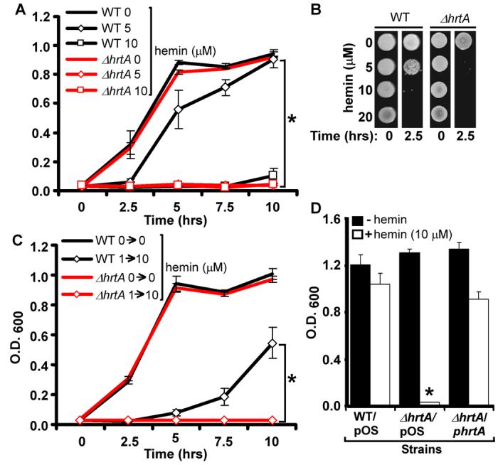
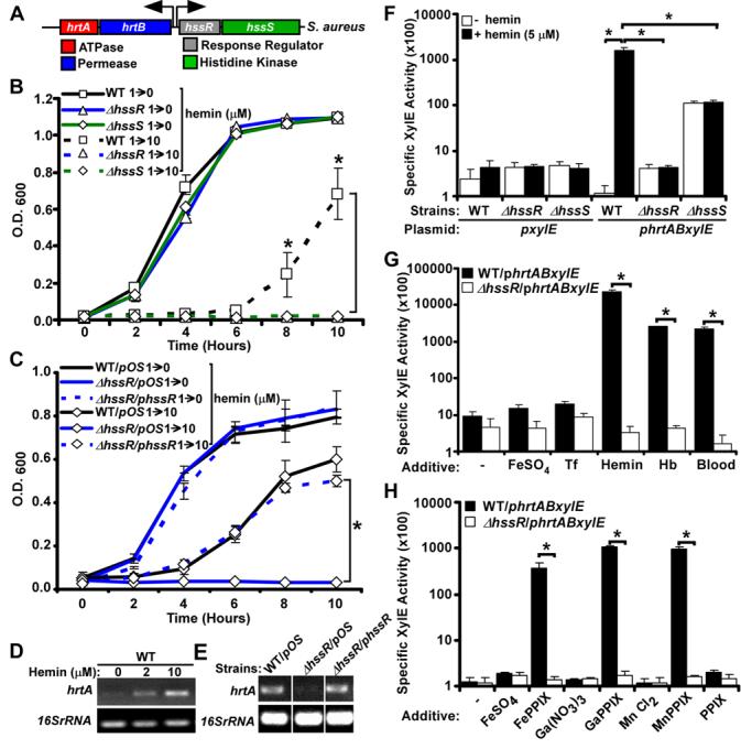
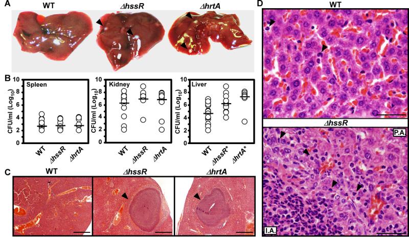
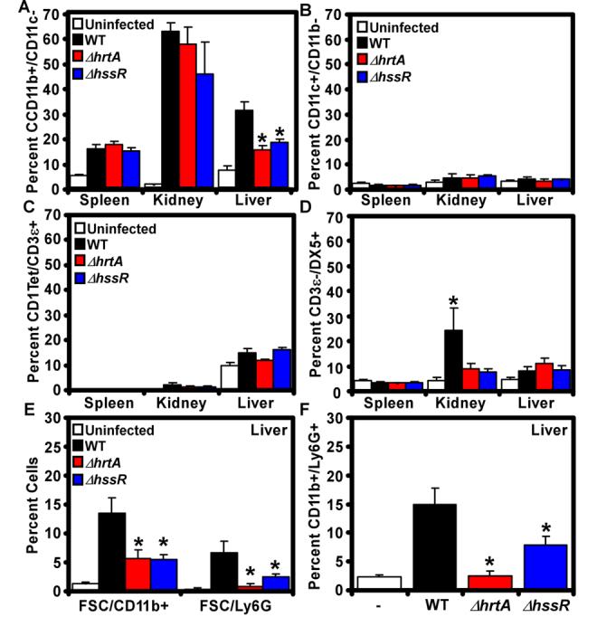
Staphylococcus aureus, a bacterium responsible for tremendous morbidity and mortality, exists as a harmless commensal in approximately 25% of humans. Identifying the molecular machinery activated upon infection is central to understanding staphylococcal pathogenesis. We describe the heme sensor system (HssRS) that responds to heme exposure and activates expression of the heme-regulated transporter (HrtAB). Inactivation of the Hss or Hrt systems leads to increased virulence in a vertebrate infection model, a phenotype that is associated with an inhibited innate immune response. We suggest that the coordinated activity of Hss and Hrt allows S. aureus to sense internal host tissues, resulting in tempered virulence to avoid excessive host tissue damage. Further, genomic analyses have identified orthologous Hss and Hrt systems in Bacillus anthracis, Listeria monocytogenes, and Enterococcus faecalis, suggesting a conserved regulatory system by which Gram-positive pathogens sense heme as a molecular marker of internal host tissue and modulate virulence.


Figures

Figure 1
Figure 1. S. aureus adapt to avoid the toxic effects of hemin
A) S. aureus wildtype and ΔsrtA were grown O/N in TSB and then subcultured into TSB supplemented with 0, 5, 10, or 20 μM hemin. Growth rates were determined by measuring the absorbance (O.D. 600) at the indicated time points. B) S. aureus wildtype was grown O/N in TSB supplemented with 0 or 1 μM hemin (1→) and then subcultured into TSB supplemented with 0 or 10 μM hemin (→0 or →10). The results represent the mean ± S.D. from triplicate experiments. Asterisks denote statistically significant differences as determined by Student's t test (p≤0.05).
Figure 2
Figure 2. The HrtAB system is required for staphylococcal heme adaptation
A) S. aureus wildtype and ΔhrtA were grown O/N in TSB and then subcultured into TSB supplemented with 0, 5, or 10 μM hemin. Growth rates were determined by measuring the absorbance (O.D. 600) at the indicated time points. B) S. aureus wildtype and ΔhrtA were grown O/N in TSB and then subcultured for 2.5 hrs into TSB supplemented with 0, 5, 10 or 20 μM hemin. The cultures were then serially diluted and plated on TSA plates. C) S. aureus wildtype and ΔhrtA were grown O/N in TSB and TSB supplemented with 1 μM hemin (1→) and then subcultured into TSB supplemented with 0 or 10 μM hemin (→0 or →10). Growth rates were determined by measuring the absorbance (O.D. 600) at the indicated time points (hrs). D) S. aureus wildtype and ΔhrtA transformed with vector alone (WT/pOS and ΔhrtA/pOS) and ΔhrtA transformed with vector containing a wildtype copy of hrtA (ΔhrtA/phrtA) were grown in TSB supplemented with 1 μM hemin and then subcultured into TSB supplemented with +/− 10 μM hemin and grown for 15 hrs. The results represent the mean ± S.D. from triplicate experiments. Asterisks denote statistically significant differences as determined by Student's t test (p≤0.05).
Figure 3
Figure 3. HssRS is required for hrtA expression upon exposure to hemin
A) Schematic representation of the hrtAB and hssRS loci in S. aureus. B-C) Listed staphylococcal strains were grown O/N in TSB supplemented with 1 μM heme (1→) and then subcultured into TSB supplemented with 0 or 10 μM heme (→0 or →10). Growth rates were determined by measuring the absorbance (O.D.600) at the indicated time points (hrs). D-E) RT-PCR analyses. D) Total RNA was extracted from O/N cultures of S. aureus wildtype grown in TSB supplemented with 0, 2 or 10 μM hemin. cDNA was synthesized as described in Experimental Procedures and transcription of the hrtA gene and the 16sRNA (loading control) was assessed by PCR. E) Total RNA was extracted from O/N cultures of wildtype (WT/pOS), ΔhssR/pOS, and the complemented ΔhssR strain (ΔhssR/phssR) grown in TSB supplemented with 2 μM hemin. The cDNA was synthesized as described above and transcription of the hrtA gene and the 16sRNA (loading control) was determined as in panel D. Differences in the relative level of RTPCR product between panels D and E are likely a result of the required inclusion of chloramphenicol to the growth media in experiments shown in panel E. F-H) XylE fusion reporter assay. F) WT, ΔhssR, and ΔhssS transformed with the phrtABxylE or the pxylE plasmid were grown 2 hrs in TSB supplemented with 0 or 5 μM hemin and XlyE activity was determined as described in Experimental Procedures. G) Wildtype and ΔhssR harboring the phrtABxylE reporter plasmid were grown 2 hrs in TSB supplemented with FeSO4 (8 μM), transferrin (Tf; 8 μM), hemin (8 μM), hemoglobin (Hb; 2 μM), or mouse blood and XlyE activity was determined as in panel F. H) Wildtype and ΔhssR harboring the phrtABxylE reporter plasmid were grown 2 hrs in TSB supplemented with 1 μM of the indicated additives and the XlyE activity determined as in panel F. The results represent the mean ± S.D. from at least triplicate experiments. Asterisks denote statistically significant differences as determined by Student's t test (p<0.05).
Figure 4
Figure 4. S. aureus ΔhssR and ΔhrtA exhibit liver-specific hypervirulence
A) Photographs of livers dissected from BALB/c mice infected with wildtype and ΔhssR or ΔhrtA (1 × 10⁶ CFUs for all strains) 96 hours post infection. Arrowheads mark ΔhssR and ΔhrtA-induced hepatic abscesses. Photographs are representative of all livers analyzed. Abscesses were visible in virtually all livers from ΔhssR and ΔhrtA infected mice, while none were found in wildtype infected mice. B) S. aureus multiplication in infected mouse organs as measured by tissue homogenization, dilution, and colony formation on agar media 96 hours post infection. Each symbol represents data from one infected animal. The limit of detection in these experiments is 100 CFUs. The horizontal line denotes the mean of the log and the asterisks denote statistically significant differences from wildtype as determined by Student's t test (p≤0.05). C) Representative Hematoxylin and Eosin (H&E) staining of liver sections infected with WT, ΔhrtA, or ΔhssR strains at 40X magnification. Arrowheads mark ΔhssR- and ΔhrtA-induced hepatic abscesses. D) Representative H&E staining of liver sections infected with WT or ΔhssR strains at 1,000X magnification. Arrowheads mark PMNs in the tissues. P.A; proximal to the abscess and I.A; inside the abscess.
Figure 5
Figure 5. Infection with S. aureus ΔhssR or ΔhrtA inhibits innate immune responses
BALB/c animals were left uninfected or infected with wildtype, ΔhssR or ΔhrtA. Four days postinfection (A-D) or two days post infection (E-F) organs were dissected, homogenized, and the infiltration of the indicated immune cells was determined by multiparametric FACS analysis as described in Experimental Procedures. Isolated cells were stained for the detection of: A) phagocytes (B220/CD11b+/CD11c), B) dendritic cells (D11c+/CD11b), C) invariant natural killer T cells (iNKT: CD1 Tetramer (tet)+/B220/CD3ε+), D) natural killer cells (DX5+/B220/CD3ε), E) large CD11b+ and Ly6G+ cells (FSC/CD11b+ and FSC/Ly6G+), and F) granulocytes (B220/CD11b+/Ly6G+). Results represent the mean ± S.E. from at least three independent animals. Asterisks denote a statistically significant reduction in the detected cells compared to animals infected with the wildtype strain as determined by Student's t test (p<0.05).
Figure 6
Figure 6. Inactivation of the HrtAB system results in increased expression of secreted virulence factors
A) S. aureus wildtype (WT/pOS1), ΔhrtA mutant (ΔhrtA/pOS1), or ΔhrtA complemented strain (ΔhrtA/phrtA) were grown O/N at 37 °C with aeration for 15 hrs in RPMI supplemented with 0 or 1 μM hemin. Culture supernatants were collected, filtered, precipitated, and separated on 15% SDS-PAGE gels. Proteins were stained with colloidal blue. B) Indicated proteins were excised from the gel and subjected to mass spectrometry-based identification. The identities of the proteins are indicated with corresponding gene numbers from S. aureus strain COL shown in parentheses. C) Fold induction of the indicated genes as determined by transcriptional analyses comparing changes between ΔhrtA grown in the presence or absence of 1 μM hemin. Transcript levels not determined due to saturating levels of expression marked with N.D.. Experiments were performed in triplicate and asterisks denote statistical significance as determined by Student's t test (p<0.05).
Figure 7
Figure 7. Model for the role of HrtAB and HssRS in S. aureus pathogenesis
A) The Hrt and Hss systems are conserved across several Gram positive bacteria. Alignment of genomic sequences among Gram positive bacteria that contain orthologous hrtAB and hssRS systems. The numbers within each box represent corresponding gene numbers in the listed annotation. The numbers underneath each gene correspond to the percent amino acid identity to the representative S. aureus genes. Arrows denote the predicted direction of transcription. B) In S. aureus, heme internalized through cell wall anchored proteins (i), is sensed by HssS which subsequently activates HssR (ii). HssR then binds the promoter region upstream of hrtAB (iii), leading to increased expression and elaboration of the HrtAB efflux pump (iv). HrtAB then pumps surplus cytoplasmic heme out of the bacterium. C) Inactivation of hrtAB leads to the cytoplasmic accumulation of heme which increases cellular stress (v). Staphylococcal stress sensing systems are activated leading to an increase in the expression and/or secretion of virulence factors including exotoxins 3, 5 and 8, Map-w, fibronectin binding protein and FLIPr (vi), which increase liver-specific hypervirulence through inhibiting immune cell recruitment.

Comment in

Similar articles

Cited by

References

    1. Anderson KL, Roberts C, Disz T, Vonstein V, Hwang K, Overbeek R, Olson PD, Projan SJ, Dunman PM. Characterization of the Staphylococcus aureus heat shock, cold shock, stringent, and SOS responses and their effects on log-phase mRNA turnover. J. Bacteriol. 2006;188:6739–6756. - PMC - PubMed
    1. Bader MW, Sanowar S, Daley ME, Schneider AR, Cho U, Xu W, Klevit RE, Le Moual H, Miller SI. Recognition of antimicrobial peptides by a bacterial sensor kinase. Cell. 2005;122:461–472. - PubMed
    1. Bae T, Banger AK, Wallace A, Glass EM, Aslund F, Schneewind O, Missiakas DM. Staphylococcus aureus virulence genes identified by bursa aurealis mutagenesis and nematode killing. Proc. Natl. Acad. Sci. U S A. 2004;101:12312–12317. - PMC - PubMed
    1. Bae T, Schneewind O. Allelic replacement in Staphylococcus aureus with inducible counter-selection. Plasmid. 2005 - PubMed
    1. Beier D, Gross R. Regulation of bacterial virulence by two-component systems. Curr. Opin. Microbiol. 2006;9:143–152. - PubMed

Publication types